Catching It Early: The Silent Warning Signs of Pancreatic Cancer

warning signs of pancreatic cancer

Catching It Early: The Silent Warning Signs of Pancreatic Cancer

Learn the silent warning signs of pancreatic cancer, early symptoms, risk factors, diagnosis, and treatment options to improve the chances of early detection.

Introduction

Pancreatic cancer is often referred to as the “silent disease” since it starts developing without noticeable symptoms. The lack of early symptoms often leads to late diagnosis of this condition when the patient experiences pain or other complications associated with an already advanced stage of cancer development. Recognizing early warning signs and knowing about possible risk factors will allow diagnosing this cancer at the earliest stage, which would help a lot with choosing the best treatment strategy and increasing chances of successful recovery.

Even though pancreatic cancer is rather uncommon among all cancer types, this disease ranks among the most deadly cancerous conditions. Progress in medical sciences and new diagnostic methods, personalized treatments, and targeted therapies allow gradually improving survival rates for this type of cancer.

The current paper discusses in detail pancreatic cancer, including its causes, anatomy, symptoms, diagnosis, treatment, and the life of patients who deal with this disease.

What Is Pancreatic Cancer?

Pancreatic cancer represents a condition when some cells start growing uncontrollably and forming tumors in the pancreas. Cancerous cells not only invade surrounding tissues but may metastasize and affect other parts of the body, for example, liver, lungs, or abdomen.

The pancreas is an important organ placed behind the stomach. This organ serves two major functions:

  • Production of enzymes that help to digest food
  • Production of hormones, such as insulin and glucagon, that help to control the level of blood sugar

The appearance of cancer in the pancreas may influence both digestion and regulation of blood sugar levels.

One of the main features of pancreatic cancer is the absence of early symptoms.

Types of Pancreatic Cancer

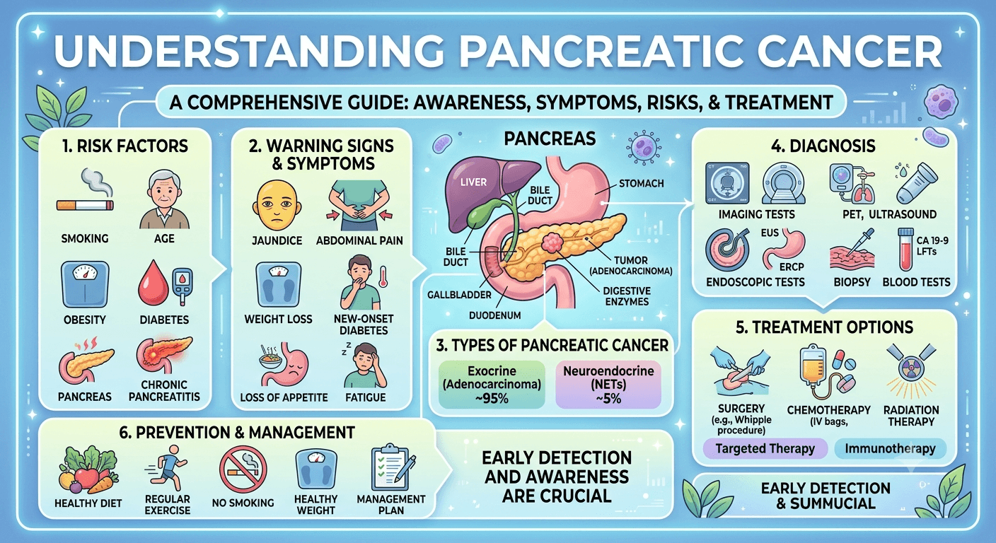
Pancreatic cancer is broadly classified into two major categories.

1. Exocrine Pancreatic Cancer

Approximately 90–95% of pancreatic cancers originate from the exocrine cells that produce digestive enzymes.

The most common type is:

Pancreatic Ductal Adenocarcinoma (PDAC)

  • Begins in the ducts carrying digestive enzymes
  • Most aggressive form
  • Responsible for the majority of pancreatic cancer deaths

Other rare exocrine cancers include:

  • Acinar cell carcinoma
  • Adenosquamous carcinoma
  • Squamous cell carcinoma

2. Neuroendocrine Pancreatic Tumors (PNETs)

These cancers develop from hormone-producing endocrine cells.

Some produce excessive hormones, causing noticeable symptoms, while others remain non-functional.

Examples include:

  • Insulinomas
  • Gastrinomas
  • Glucagonomas
  • VIPomas

Neuroendocrine tumors generally grow more slowly and often have a better prognosis than pancreatic adenocarcinoma.

Anatomy of the Pancreas
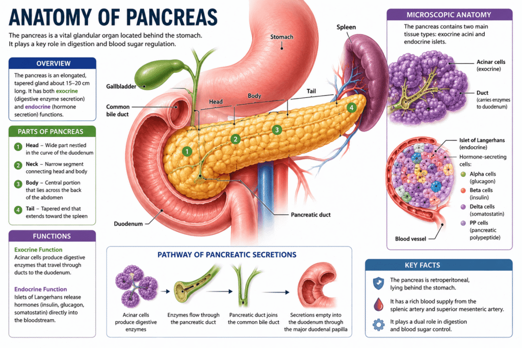
Anatomy of Pancreas

The pancreas is a long, flat gland measuring about 15–20 cm (6–8 inches) in length.

It consists of three major sections:

Head

  • Located near the small intestine (duodenum)
  • Most pancreatic cancers develop here

Body

  • Middle portion extending across the abdomen

Tail

  • Extends toward the spleen
  • Tumors here are often detected later because they cause fewer early symptoms

Functions of the Pancreas

Exocrine Function

Produces digestive enzymes including:

  • Amylase
  • Lipase
  • Proteases

These enzymes help digest carbohydrates, fats, and proteins.

Endocrine Function

Produces hormones including:

  • Insulin
  • Glucagon
  • Somatostatin

These regulate blood glucose and metabolism.

Pathophysiology

Pancreatic cancer develops through a gradual accumulation of genetic mutations that transform healthy pancreatic cells into malignant ones.

The process often begins with precancerous lesions known as:

  • PanIN (Pancreatic Intraepithelial Neoplasia)
  • IPMN (Intraductal Papillary Mucinous Neoplasm)
  • MCN (Mucinous Cystic Neoplasm)

Over time, mutations in genes such as:

  • KRAS
  • TP53
  • CDKN2A
  • SMAD4

allow cells to:

  • Divide uncontrollably
  • Resist normal cell death
  • Invade nearby tissues
  • Spread through blood vessels and lymphatics

Pancreatic tumors also create a dense fibrous environment that limits blood supply and makes chemotherapy less effective.

Silent Warning Signs of Pancreatic Cancer

One reason pancreatic cancer is so dangerous is that symptoms may initially seem mild or unrelated.

Persistent Upper Abdominal Pain

Pain often develops in the upper abdomen and may radiate to the back.

It may worsen:

  • After eating
  • While lying flat

Unexplained Weight Loss

Unexpected weight loss is one of the earliest warning signs.

Patients often notice:

  • Loss of appetite
  • Feeling full quickly
  • Muscle wasting

Jaundice

Tumors in the pancreatic head can block the bile duct.

Symptoms include:

  • Yellow skin
  • Yellow eyes
  • Dark urine
  • Pale stools
  • Itchy skin

Digestive Problems

Many patients develop:

  • Nausea
  • Vomiting
  • Indigestion
  • Bloating
  • Fatty stools
  • Diarrhea

New-Onset Diabetes

Suddenly developing diabetes after age 50 without obvious risk factors can occasionally be an early indicator of pancreatic cancer.

Fatigue

Persistent tiredness often accompanies cancer progression.

Blood Clots

Some patients experience unexplained blood clots before their cancer diagnosis.

Back Pain

Pain spreading into the middle or lower back may occur as tumors enlarge.

Causes and Risk Factors

The exact cause of pancreatic cancer is not always known. It develops when genetic mutations cause pancreatic cells to grow uncontrollably, forming a tumor. Several lifestyle, environmental, and inherited factors can increase the risk of developing the disease.

Common risk factors include:

  • Smoking: The leading preventable risk factor, significantly increasing the likelihood of pancreatic cancer.
  • Older age: Most cases occur in people over 60 years of age.
  • Family history: Having close relatives with pancreatic cancer raises the risk.
  • Inherited genetic syndromes: Mutations in genes such as BRCA1, BRCA2, PALB2, CDKN2A, STK11, and PRSS1 can increase susceptibility.
  • Chronic pancreatitis: Long-term inflammation of the pancreas increases cancer risk, particularly hereditary pancreatitis.
  • Diabetes mellitus: Long-standing type 2 diabetes and newly diagnosed diabetes in older adults may be associated with pancreatic cancer.
  • Obesity: Excess body weight contributes to chronic inflammation and metabolic changes that promote cancer development.
  • Unhealthy diet: Diets high in processed meats, red meat, and saturated fats and low in fruits and vegetables may increase risk.
  • Heavy alcohol consumption: Excessive alcohol intake can lead to chronic pancreatitis, an established risk factor.
  • Physical inactivity: A sedentary lifestyle contributes indirectly through obesity and insulin resistance.
  • Occupational exposure: Long-term exposure to certain chemicals used in the petroleum, metalworking, and pesticide industries may increase risk.
  • Race and ethnicity: Incidence is higher in some populations, including people of African ancestry.
  • Male sex: Men have a slightly higher risk than women, partly due to higher rates of smoking and occupational exposures.

Diagnosis

Because symptoms are often vague, multiple diagnostic tools are used.

Medical History and Physical Examination

Doctors evaluate:

  • Symptoms
  • Weight changes
  • Family history
  • Risk factors

Blood Tests

May include:

  • Liver function tests
  • RBS, HbA1c
  • CRP, ESR
  • Kidney function test
  • S. Amylase and S. Lipase
  • Blood sugar levels
  • CA 19-9 tumor marker

CA 19-9 alone cannot diagnose pancreatic cancer but helps monitor treatment.

Imaging Studies

Common imaging tests include:

  • CT scan
  • MRI
  • MRCP
  • PET scan
  • Ultrasound

Endoscopic Ultrasound (EUS)

Allows detailed imaging while obtaining tissue samples through biopsy.

Biopsy

A biopsy confirms the diagnosis by examining tissue under a microscope.

Treatment Options

Treatment depends on:

  • Stage
  • Tumor location
  • Overall health
  • Cancer type

Surgery

Surgery offers the best chance for cure when cancer is localized.

Common procedures include:

Whipple Procedure

Removes:

  • Pancreatic head
  • Duodenum
  • Gallbladder
  • Part of the bile duct

Distal Pancreatectomy

Removes:

  • Pancreatic body
  • Tail
  • Sometimes the spleen

Total Pancreatectomy

Entire pancreas removed.

Patients require lifelong insulin and digestive enzyme replacement.

Therapy

Chemotherapy

Common chemotherapy regimens include:

  • FOLFIRINOX
  • Gemcitabine
  • Gemcitabine plus nab-paclitaxel

Chemotherapy may be given:

  • Before surgery
  • After surgery
  • For advanced disease

Radiation Therapy

High-energy radiation destroys cancer cells.

It may:

  • Shrink tumors
  • Reduce pain
  • Improve surgical outcomes

Targeted Therapy

Patients with specific genetic mutations may benefit from targeted medications.

Examples include:

  • PARP inhibitors for BRCA mutations
  • NTRK inhibitors for rare gene fusions

Immunotherapy

Although only effective in a small percentage of patients, immunotherapy can be beneficial for tumors with specific genetic characteristics such as mismatch repair deficiency.

Medications

Several medications help manage symptoms and improve quality of life.

These include:

Pain medications

  • Acetaminophen
  • NSAIDs
  • Opioids

Pancreatic enzyme replacement

Improves digestion and nutrient absorption.

Insulin

For diabetes caused by pancreatic dysfunction.

Anti-nausea medications

Help reduce chemotherapy-related nausea.

Appetite stimulants

May improve nutritional intake.

Anticoagulants

Used if blood clots develop.

Living with Pancreatic Cancer

The condition influences one’s physical, psychological, social and economic aspects of life. However, people are able to live an enriched life, retain their connections and relationships, and feel well with proper treatment and assistance.

Eating Right

It is hard to eat properly due to problems with digestion and lack of appetite.

Useful measures comprise:

  • Eating frequently and in small portions
  • Foods with high amount of protein
  • Taking pancreatic enzyme preparations
  • Hydration
  • Meeting with a nutritionist

Dealing with Fatigue

Fatigue is a typical problem.

There are some things that patients may find useful:

  • Smooth physical activity
  • Getting enough rest
  • Conserving energy
  • Balanced nutrition

Being Mentally Healthy

Anxiety, sadness, or fear may be felt. One may use counseling services, take part in discussion groups, engage in meditation and speak to relatives and friends.

Palliative Treatment

It helps to alleviate symptoms, control pain, and improve life quality. It can be used not only when cancer is terminal but also alongside active treatment.

Regular Check-Ups

They help to evaluate one’s progress in treatment, handle side effects and detect early signs of disease recurrence.

Frequently Asked Questions (FAQs)

Is pancreatic cancer hereditary?

Most cases are not inherited, but approximately 10% are associated with inherited genetic mutations or strong family history.

Can pancreatic cancer be cured?

When detected early and completely removed surgically, some patients can achieve long-term survival. However, many cases are diagnosed at an advanced stage.

Who is most at risk?

People over 60 years of age, smokers, individuals with obesity, chronic pancreatitis, diabetes, or a family history of pancreatic cancer have a higher risk.

Does pancreatic cancer always cause pain?

No. Early pancreatic cancer may not cause pain or any noticeable symptoms.

Can lifestyle changes reduce risk?

Maintaining a healthy weight, avoiding tobacco, limiting alcohol consumption, eating a balanced diet, and staying physically active may reduce the risk.

What are the survival rates?

Survival depends on the stage at diagnosis. Patients diagnosed when the disease is localized generally have significantly better outcomes than those diagnosed after the cancer has spread.

Conclusion

Pancreatic cancer continues to be one of the toughest cancers because it develops subtly with symptoms that could be misunderstood as something else. However, increased awareness of its hidden signs including sudden weight loss, constant abdominal or back pain, yellow skin or eyes, digestive problems, and diabetes could help people visit their doctor earlier and perhaps even save their lives.

With the development of better surgical procedures, chemotherapies, radiation therapies, targeted therapies, supportive care, and personalized medicine, many people are surviving longer. Nutritional support, pain relief, mental counseling, and palliative care are equally important during treatment.

If you have symptoms or some risk factors for developing the disease, it is crucial to visit your doctor without delay. Early detection, timely diagnosis, and comprehensive treatment are the best way of fighting this type of cancer.

Read More

Disclaimer: The information provided in this blog post is for educational and informational purposes only and should not be considered medical advice. It is not intended to replace professional medical consultation, diagnosis, or treatment.
Blood in Vomit: 10 Causes, Risks, and Warning Signs

Blood in vomit

Blood in Vomit: 10 Causes, Risks, and Warning Signs

Is blood in vomit an emergency? Learn 10 potential causes, understand the risks, and find out exactly which warning signs require immediate medical attention.

Introduction

Bleeding in vomit may cause considerable anxiety in many patients. It may appear in different forms such as bright red, dark clots or like coffee grounds and it always indicates the development of a bleed somewhere in the upper gastrointestinal tract. Though sometimes bleeding is rather benign, in other cases, it may indicate serious conditions and requires immediate medical intervention.

As a healthcare specialist, I usually make my patients understand that it is essential to know about possible causes and warning signs to obtain adequate treatment. From the viewpoint of a medical student, hematemesis is an important symptom which may indicate certain disorders in the esophagus, stomach, or duodenum.

This paper will consider 10 main causes of blood in vomit, their risks, warning signs, methods of diagnostics and treatment.

Disclaimer: The information provided in this blog post is for educational and informational purposes only and should not be considered medical advice.

Understanding Blood in Vomit

Blood in vomit occurs when bleeding develops within the:

  • Esophagus
  • Stomach
  • Duodenum (first part of the small intestine)
  • Occasionally the mouth, throat, or respiratory tract

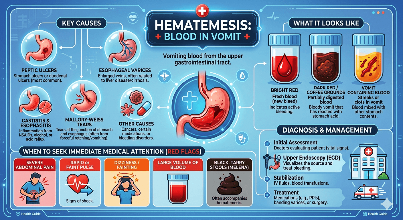
The appearance of blood can provide important diagnostic clues:

AppearancePossible Meaning
Bright red bloodActive or recent bleeding
Dark red bloodSlower bleeding
Coffee-ground appearanceBlood exposed to stomach acid
Blood mixed with foodStomach-related bleeding

Not all red-colored vomit contains blood. Certain foods, drinks, or medications may mimic the appearance of blood, but any suspicion of true bleeding warrants medical evaluation.

10 Causes of Blood in Vomit

1. Peptic Ulcers

Peptic ulcers are among the most common causes of hematemesis.

These sores develop in the lining of the:

  • Stomach
  • Duodenum

Common causes include:

  • Infection with Helicobacter pylori
  • Long-term use of NSAIDs (such as ibuprofen or naproxen)
  • Excessive smoking
  • Chronic alcohol consumption

Why Ulcers Bleed

An ulcer can erode into nearby blood vessels. When this happens, bleeding may range from mild to severe.

Symptoms

  • Burning abdominal pain
  • Nausea
  • Bloating
  • Black stools
  • Vomiting blood

2. Gastritis

Gastritis refers to inflammation of the stomach lining.

When severe, inflamed tissues may bleed, resulting in blood-stained vomit.

Common Causes

  • Alcohol abuse
  • NSAID medications
  • Stress-related illness
  • Autoimmune disorders
  • Infections

Symptoms

  • Upper abdominal discomfort
  • Nausea
  • Loss of appetite
  • Vomiting
  • Blood in vomit

Mild gastritis may resolve with treatment, but severe bleeding requires urgent care.

3. Esophageal Varices

Esophageal varices are enlarged veins within the esophagus.

They commonly occur in patients with:

Why They Are Dangerous

These veins have thin walls and can rupture suddenly.

Clinical Features

  • Massive vomiting of blood
  • Dizziness
  • Rapid heart rate
  • Low blood pressure
  • Shock

This condition represents a true medical emergency and carries significant mortality if untreated.

4. Mallory-Weiss Tear

A Mallory-Weiss tear is a laceration at the junction of the esophagus and stomach.

It often occurs after:

  • Severe vomiting
  • Persistent retching
  • Excessive coughing
  • Heavy alcohol use

Symptoms

  • Bright red blood in vomit
  • Chest discomfort
  • Abdominal pain
  • History of repeated vomiting

Most tears heal naturally, though severe bleeding occasionally requires endoscopic treatment.

5. Esophagitis

Esophagitis is inflammation of the esophagus.

Potential Causes

  • Acid reflux disease (GERD)
  • Certain medications
  • Infections
  • Radiation therapy

Chronic irritation may damage blood vessels and lead to bleeding.

Symptoms

  • Painful swallowing
  • Heartburn
  • Chest discomfort
  • Blood in vomit

6. Stomach Cancer

Although less common than ulcers or gastritis, stomach cancer can cause gastrointestinal bleeding.

Risk Factors

  • Smoking
  • Family history
  • Chronic gastritis
  • H. pylori infection

Warning Signs

  • Unexplained weight loss
  • Persistent stomach pain
  • Early fullness after meals
  • Fatigue
  • Blood in vomit

Early diagnosis significantly improves outcomes.

7. Esophageal Cancer

Cancer of the esophagus may cause progressive bleeding as tumors invade nearby tissues.

Symptoms

  • Difficulty swallowing
  • Weight loss
  • Chest pain
  • Chronic reflux
  • Blood in vomit

Bleeding is often gradual but can occasionally become severe.

8. Severe Gastroenteritis

Intense vomiting caused by viral or bacterial infections may damage the lining of the upper gastrointestinal tract.

Repeated forceful vomiting can:

  • Irritate tissues
  • Cause minor tears
  • Trigger bleeding

Associated Symptoms

  • Diarrhea
  • Fever
  • Dehydration
  • Abdominal cramps

Small streaks of blood may appear after repeated episodes of vomiting.

9. Swallowed Blood

Sometimes the blood originates outside the digestive tract.

Examples Include

  • Nosebleeds
  • Dental bleeding
  • Oral injuries
  • Throat injuries

Blood may be swallowed unknowingly and later appear in vomit.

Healthcare providers distinguish this from true gastrointestinal bleeding through history-taking and examination.

10. Blood-Clotting Disorders

Conditions affecting normal blood clotting can increase bleeding risk throughout the body, including the digestive tract.

Examples

  • Hemophilia
  • Liver failure
  • Low platelet counts
  • Anticoagulant medication use

Even minor irritation may produce noticeable bleeding in susceptible individuals.

Risk Factors for Vomiting Blood

Several factors increase the likelihood of hematemesis.

Lifestyle Factors

  • Heavy alcohol use
  • Smoking
  • Poor diet
  • Chronic NSAID use

Medical Conditions

  • Liver disease
  • Portal Hypertension
  • Type 2 Diabetes
  • Acid reflux
  • Peptic ulcer disease
  • Blood-clotting disorders

Medication Risks

Certain drugs increase bleeding risk:

  • Aspirin
  • Ibuprofen
  • Naproxen
  • Warfarin
  • Apixaban
  • Rivaroxaban

Medical students should always obtain a detailed medication history when evaluating gastrointestinal bleeding.

Warning Signs That Require Immediate Medical Attention

Some symptoms indicate potentially severe blood loss and should never be ignored.

Seek Emergency Care If Blood in Vomit Is Accompanied By:

  • Large amounts of blood
  • Black, tarry stools
  • Severe abdominal pain
  • Chest pain
  • Difficulty breathing
  • Fainting
  • Confusion
  • Pale skin
  • Rapid heartbeat
  • Low blood pressure

These symptoms may indicate significant internal bleeding requiring urgent intervention.

How Doctors Diagnose the Cause

Accurate diagnosis requires a systematic approach.

Medical History

Doctors typically ask about:

  • Timing of bleeding
  • Quantity of blood
  • Associated symptoms
  • Medication use
  • Alcohol consumption

Physical Examination

Assessment includes:

  • Vital signs
  • Abdominal examination
  • Signs of liver disease
  • Evidence of anemia

Laboratory Tests

Common investigations include:

  • Complete blood count (CBC)
  • Coagulation studies
  • Liver function tests
  • Lipid profile
  • LDH
  • KFT
  • Viral markers for Hepatitis
  • Blood chemistry panels

Upper Endoscopy

Upper gastrointestinal endoscopy remains the most valuable diagnostic tool.

It allows physicians to:

  • Visualize the bleeding source
  • Diagnose ulcers
  • Identify varices
  • Obtain biopsies
  • Deliver treatment

Treatment Options

Treatment depends on the underlying cause and severity.

Emergency Stabilization

For significant bleeding:

  • Intravenous fluids
  • Blood transfusions
  • Oxygen support
  • Monitoring in hospital
  • Endoscopic Band Ligation (EVL)

Medications

Doctors may prescribe:

  • Proton pump inhibitors
  • Beta Blockers (carvedilol, nadolol)
  • Antibiotics for H. pylori
  • Acid-reducing medications
  • Drugs to control portal hypertension (beta blockers)
  • Vasoactive Drugs (terlipressin or Octreotide)

Endoscopic Therapy

Endoscopy can stop bleeding through:

  • Clipping
  • Injection therapy
  • Thermal coagulation
  • Band ligation of varices

Surgery

Surgery may be necessary when:

  • Endoscopic treatment fails
  • Cancer is present
  • Severe ulcers continue to bleed

Frequently Asked Questions (FAQs)

Is blood in vomit always serious?

Not always. Small amounts of blood may result from irritation or minor tears. However, because it can signal severe bleeding, medical evaluation is recommended.

What does coffee-ground vomit mean?

Coffee-ground vomit usually indicates blood that has been partially digested by stomach acid, suggesting upper gastrointestinal bleeding.

Can stress cause blood in vomit?

Stress alone rarely causes bleeding, but severe illness-related stress can contribute to gastritis and ulcer formation.

When should I go to the emergency room?

Seek emergency care immediately if vomiting blood is accompanied by dizziness, fainting, rapid heartbeat, breathing difficulty, or large-volume bleeding.

Can acid reflux cause blood in vomit?

Yes. Severe reflux can cause esophageal inflammation and bleeding, although major bleeding is uncommon.

How is vomiting blood different from coughing up blood?

Vomiting blood originates from the digestive tract, while coughing up blood usually comes from the lungs or airways.

Can children vomit blood?

Yes. Causes may include swallowed blood, infections, gastritis, or rare gastrointestinal disorders. Pediatric evaluation is important.

Is blood in vomit a sign of cancer?

Sometimes, but cancer is a less common cause than ulcers, gastritis, or tears. Persistent symptoms require investigation.

Conclusion

Bloody vomit is an alarming sign that must not be taken lightly. Whereas gastritis, small tears, or swallowed blood might lead to minor bleeding, peptic ulcers, esophageal varices, liver diseases, and gastrointestinal cancers are among some serious illnesses.

For patients, the most critical point is that any hematemesis is to be checked immediately, especially in case of accompanying weakness, dizzy spells, black stools, or ongoing bleeding. For medical students, hematemesis will always be one of the critical clinical presentations.

Timely diagnosis and therapy are vital, which means knowing causes, risks, and warning signs is essential for everyone.

Read More

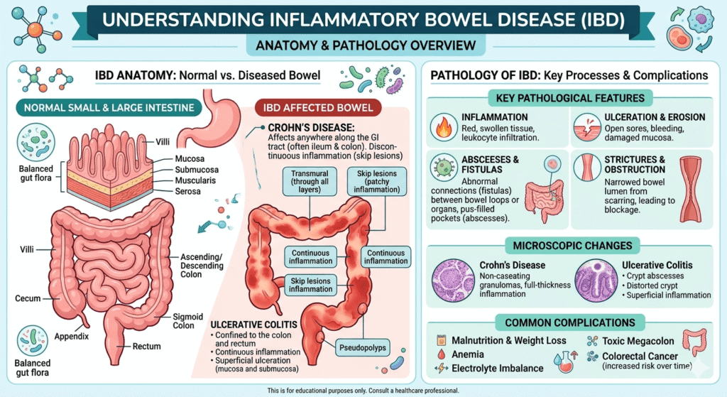
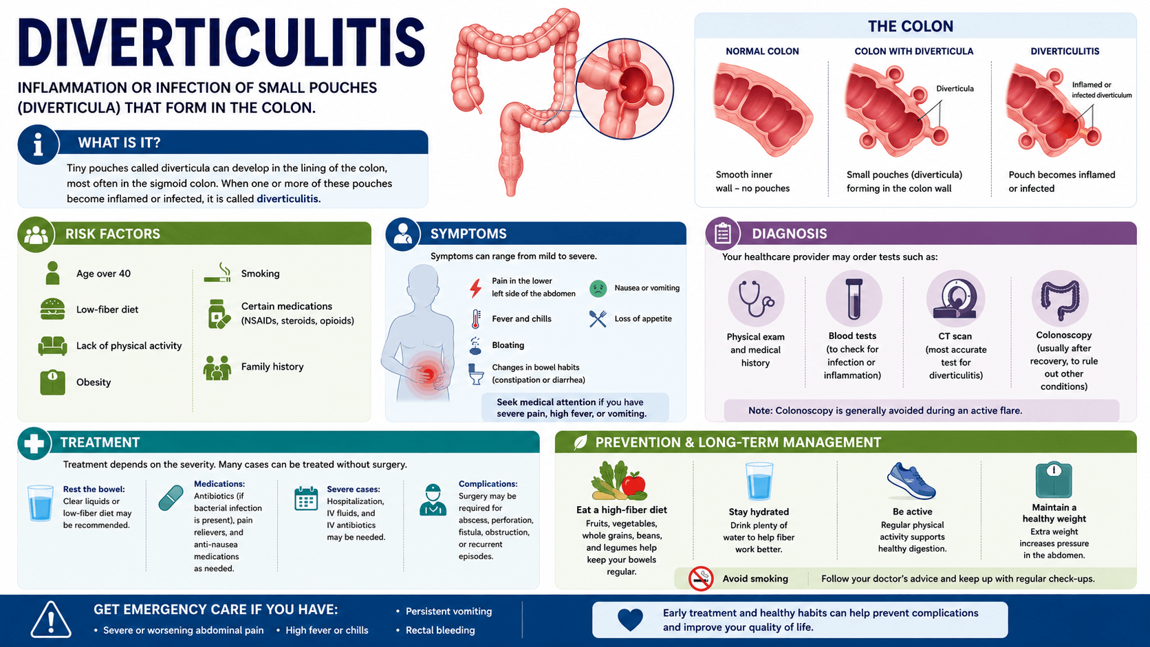
What Is Diverticulitis? Signs, Risk Factors, and Treatment options

what is Diverticulitis

What Is Diverticulitis? Signs, Risk Factors, and Treatment options

Learn about what is diverticulitis, including its symptoms, causes, diagnosis, treatment options, complications, and prevention strategies in this comprehensive guide.

Introduction

Diverticulitis is a digestive disease that can result in significant pain in the abdomen and, at times, lead to severe complications. In patients, knowledge of the disease helps minimize their anxiety levels and directs them towards the right treatment. In medical students, on the other hand, diverticulitis is a good example of how structural changes in the colon can lead to inflammation and infection.

Learn about diverticulitis, including its causes, symptoms, risk factors, diagnosis, treatment options, and prevention strategies. Discover when to seek medical care and how lifestyle changes can help reduce the risk of complications.

As a health practitioner, I have observed that, although most patients know what diverticulitis is, they do not understand its nature, causative agents, and treatment measures. This article presents an in-depth overview of diverticulitis, covering aspects such as causes, symptoms, risk factors, diagnosis, treatment modalities, and preventive measures for the disease.

Understanding Diverticulitis

Diverticulitis develops when small pouches forming on the wall of the colon become inflamed or infected.

For you to understand diverticulitis better, it is important to learn about diverticulosis.

What Is Diverticulosis?

Diverticulosis is a disease characterized by the formation of small, bulging sacs referred to as diverticula along the lining of the large intestine (colon). These sacs are most prevalent in the sigmoid colon, the lower section of the large intestine.

Diverticulosis is a very common disease and becomes more prevalent as people age. Most individuals with diverticula do not experience any symptoms.

What Is Diverticulitis?

If there is inflammation or infection of the diverticula, the condition is referred to as diverticulitis.

There may be different stages of the inflammation, ranging from simple irritation to a serious infection; complications of the condition include abscess formation, perforation, or blockage of the intestines.

Anatomy and Pathophysiology

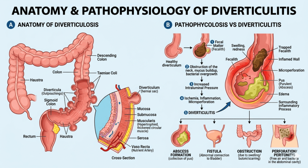
Anatomy and pathophysiology of diverticulitis

To understand diverticulitis, it is essential to first understand the anatomy of the colon.

The colon (large intestine), is the final portion of the gastrointestinal tract. It consists of:

  • Cecum
  • Ascending colon
  • Transverse colon
  • Descending colon
  • Sigmoid colon
  • Rectum

Diverticulitis most commonly affects the sigmoid colon, the S-shaped segment located just before the rectum. This region experiences relatively high intraluminal pressure during bowel movements, making it particularly susceptible to the formation of diverticula.

Development of Diverticula

Diverticula form due to herniation of the colon wall through weak spots at high pressure.

Increased intra-colonic pressure plays an important role in the development of the disease.

Usually, they are formed in places where there is a penetration of blood vessels into the intestinal wall.

Mechanism of Developing Diverticulitis

Although the mechanism of development is unknown, several factors could be contributing:

  • Obstruction of the diverticulum with stools;
  • Inflammation;
  • Gut microflora changes;
  • Tiny hole formation in the wall of the diverticulum;
  • Decreased blood supply to the damaged area.

Signs and Symptoms of Diverticulitis

The presentation of diverticulitis can vary from mild to severe.

Common Symptoms

The most frequent symptoms include:

  • Abdominal pain
  • Tenderness over the affected area
  • Fever
  • Nausea
  • Vomiting
  • Loss of appetite
  • Changes in bowel habits

Characteristic Pain Location

In Western populations, diverticulitis most commonly affects the sigmoid colon, causing:

However, right-sided diverticulitis may occur and is relatively more common in some Asian populations.

Gastrointestinal Symptoms

Patients may experience:

  • Constipation
  • Diarrhea
  • Bloating
  • Excessive gas
  • Difficulty passing stool

Systemic Symptoms

More significant inflammation may produce:

  • Fever
  • Chills
  • Fatigue
  • General malaise

When Symptoms Suggest a Medical Emergency

Certain symptoms may indicate serious complications and require immediate medical attention.

These include:

  • Severe abdominal pain
  • High fever
  • Persistent vomiting
  • Signs of sepsis
  • Abdominal rigidity
  • Inability to pass stool or gas
  • Rectal bleeding
  • Dizziness or fainting

Prompt evaluation is essential because delayed treatment can increase the risk of life-threatening complications.

Risk Factors for Diverticulitis

Not everyone with diverticulosis develops diverticulitis. Several factors increase the likelihood of inflammation.

Age

Risk increases with advancing age.

Although diverticulosis is increasingly recognized in younger adults, it remains most common in individuals over 50 years old.

Dietary Factors

A diet low in dietary fiber may contribute to:

  • Increased colonic pressure
  • Altered bowel habits
  • Changes in gut microbial composition

Low consumption of fruits, vegetables, legumes, and whole grains has been associated with a greater risk of diverticular disease.

Obesity

Excess body weight is a significant risk factor.

Obesity is associated with:

  • Increased inflammation throughout the body
  • Higher rates of complicated diverticulitis
  • Increased likelihood of recurrence

Physical Inactivity

Regular exercise supports healthy bowel function and may reduce the risk of diverticular disease complications.

Sedentary lifestyles are associated with a higher incidence of diverticulitis.

Smoking

Smoking appears to increase:

  • Risk of diverticulitis
  • Risk of perforation
  • Risk of recurrent disease

Medication Use

Certain medications may increase susceptibility, including:

  • Nonsteroidal anti-inflammatory drugs (NSAIDs)
  • Corticosteroids
  • Opioids
  • Some immunosuppressive agents

Genetic Factors

Family history plays a role.

Studies suggest that genetic predisposition contributes significantly to the development of diverticular disease and its complications.

Complications of Diverticulitis

While many cases are uncomplicated, some patients develop serious complications.

Abscess

An abscess is a localized collection of pus near the infected diverticulum.

Symptoms may include:

  • Persistent fever
  • Ongoing abdominal pain
  • Elevated inflammatory markers

Perforation

A perforation occurs when the intestinal wall ruptures.

This can lead to:

  • Peritonitis
  • Severe infection
  • Septic shock

Perforation is a medical emergency.

Fistula Formation

A fistula is an abnormal connection between the colon and another structure.

Common examples include:

  • Colon-to-bladder fistula
  • Colon-to-vagina fistula
  • Colon-to-skin fistula

Patients may experience recurrent infections or passage of gas through unusual routes.

Bowel Obstruction

Repeated inflammation can cause scarring and narrowing of the colon, resulting in partial or complete obstruction.

Recurrent Diverticulitis

Some patients experience repeated episodes, which may affect quality of life and influence treatment decisions.

How Diverticulitis Is Diagnosed

Accurate diagnosis is important because many other conditions can mimic diverticulitis.

Medical History

Clinicians assess:

  • Location of pain
  • Duration of symptoms
  • Previous episodes
  • Associated gastrointestinal symptoms
  • Medication use

Physical Examination

The examination often reveals:

  • Localized abdominal tenderness
  • Guarding
  • Signs of systemic infection

Laboratory Tests

Common investigations include:

  • Complete blood count (CBC)
  • C-reactive protein (CRP)
  • ESR
  • Electrolytes
  • Kidney function tests

Elevated white blood cell counts and inflammatory markers often support the diagnosis.

Computed Tomography (CT) Scan

CT imaging is considered the gold standard for diagnosing diverticulitis.

A CT scan can identify:

  • Inflamed diverticula
  • Abscesses
  • Perforations
  • Obstruction
  • Fistulas

It also helps determine disease severity and guides treatment planning.

Colonoscopy

Colonoscopy is generally avoided during an acute attack because of the risk of perforation.

However, it is often recommended several weeks after recovery to:

  • Exclude colorectal cancer
  • Evaluate the extent of diverticular disease
  • Investigate persistent symptoms

Classification of Diverticulitis

Uncomplicated Diverticulitis

This form involves localized inflammation without major complications.

Characteristics include:

  • Mild to moderate symptoms
  • No abscess
  • No perforation
  • No obstruction

Most cases fall into this category.

Complicated Diverticulitis

Complicated disease includes:

  • Abscess formation
  • Perforation
  • Fistula
  • Obstruction
  • Generalized peritonitis

These patients often require hospitalization and more aggressive treatment.

Treatment Options for Diverticulitis

Treatment depends on disease severity, patient characteristics, and the presence of complications.

Management of Mild Uncomplicated Diverticulitis

Many patients can be treated at home.

Treatment may include:

  • Rest
  • Adequate hydration
  • Pain management
  • Temporary dietary modifications

Current evidence suggests that antibiotics may not always be necessary for carefully selected patients with mild uncomplicated diverticulitis.

Clinical judgment remains essential.

Dietary Recommendations During Recovery

During acute symptoms, physicians may recommend:

  1. Clear liquids initially
  2. Gradual advancement to low-fiber foods
  3. Return to a balanced high-fiber diet after recovery

Examples of clear liquids include:

  • Water
  • Broth
  • Gelatin
  • Oral rehydration solutions

Antibiotic Therapy

Antibiotics may be prescribed when:

  • Symptoms are moderate to severe
  • Patients are immunocompromised
  • Significant systemic infection is present
  • Complications are suspected

The choice of antibiotic depends on local guidelines and patient-specific factors.

Hospital Treatment

Hospitalization may be necessary for patients with:

  • Severe pain
  • High fever
  • Inability to tolerate oral intake
  • Significant comorbidities
  • Complicated diverticulitis

Hospital-based treatment often includes:

  • Intravenous fluids
  • Intravenous antibiotics
  • Pain control
  • Monitoring for complications

Interventional Procedures

Percutaneous Abscess Drainage

For larger abscesses, radiologists may insert a drainage catheter under CT or ultrasound guidance.

Benefits include:

  • Control of infection
  • Reduced need for emergency surgery

Faster recovery

Surgical Treatment

Surgery is reserved for selected situations.

Indications for Surgery

Potential indications include:

  • Perforation
  • Generalized peritonitis
  • Large abscesses not responding to drainage
  • Recurrent severe attacks
  • Obstruction
  • Fistula formation

Types of Surgical Procedures

Colon Resection

The diseased segment of colon is removed and healthy sections are reconnected.

Hartmann Procedure

In emergency situations, surgeons may:

  • Remove the affected colon segment
  • Create a temporary colostomy
  • Restore bowel continuity later if feasible

The specific operation depends on patient stability and disease severity.

Frequently Asked Question (FAQ)

What is the difference between diverticulosis and diverticulitis?

Diverticulosis refers to the presence of small pouches (diverticula) in the wall of the colon. Most people with diverticulosis have no symptoms. Diverticulitis occurs when one or more of these pouches become inflamed or infected, leading to symptoms such as abdominal pain, fever, and digestive disturbances.

What are the most common symptoms of diverticulitis?

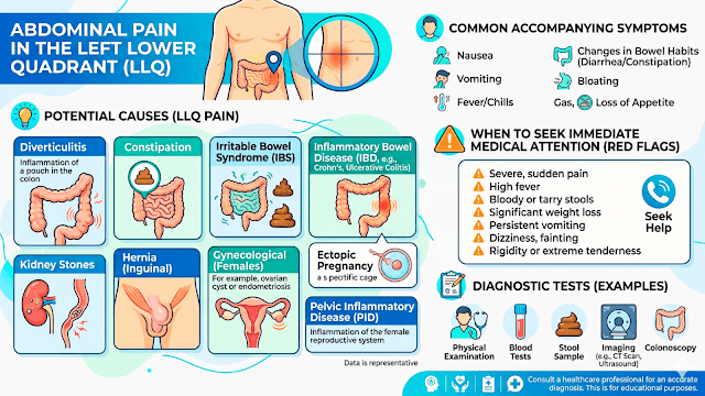
The most common symptom is persistent pain in the lower left side of the abdomen. Other symptoms may include:
Fever and chills
Nausea or vomiting
Constipation or diarrhea
Abdominal tenderness
Bloating and gas
Symptoms can range from mild discomfort to severe illness requiring hospitalization.

What causes diverticulitis and who is at risk?

The exact cause is not fully understood, but factors that increase the risk include:
Age over 40 years
A low-fiber diet
Obesity
Physical inactivity
Smoking
Regular use of certain medications, such as NSAIDs and steroids
A family history of diverticular disease
These factors may contribute to inflammation or infection of diverticula.

How is diverticulitis diagnosed?

Doctors diagnose diverticulitis using a combination of:
Medical history and physical examination
Blood tests to look for signs of infection or inflammation
Imaging studies, especially a CT scan of the abdomen and pelvis, which is the most accurate test for confirming diverticulitis and identifying complications
A colonoscopy is usually performed after recovery to evaluate the colon and rule out other conditions.

What are the treatment options for diverticulitis?

Treatment depends on the severity of the condition:
Mild cases: Rest, increased fluid intake, and dietary modifications
Moderate cases: Oral antibiotics may be prescribed when appropriate
Severe cases: Hospitalization, intravenous antibiotics, and close monitoring
Complicated cases: Surgery may be necessary if there is an abscess, perforation, fistula, or recurrent severe attacks

Conclusion

Fortunately, most patients are diagnosed with uncomplicated diverticulitis and manage to get cured without serious complications. New scientific research helps to understand the condition better and allows individual approaches to treatment with the use of medication, imaging tests, and surgery. Early diagnosis, appropriate evaluation, and a healthy way of life are considered the keys to success.

Disclaimer: The information provided in this blog post is for educational and informational purposes only and should not be considered medical advice. It is not intended to replace professional medical consultation, diagnosis, or treatment.
Read more https://www.niddk.nih.gov/health-information/digestive-diseases/diverticulosis-diverticulitis

Dysphagia: Causes, Symptoms, Diagnosis, Treatment, and When to Worry

dysphagia

Dysphagia: Causes, Symptoms, Diagnosis, Treatment, and When to Worry

This article describes the phenomenon of dysphagia in detail, explaining its causes, possible symptoms, diagnostic methods, potential complications, available treatments, and the criteria requiring immediate medical assistance.

Introduction

Dysphagia is an extremely frightening condition for many people. Indeed, most people can relate to occasionally feeling like food went down the wrong pipe or getting stuck in their throats. On the other hand, the repeated occurrence, progression, or worsening of swallowing difficulties may suggest dysphagia.

As a healthcare professional, I would want to assure my patients that dysphagia is a condition rather than a disease. This condition may appear because of various conditions ranging from minor issues, such as gastroesophageal reflux disease, to major neurological disorders or cancers. Being aware of dysphagia is especially crucial since swallowing is a complicated process that one may underestimate until experiencing some issues.

This article describes the phenomenon of dysphagia in detail, explaining its causes, possible symptoms, diagnostic methods, potential complications, available treatments, as well as the criteria requiring immediate medical assistance. Thus, this article will not only provide patients with essential information concerning dysphagia but also give medical students a comprehensive foundation of knowledge.

What Is Dysphagia?

https://www.youtube.com/watch?v=d6Hmq3prOIM

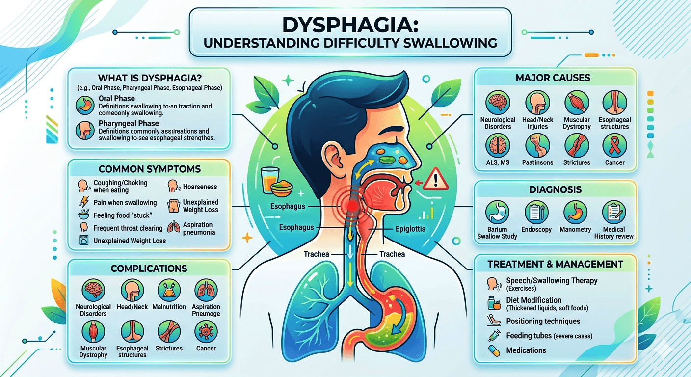
The word dysphagia means difficulty swallowing. This condition implies any problems related to the movement of food, beverages, and even saliva from the mouth to the stomach.

Swallowing involves the coordination of more than 30 muscles and several nerves, which makes this action a rather complex process that might be disrupted, leading to dysphagia.

People with dysphagia may experience numerous symptoms, including:

  • Food being trapped in the throat area
  • Sticking of food in the area below the breastbone
  • Difficulties starting swallowing
  • Choking on food
  • Coughing during meals
  • Feeling of pain during swallowing
  • Ground-up regurgitation of food

Although anyone can have dysphagia, this problem usually affects older people owing to age-specific factors and higher prevalence of neurological diseases among older adults.

Types of Dysphagia

Healthcare providers generally classify dysphagia into two major categories.

Oropharyngeal Dysphagia

This type involves difficulty initiating a swallow.

Patients may experience:

  • Coughing during meals
  • Choking episodes
  • Nasal regurgitation
  • Difficulty moving food from the mouth into the throat

Common causes include neurological disorders and muscle diseases.

Esophageal Dysphagia

This occurs when food has difficulty passing through the esophagus into the stomach.

Patients often describe:

  • Food sticking in the chest
  • Pressure behind the breastbone
  • Regurgitation after swallowing

Structural abnormalities and esophageal motility disorders are common causes.

Common Causes of Dysphagia

The causes of dysphagia can be divided into neurological, muscular, structural, inflammatory, and functional disorders.

Neurological Causes

The brain and nervous system play a crucial role in swallowing.

Common neurological causes include:

  • Stroke
  • Parkinson’s disease
  • Multiple sclerosis
  • Dementia
  • Amyotrophic lateral sclerosis (ALS)
  • Brain tumors
  • Traumatic brain injury

Stroke remains one of the most common causes of dysphagia worldwide.

Muscular Disorders

Muscles responsible for swallowing may become weak or dysfunctional.

Examples include:

  • Myasthenia gravis
  • Muscular dystrophy
  • Polymyositis
  • Dermatomyositis

These conditions often cause fatigue-related swallowing difficulties.

Structural Causes

Physical narrowing or blockage of the swallowing pathway can impair food passage.

Examples include:

  • Esophageal strictures
  • Esophageal rings
  • Webs of tissue
  • Tumors
  • Enlarged thyroid gland
  • Cervical osteophytes (bone spurs)

Gastroesophageal Reflux Disease (GERD)

Chronic acid reflux can damage the esophagus.

Repeated inflammation may lead to:

  • Scarring
  • Narrowing (stricture formation)
  • Difficulty swallowing solid foods

Achalasia

Achalasia is a rare disorder in which the lower esophageal sphincter fails to relax properly.

Symptoms include:

  • Difficulty swallowing solids and liquids
  • Regurgitation
  • Chest discomfort
  • Weight loss

Esophageal Cancer

Cancer of the esophagus is an important cause of progressive dysphagia.

Typically:

  • Difficulty swallowing solids appears first
  • Progresses to difficulty swallowing liquids
  • Significant weight loss may occur

Any progressively worsening dysphagia should be investigated promptly.

Eosinophilic Esophagitis

This chronic inflammatory condition is increasingly recognized, especially among younger individuals.

Patients often report:

  • Food sticking in the esophagus
  • Recurrent food impactions
  • History of allergies or asthma

Risk Factors for Dysphagia

Several factors increase the likelihood of developing swallowing difficulties.

Age

Older adults are more vulnerable due to:

  • Reduced muscle strength
  • Neurological disease
  • Medication side effects

Neurological Disease

Conditions affecting the brain and nerves significantly increase risk.

Head and Neck Cancer

Both cancer and its treatment can impair swallowing.

Radiation Therapy

Radiation can cause scarring and stiffness in swallowing structures.

Chronic Reflux

Long-standing GERD can lead to strictures and esophageal damage.

Symptoms of Dysphagia

Symptoms vary depending on the underlying cause.

Typical Symptoms

  • Difficulty swallowing
  • Sensation of food sticking
  • Choking during meals
  • Coughing while eating
  • Excessive throat clearing
  • Regurgitation of food
  • Drooling
  • Hoarseness

Associated Symptoms

Some symptoms provide clues to the underlying diagnosis.

SymptomPossible Cause
HeartburnGERD
Weight lossCancer, severe dysphagia
Nasal regurgitationNeurological disorders
Chest painEsophageal spasm, achalasia
Progressive symptomsStructural blockage or cancer
Recurrent pneumoniaAspiration

Complications of Dysphagia

Untreated dysphagia can lead to serious health problems.

Aspiration

Food or liquid enters the airway instead of the esophagus.

This may result in:

  • Coughing
  • Breathing difficulties
  • Lung infections

Aspiration Pneumonia

Aspiration pneumonia is one of the most dangerous complications.

It occurs when bacteria-containing material enters the lungs.

Symptoms include:

Malnutrition

Patients may avoid eating due to fear of choking.

Consequences include:

  • Weight loss
  • Muscle wasting
  • Weakness
  • Vitamin deficiencies

Dehydration

Difficulty swallowing liquids can result in inadequate fluid intake.

How Dysphagia Is Diagnosed

A careful evaluation is essential.

Medical History

The clinician will ask questions such as:

  • When did symptoms begin?
  • Are solids, liquids, or both affected?
  • Is swallowing painful?
  • Is there weight loss?
  • Are choking episodes occurring?

The answers often narrow the diagnostic possibilities.

Physical Examination

Examination focuses on:

  • Neurological function
  • Oral cavity
  • Neck
  • Voice quality
  • Signs of malnutrition

Diagnostic Tests

Barium Swallow Study

The patient swallows a contrast material while X-rays are taken.

This test can identify:

  • Narrowing
  • Obstructions
  • Motility disorders

Videofluoroscopic Swallow Study

Often performed by speech-language pathologists.

This test evaluates:

  • Oral phase function
  • Aspiration risk
  • Coordination of swallowing muscles

Upper Endoscopy (EGD)

A flexible camera is inserted through the mouth into the esophagus.

It allows physicians to identify:

  • Inflammation
  • Ulcers
  • Tumors
  • Strictures

Biopsies can be obtained if needed.

Esophageal Manometry

This test measures pressure and muscle contractions within the esophagus.

It is particularly useful for diagnosing:

  • Achalasia
  • Esophageal spasms
  • Motility disorders

CT Scan or MRI

Imaging may be necessary when tumors or neurological disorders are suspected.

Treatment of Dysphagia

Treatment depends entirely on the underlying cause.

Swallowing Therapy

Speech-language pathologists play a central role.

Therapy may include:

  • Swallowing exercises
  • Postural techniques
  • Compensatory strategies
  • Muscle-strengthening exercises

Dietary Modifications

Food consistency may be adjusted.

Examples include:

  • Pureed foods
  • Soft foods
  • Thickened liquids

These modifications help reduce aspiration risk.

Medications

Certain medications may improve symptoms.

Examples:

  • Proton pump inhibitors for GERD
  • Steroids for eosinophilic esophagitis
  • Medications targeting muscle disorders

Endoscopic Procedures

Some structural abnormalities can be treated endoscopically.

Examples include:

  • Dilation of strictures
  • Removal of food impactions
  • Treatment of esophageal rings

Surgery

Surgical intervention may be required for:

  • Cancer
  • Severe achalasia
  • Large diverticula
  • Certain structural abnormalities

Feeding Tubes

In severe cases, temporary or permanent feeding tubes may be necessary to maintain nutrition and hydration.

Dysphagia in Older Adults

Swallowing difficulties are particularly common among elderly individuals.

Contributing factors include:

  • Reduced muscle strength
  • Frailty
  • Stroke
  • Parkinson’s disease
  • Dementia

Management often requires a multidisciplinary approach involving physicians, speech therapists, dietitians, and caregivers.

Dysphagia in Children

Children can also experience dysphagia.

Possible causes include:

  • Congenital abnormalities
  • Neurological disorders
  • Developmental delays
  • Gastroesophageal reflux

Warning signs include:

  • Poor weight gain
  • Feeding difficulties
  • Frequent respiratory infections

Early evaluation is essential.

When to Worry About Dysphagia

Many cases of dysphagia require medical evaluation, but certain symptoms demand urgent attention.

Seek immediate medical care if you experience:

Food Completely Stuck in the Esophagus

This may represent an emergency requiring endoscopic removal.

Difficulty Breathing

Breathing problems combined with swallowing difficulties require urgent assessment.

Rapid Weight Loss

Unexplained weight loss may indicate a serious underlying condition.

Progressively Worsening Symptoms

Increasing difficulty swallowing over weeks or months should never be ignored.

Recurrent Aspiration or Pneumonia

Repeated lung infections suggest food or liquid is entering the airway.

Blood in Vomit or Stools

These symptoms may indicate significant gastrointestinal disease.

Persistent Pain While Swallowing

Painful swallowing can signal infection, inflammation, or malignancy.

Prevention and Lifestyle Measures

Although not all cases can be prevented, certain strategies may reduce risk.

Eat Slowly

Taking smaller bites improves swallowing safety.

Chew Thoroughly

Proper chewing reduces choking risk.

Stay Upright During Meals

Remain seated upright while eating and for at least 30 minutes afterward.

Manage Acid Reflux

Early treatment of GERD can prevent complications.

Maintain Good Oral Hygiene

This reduces bacterial load and lowers aspiration pneumonia risk.

Regular Medical Follow-Up

Patients with neurological disorders should undergo periodic swallowing assessments.

Frequently Asked Questions (FAQs)

Is dysphagia a disease or a symptom?

Dysphagia is a symptom indicating an underlying problem affecting swallowing.

Can stress cause dysphagia?

Stress and anxiety can contribute to a sensation of difficulty swallowing, often called globus sensation, but true dysphagia requires medical evaluation.

Is dysphagia always serious?

Not always. Some causes are relatively benign, but persistent or worsening symptoms should be assessed by a healthcare professional.

Can dysphagia be cured?

Many causes are treatable, and some are completely reversible. Outcomes depend on the underlying condition.

Which specialist treats dysphagia?

Depending on the cause, treatment may involve gastroenterologists, neurologists, otolaryngologists (ENT specialists), speech-language pathologists, and surgeons.

Can acid reflux cause swallowing problems?

Yes. Chronic acid reflux can damage the esophagus and lead to narrowing that makes swallowing difficult.

What foods are easier to swallow?

Soft foods such as yogurt, mashed potatoes, soups, oatmeal, and pureed foods are often easier for people with dysphagia.

Can dysphagia lead to pneumonia?

Yes. Aspiration of food or liquids into the lungs can cause aspiration pneumonia, a potentially serious complication.

Conclusion

Prompt diagnosis is crucial. In cases of coughing, difficulty in passing solid foods through the chest area, recurrent pneumonia, unexplained weight loss, and other possible symptoms, one needs to undergo an examination by a professional as soon as possible. Thankfully, nowadays, it is possible to treat many patients successfully, with the help of swallowing therapy, drugs, changes in diet, endoscopy, or surgery.

In terms of patients, knowing about dysphagia is necessary to receive the needed assistance. In terms of medical students, dysphagia should be considered an interesting clinical symptom combining gastroenterology, neurology, otorhinolaryngology, and physical rehabilitation medicine. No matter what caused this issue, the proper approach is always required.

Disclaimer: The information provided in this blog post is for educational and informational purposes only and should not be considered medical advice. It is not intended to replace professional medical consultation, diagnosis, or treatment.
Gastropathy vs. Gastritis: Key Differences Explained

Gastropathy vs. Gastritis

Gastropathy vs. Gastritis: Key Differences Explained

Gastropathy vs. Gastritis: Key Differences Explained clears up the frequent confusion between these two stomach conditions. While they share similar symptoms like nausea and pain, gastritis involves active stomach lining inflammation (often from infections), whereas gastropathy refers to lining damage without significant inflammation (typically caused by NSAIDs or alcohol).

Introduction

The terms Gastropathy and Gastritis are frequently used interchangeably in clinical practice and by patients, but they represent fundamentally different pathological processes. Understanding the distinction is important because the causes, microscopic findings, treatment strategies, and long-term implications may differ significantly.

Gastropathy vs. Gastritis: Key Differences Explained clears up the frequent confusion between these two stomach conditions. While they share similar symptoms like nausea and pain.

Gastritis refers specifically to inflammation of the gastric (stomach) mucosa, characterized by inflammatory cell infiltration within the stomach lining. It is therefore a disease process defined by inflammation.

Gastropathy, in contrast, refers to damage or injury to the gastric mucosa with little or no significant inflammation. It is primarily a morphological (structural) description based on endoscopic and histological findings rather than a specific inflammatory disease.

The purpose of this explanation is to clarify the key differences between gastropathy and gastritis, including their pathophysiology, causes, symptoms, diagnosis, and management.

Anatomy & Pathophysiology

Stomach Anatomy

Normal Anatomy of the Stomach

The stomach is a muscular organ located between the esophagus and the small intestine. Its primary functions include:

  • Storage of food
  • Mechanical digestion
  • Secretion of gastric acid and digestive enzymes
  • Regulation of food passage into the duodenum

The stomach wall consists of several layers:

  1. Mucosa—the innermost layer containing epithelial cells, gastric glands, and protective mucus-producing cells.
  2. Submucosa—contains blood vessels, lymphatics, and nerves.
  3. Muscularis propria—responsible for stomach contractions.
  4. Serosa—the outer covering.

The gastric mucosa is protected by a sophisticated defense system that includes:

  • Mucus secretion
  • Bicarbonate production
  • Adequate blood flow
  • Tight epithelial junctions
  • Rapid cell regeneration

When these protective mechanisms fail, mucosal injury develops.

YouTube

Pathophysiology of Gastritis

Gastritis is fundamentally an inflammatory disorder.

When the gastric mucosa is exposed to damaging stimuli, the immune system responds by recruiting inflammatory cells such as:

  • Neutrophils
  • Lymphocytes
  • Plasma cells
  • Macrophages

The resulting inflammation may be:

Acute Gastritis

Characterized by:

  • Sudden onset
  • Neutrophilic infiltration
  • Mucosal edema
  • Surface erosions
  • Potential bleeding

Chronic Gastritis

Characterized by:

  • Persistent inflammation
  • Lymphocyte and plasma cell infiltration
  • Progressive glandular damage
  • Mucosal atrophy
  • Intestinal metaplasia in some cases

Chronic inflammation can increase the risk of gastric ulcers and, in some patients, gastric cancer.

Pathophysiology of Gastropathy

Gastropathy involves epithelial injury and mucosal damage with little or no inflammatory response.

Instead of inflammation, pathological findings often include:

  • Foveolar hyperplasia (elongation of gastric pits)
  • Mucosal edema
  • Vascular congestion
  • Surface epithelial degeneration
  • Capillary dilation

The injury typically results from:

  • Chemical irritation
  • Bile reflux
  • Medications
  • Alcohol
  • Vascular abnormalities

The classic example is reactive (chemical) gastropathy, where the stomach lining becomes damaged but inflammatory cells are largely absent. Thus, the central distinction is

GastritisGastropathy
Inflammatory processNon-inflammatory mucosal injury
Inflammatory cells presentMinimal or absent inflammation
Disease diagnosisMorphological description
Often immune-mediated or infectiousOften chemical, vascular, or mechanical

Causes

Causes of Gastritis

Helicobacter pylori Infection

The most common cause worldwide is infection with the bacterium:

Helicobacter pylori Infection

This organism colonizes the gastric mucosa and triggers chronic inflammation.

Autoimmune Gastritis

An autoimmune disorder in which antibodies attack:

  • Parietal cells
  • Intrinsic factor

This can lead to:

  • Vitamin B12 deficiency
  • Pernicious anemia
  • Gastric atrophy

Nonsteroidal Anti-inflammatory Drugs (NSAIDs)

Examples include:

  • Aspirin
  • Ibuprofen
  • Naproxen

These medications impair mucosal protection and may induce inflammation.

Alcohol

Heavy alcohol consumption can directly damage the gastric mucosa and provoke gastritis.

Stress-Related Gastritis

Seen in critically ill patients with:

  • Severe burns
  • Trauma
  • Sepsis
  • Major surgery

Radiation and Chemotherapy

These therapies may injure gastric tissues and trigger inflammation.

Less Common Causes

  • Viral infections
  • Fungal infections
  • Parasitic infections
  • Crohn’s disease
  • Sarcoidosis
  • Eosinophilic disorders

Causes of Gastropathy

Reactive (Chemical) Gastropathy

The most common form.

Typically caused by:

  • Bile reflux into the stomach
  • Chronic NSAID exposure
  • Alcohol use

Portal Hypertensive Gastropathy

Occurs in patients with:

Portal Hypertension

Most often due to:

  • Cirrhosis
  • Advanced liver disease

The increased pressure alters gastric blood flow and causes characteristic mucosal changes.

Congestive Gastropathy

Associated with:

  • Heart failure
  • Venous congestion

Radiation Gastropathy

Develops after radiation exposure to the stomach.

Ischemic Gastropathy

Results from reduced blood supply to gastric tissues.

Medication-Induced Gastropathy

Can occur with:

  • NSAIDs
  • Potassium supplements
  • Iron tablets
  • Certain chemotherapy agents

Symptoms

Symptoms of Gastritis

Many patients are asymptomatic, but common symptoms include:

  • Upper abdominal pain
  • Burning epigastric discomfort
  • Nausea
  • Vomiting
  • Bloating
  • Loss of appetite
  • Early satiety

More severe cases may present with:

Chronic gastritis may also lead to:

  • Fatigue
  • Vitamin B12 deficiency
  • Anemia

Symptoms of Gastropathy

Symptoms can overlap considerably with gastritis.

Common manifestations include:

  • Indigestion
  • Upper abdominal discomfort
  • Bloating
  • Nausea
  • Early fullness after meals
  • Mild epigastric pain

Some patients experience no symptoms at all.

Portal hypertensive gastropathy may present with:

  • Chronic blood loss
  • Iron-deficiency anemia
  • Occult gastrointestinal bleeding

Acute bleeding can occur in severe cases.

Diagnosis

Clinical Evaluation

Diagnosis begins with:

  • Medical history
  • Medication review
  • Alcohol intake assessment
  • Evaluation of symptoms
  • Physical examination

However, symptoms alone cannot reliably distinguish gastritis from gastropathy.

Laboratory Testing

Depending on the clinical situation, testing may include:

  • Complete blood count (CBC)
  • Iron studies
  • Vitamin B12 levels
  • Vitamin D3
  • Liver function tests
  • LDH
  • Lipid profile
  • Viral marker
  • H. pylori testing

Methods for detecting H. pylori include:

  • Urea breath test
  • Stool antigen testing
  • Biopsy testing
  • Blood antibody testing

Upper Endoscopy

The most important diagnostic procedure is:

Esophagogastroduodenoscopy (EGD)

During endoscopy, physicians can evaluate:

  • Erythema
  • Erosions
  • Ulcers
  • Bleeding
  • Mucosal abnormalities

Visual appearance alone may not distinguish gastritis from gastropathy.

Histopathological Examination

Biopsy is the gold standard for differentiation.

Gastritis Biopsy Findings

Demonstrates:

  • Neutrophils
  • Lymphocytes
  • Plasma cells
  • Mucosal inflammation
  • Atrophy or metaplasia

Gastropathy Biopsy Findings

Shows:

  • Foveolar hyperplasia
  • Mucosal edema
  • Vascular congestion
  • Surface epithelial damage

Inflammatory cells are absent or minimal.

This histological distinction is what definitively separates gastritis from gastropathy.

Treatment

Treatment of Gastritis

Treatment depends on the underlying cause.

H. pylori Eradication

Patients with H. pylori infection require antibiotic therapy combined with acid suppression.

Acid Suppression

Common medications include:

  • Proton pump inhibitors (PPIs)
  • Histamine-2 receptor blockers

Examples include:

  • Omeprazole
  • Pantoprazole

Autoimmune Gastritis Management

Treatment focuses on:

  • Vitamin B12 replacement
  • Monitoring for complications
  • Surveillance when indicated

Lifestyle Measures

Patients are advised to:

  • Avoid excessive alcohol
  • Stop smoking
  • Limit gastric irritants
  • Avoid unnecessary NSAID use

Treatment of Gastropathy

Management focuses on removing or correcting the underlying source of injury.

Reactive Gastropathy

Treatment includes:

  • Discontinuing offending medications
  • Reducing alcohol intake
  • Managing bile reflux
  • Acid suppression therapy when appropriate

Portal Hypertensive Gastropathy

Management focuses on reducing portal pressure.

Treatment options may include:

  • Nonselective beta-blockers
  • Endoscopic therapy
  • Management of underlying liver disease

Ischemic or Congestive Gastropathy

Treatment addresses:

  • Cardiovascular disease
  • Blood flow abnormalities
  • Underlying systemic conditions

Supportive Measures

General recommendations include:

  • Balanced nutrition
  • Avoidance of irritants
  • Monitoring for bleeding
  • Treatment of anemia if present

Living with Gastropathy & Gastritis

Living with either condition often requires long-term attention to digestive health.

Dietary Considerations

Patients may benefit from:

  • Smaller, more frequent meals
  • Avoiding spicy foods if symptoms worsen
  • Limiting alcohol
  • Reducing highly acidic foods when poorly tolerated
  • Maintaining adequate hydration

Medication Awareness

Patients should discuss chronic use of:

  • Aspirin
  • NSAIDs
  • Steroids

with their healthcare provider.

Monitoring Symptoms

Seek medical attention if any of the following occur:

  • Vomiting blood
  • Black stools
  • Unintentional weight loss
  • Persistent vomiting
  • Severe abdominal pain
  • Progressive anemia

Emotional and Quality-of-Life Impact

Chronic digestive symptoms can affect:

  • Appetite
  • Sleep
  • Daily functioning
  • Social activities

Working closely with a gastroenterologist can help optimize symptom control and prevent complications.

Key Differences Summary

FeatureGastritisGastropathy
DefinitionInflammation of gastric mucosaGastric mucosal injury without significant inflammation
NatureDisease processMorphological/histological description
Inflammatory CellsPresentMinimal or absent
Common CausesH. pylori, autoimmune disease, infectionsNSAIDs, bile reflux, alcohol, portal hypertension
HistologyInflammatory infiltratesEpithelial injury and reactive changes
Cancer RiskMay increase in chronic casesUsually lower unless associated with another disorder
Treatment FocusEliminate inflammation causeRemove the source of mucosal injury.

Conclusion

Although the terms are sometimes used interchangeably, gastritis and gastropathy are distinct entities with different pathological foundations. Accurate diagnosis, frequently requiring endoscopic biopsy, is essential because treatment strategies depend on identifying the underlying cause. Recognizing these differences enables clinicians to provide targeted therapy, reduce complications, and improve long-term gastrointestinal health and quality of life for affected patients.

The Ultimate Guide to Fibromyalgia for Patients and Caregivers

Fibromyalgia

The Ultimate Guide to Fibromyalgia for Patients and Caregivers

Empower your journey with the definitive resource for managing fibromyalgia. Written for patients and caregivers, this comprehensive guide demystifies chronic pain, fatigue, and brain fog. Discover actionable management strategies, evidence-based treatments, and daily coping mechanisms designed to improve quality of life and restore hope. Your path to wellness starts here.

Introduction

Fibromyalgia is indeed an example of a chronic illness that afflicts many individuals across the globe. Symptoms associated with fibromyalgia include constant pain, extreme fatigue, sleeping problems, and cognitive impairments. Fibromyalgia has received attention from many medical practitioners but is still poorly understood by many as being one of the chronic pains out there.

Get equipped with the best guide that will guide you through your fight with fibromyalgia. This guide has been written for patients and caregivers to empower them with knowledge on how they can cope with fibromyalgia. The guide discusses effective ways of dealing with fibromyalgia, including symptom management, treatment, and coping techniques.

It can be quite difficult for patients to deal with fibromyalgia. On the other hand, it can be quite tricky for a caregiver to understand their loved one who suffers from fibromyalgia due to its complex symptoms that cannot be seen by the naked eye.

Understanding Fibromyalgia

https://www.youtube.com/watch?v=QJ3utlgO2Ko

What Is Fibromyalgia?

Fibromyalgia is a chronic pain disorder characterized by:

  • Widespread musculoskeletal pain
  • Persistent fatigue
  • Sleep disturbances
  • Cognitive difficulties (fibro fog)
  • Heightened sensitivity to sensory stimuli

Unlike conditions such as rheumatoid arthritis or osteoarthritis, fibromyalgia does not cause visible joint damage or inflammation. Instead, it is believed to involve changes in how the brain and nervous system process pain signals.

Fibromyalgia is recognized by major medical organizations worldwide and is classified as a legitimate neurological and pain-processing disorder.

Who Is Most Commonly Affected?

  • Women are diagnosed more frequently than men.
  • Symptoms commonly appear between ages 20 and 60.
  • Children and older adults can also develop fibromyalgia.
  • Individuals with autoimmune disorders may have a higher risk.

Impact on Daily Life

Fibromyalgia affects much more than physical comfort.

Common challenges include:

Physical Impact

  • Difficulty performing household tasks
  • Reduced stamina
  • Exercise intolerance
  • Frequent pain flares

Emotional Impact

  • Frustration
  • Anxiety
  • Depression
  • Feelings of isolation

Occupational Impact

  • Reduced productivity
  • Increased sick days
  • Difficulty maintaining full-time employment

Social Impact

  • Cancelled plans
  • Strained relationships
  • Reduced participation in activities

Many patients describe the condition as unpredictable, with symptoms fluctuating from day to day.

Understanding the Anatomy and Physiology

The Nervous System’s Role: Central Sensitization

One of the leading theories explaining fibromyalgia is a phenomenon known as central sensitization.

What Is Central Sensitization?

Central sensitization occurs when the nervous system becomes overly responsive to stimuli.

In fibromyalgia:

  • Pain signals are amplified.
  • Non-painful sensations may become painful.
  • Pain may persist long after an injury has healed.

Imagine the body’s pain volume control becoming stuck at a much higher setting than normal.

How Pain Processing Changes

Normally, the brain helps filter incoming sensory information.

In fibromyalgia:

  • Pain signals are intensified.
  • The brain becomes more sensitive to stimuli.
  • Pain inhibition systems become less effective.

As a result, patients may experience significant pain without obvious tissue damage.

Neurotransmitters Involved

Neurotransmitters are chemical messengers that allow nerve cells to communicate.

Elevated Neurotransmitters

Research suggests increased levels of:

  • Substance P
  • Glutamate
  • Nerve growth factors

These chemicals may enhance pain perception.

Reduced Neurotransmitters

Lower levels of:

  • Serotonin
  • Dopamine
  • Norepinephrine

may contribute to:

  • Increased pain sensitivity
  • Fatigue
  • Mood disturbances
  • Sleep problems

The Musculoskeletal System’s Role

Despite significant muscle and joint pain, fibromyalgia is not primarily a muscle disease.

Researchers believe:

  • Muscles may become painful because of altered pain processing.
  • Reduced physical activity can contribute to deconditioning.
  • Muscle tension may worsen symptoms.

No consistent structural abnormalities have been identified in muscles or joints that fully explain fibromyalgia pain.

Symptoms and Diagnosis

Common Symptoms of Fibromyalgia

Symptoms vary widely between individuals.

Widespread Pain

The hallmark symptom is chronic widespread pain.

Patients often describe it as:

  • Aching
  • Burning
  • Throbbing
  • Stabbing
  • Deep soreness

Pain may affect:

  • Neck
  • Shoulders
  • Back
  • Hips
  • Arms
  • Legs

Fatigue

Fibromyalgia-related fatigue goes beyond ordinary tiredness.

Patients often report:

  • Exhaustion despite adequate sleep
  • Low-energy levels
  • Reduced endurance

Many describe feeling as though they have the flu every day.

Sleep Disturbances

Sleep problems are extremely common.

These include:

  • Difficulty falling asleep
  • Frequent awakenings
  • Non-restorative sleep
  • Restless legs symptoms

Poor sleep often intensifies pain and fatigue.

Cognitive Dysfunction (Fibro Fog)

Fibro fog can include:

  • Memory difficulties
  • Concentration problems
  • Slower information processing
  • Difficulty finding words

These symptoms can affect work, education, and daily functioning.

Additional Symptoms

Many individuals also experience:

  • Headaches
  • Migraines
  • Irritable bowel syndrome (IBS)
  • Temporomandibular joint (TMJ) pain
  • Dizziness
  • Sensitivity to light and sound
  • Numbness and tingling sensations

Diagnostic Criteria and Process

How Fibromyalgia Is Diagnosed

That’s correct, There is currently no single laboratory test that can definitively diagnose fibromyalgia.

Diagnosis typically relies on:

Medical History

Healthcare providers assess:

  • Pain patterns
  • Symptom duration
  • Fatigue levels
  • Sleep quality

Physical Examination

Doctors may evaluate:

  • Areas of tenderness
  • Range of motion
  • Signs of alternative conditions

Symptom Assessment Tools

Modern diagnostic criteria emphasize:

  • Widespread pain index (WPI)
  • Symptom severity scale (SSS)

Challenges in Diagnosis

Fibromyalgia often remains undiagnosed for years.

Reasons include:

  • Symptoms overlap with many other disorders.
  • Symptoms may fluctuate.
  • Standard blood tests are usually normal.
  • Misconceptions about chronic pain persist.

Many patients see multiple healthcare providers before receiving a diagnosis.

Differential Diagnoses

Healthcare professionals may need to rule out:

  • Rheumatoid arthritis
  • Lupus
  • Hypothyroidism
  • Multiple sclerosis
  • Chronic fatigue syndrome
  • Polymyalgia rheumatica
  • Vitamin deficiencies
  • Sleep disorders

Accurate diagnosis is critical because treatment approaches differ significantly among these conditions.

Causes and Risk Factors

What Causes Fibromyalgia?

Despite decades of research, the exact cause of fibromyalgia remains unclear. Rather than being linked to a single disease process, fibromyalgia is now understood as a complex condition that develops through an interaction of genetic, biological, neurological, and environmental factors.

Genetic Factors

Fibromyalgia often runs in families, suggesting that genetics play an important role in determining who may be susceptible to the condition.

Certain inherited traits may influence:

  • How the body processes pain
  • The way an individual responds to physical and emotional stress
  • The function of neurotransmitters involved in pain regulation, such as serotonin and norepinephrine.

Physical Trauma

Some patients report the onset of symptoms following a significant physical event. Potential triggers may include:

  • Motor vehicle accidents
  • Surgical procedures
  • Sports injuries
  • Other forms of physical trauma

However, many people experience similar injuries without developing fibromyalgia, indicating that trauma is only one piece of a much larger puzzle.

Infections and Illnesses

In certain cases, fibromyalgia symptoms appear after an infection or severe illness. Researchers have observed associations with:

  • Viral infections
  • Serious bacterial illnesses
  • Prolonged inflammatory conditions

Psychological and Emotional Stress

Chronic stress can have profound effects on the body and nervous system. Long-term emotional stress has been associated with:

  • Increased nervous system sensitization
  • Disrupted sleep patterns
  • Hormonal imbalances
  • Heightened pain perception

Treatment and Management Strategies

A Personalized Approach to Care

Fibromyalgia affects each person differently. As a result, there is no single treatment that works for everyone.

The most successful management plans typically combine several approaches, including:

  • Appropriate medications
  • Regular physical activity
  • Psychological support
  • Lifestyle modifications
  • Patient education

The goal is not only to reduce symptoms but also to improve overall quality of life and daily functioning.

Medications

Pain Relievers

Some individuals may experience symptom relief with medications such as acetaminophen and certain non-opioid pain relievers. Because fibromyalgia is not primarily an inflammatory condition, traditional anti-inflammatory medications often provide limited benefit compared with their effectiveness in conditions such as arthritis.

Antidepressants

Several antidepressant medications have demonstrated benefits beyond treating depression. These medications may help improve:

  • Pain levels
  • Sleep quality
  • Fatigue
  • Overall well-being

Many of these benefits are thought to result from their effects on neurotransmitters involved in pain regulation.

Anticonvulsant Medications

Certain anticonvulsant drugs can alter nerve signaling and reduce pain sensitivity in some patients with fibromyalgia.

Potential benefits include:

  • Reduced pain intensity
  • Improved sleep quality
  • Enhanced daily functioning

Non-Pharmacological Therapy

Physical Therapy

Physical therapy is often an important component of treatment. A skilled physical therapist can help patients:

  • Improve flexibility and mobility
  • Build strength safely
  • Reduce stiffness
  • Develop personalized exercise programs

Success is usually achieved through gradual progression rather than aggressive exercise.

Occupational Therapy

Occupational therapists assist with:

  • Energy conservation
  • Workplace adaptations
  • Daily activity modification

These strategies can help reduce symptom flares.

Cognitive Behavioral Therapy (CBT)

Cognitive Behavioral Therapy is sometimes misunderstood. Its purpose is not to suggest that fibromyalgia symptoms are psychological or imaginary.

Instead, CBT helps patients:

  • Develop healthier coping strategies
  • Manage stress more effectively
  • Address unhelpful thought patterns
  • Improve overall quality of life

Research shows that psychological resilience can play a significant role in living well with chronic pain.

Lifestyle Modifications

Exercise

Exercise remains one of the most effective long-term strategies for managing fibromyalgia.

Recommended activities often include:

  • Walking
  • Swimming
  • Water aerobics
  • Cycling
  • Light resistance training

Sleep Hygiene

Because poor sleep is closely linked to symptom severity, improving sleep habits can make a meaningful difference.

Helpful strategies include:

  • Maintaining a consistent sleep schedule
  • Limiting caffeine later in the day
  • Creating a dark, quiet sleep environment
  • Reducing screen exposure before bedtime

Diet and Nutrition

No specific diet cures fibromyalgia.

However, many patients benefit from:

  • Balanced nutrition
  • Adequate hydration
  • Limiting highly processed foods
  • Monitoring personal food sensitivities

A registered dietitian can provide individualized guidance.

Stress Management

Effective stress reduction techniques include:

  • Relaxation exercises
  • Journaling
  • Counseling
  • Support groups
  • Nature exposure
  • Creative hobbies

Stress management is often an essential part of symptom control.

Conclusion

While there is currently no cure, many individuals achieve substantial symptom improvement through a combination of medical treatment, exercise, psychological support, healthy lifestyle habits, and effective self-management strategies. Because symptoms vary widely between individuals, treatment plans should always be personalized and developed in partnership with qualified healthcare professionals.

For patients, understanding fibromyalgia can foster confidence, resilience, and greater control over their health journey. For caregivers, empathy, education, and ongoing support can make a meaningful difference. With the right approach, many people living with fibromyalgia can improve their quality of life and continue pursuing fulfilling personal, professional, and social goals.

Disclaimer: The information provided in this blog post is for educational and informational purposes only and should not be considered medical advice. It is not intended to replace professional medical consultation, diagnosis, or treatment. Always seek the guidance of a qualified healthcare professional regarding any medical condition or health-related concerns.
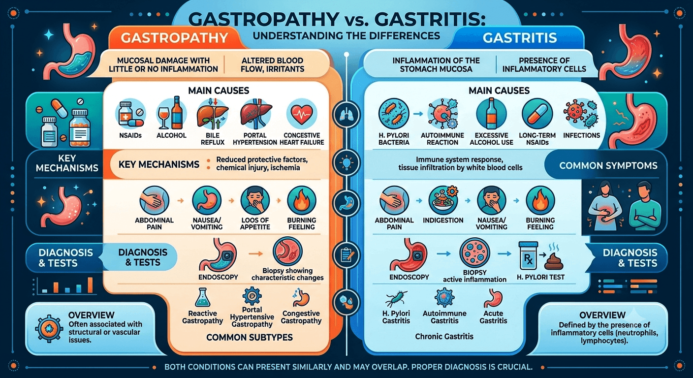
Essential Guide to What Is Visceral Hypersensitivity?

Visceral Hypersensitivity

Essential Guide to What Is Visceral Hypersensitivity?

What Is Visceral Hypersensitivity? Understanding the Hidden Cause of Chronic Gut Pain is explained in detail, including causes, symptoms, diagnosis, treatment, prognosis, and practical advice for patients and medical students.

Understanding the Hidden Cause of Chronic Gut Pain.

Among the most frustrating cases I have considered as a doctor are those where the patient suffers from some form of stomach pain, bloating, or indigestion, while all tests show normal results. Many of such people have been told that they do not have any problem while they suffer from obvious symptoms.

A possible cause of the problem could be visceral hypersensitivity, which occurs due to high sensitivity of the internal organs’ nerves to normal processes. It is thought to be one of the key causes of chronic gastrointestinal disorders, especially Irritable Bowel Syndrome (IBS).

The knowledge of the problem is beneficial for patients as it helps to understand why their symptoms are valid. For medical students, this condition shows how complex the interrelation between the gut and central nervous system really is.

Definition and Significance

What Is Visceral Hypersensitivity?

Visceral hypersensitivity is a condition where the internal organs become more sensitive to stimuli that would not otherwise be painful or uncomfortable.

In normal people:

  • The stretching of the stomach after eating a meal,
  • The passage of gases within the intestine,
  • And normal intestinal movements
  • Are usually not even noticed or only noticed mildly.

In people with visceral hypersensitivity:

  • The same normal sensations become painful.
  • A slight distention of the intestine may be severely felt.
  • Essentially normal digestive actions create considerable pain.

Such hypersensitivity occurs due to the disruption of the signaling pathway among:

  • The digestive system
  • The peripheral nerves
  • The spinal cord
  • The brain.

Visceral hypersensitivity is being understood more and more as a major source of abdominal pain.

Anatomy and Function: How Normal Gut Sensation Works

Anatomy and Pathophysiology of Visceral Hypersensitivity

The Visceral Nervous System

There are millions of neurons present in the alimentary canal, and together, they constitute the enteric nervous system, which is also referred to as the second brain.

Main elements include:

Visceral sensory receptors

This receptor responds to:

• Stretching

• Pressure

• Inflammation

• Chemical change

Peripheral nerves

These are used for carrying signals from:

• Vagal pathways

• Sympathetic pathways

• Spinal afferent nerves

Central nervous system

Here, the brain receives signals to understand whether they are:

• Normal sensation

• Discomfort

• Pain

Process in visceral hypersensitivity

There is an increase in sensitivity in the pain system.

It implies that:

• Normal sensation feels like pain.

• Mild stimulation causes disproportionate sensation of pain.

• Sensation of pain is processed by the brain on an exaggerated level.

An example of a metaphor here would be using a microphone with the volume increased.

Causes of Visceral Hypersensitivity

Visceral hypersensitivity is often multifactorial. Several mechanisms may contribute simultaneously.

1. Gastrointestinal Disorders

The most common causes include:

Irritable Bowel Syndrome (IBS)

Irritable Bowel Syndrome (IBS) is strongly associated with visceral hypersensitivity.

Research suggests many IBS patients experience:

  • Lower pain thresholds
  • Increased bowel sensitivity
  • Enhanced brain responses to gut stimulation

Functional Dyspepsia

Patients often experience:

  • Upper abdominal pain
  • Early satiety
  • Post-meal fullness

due partly to hypersensitive stomach nerves.

2. Previous Gastrointestinal Infections

Some individuals develop symptoms after:

  • Food poisoning
  • Gastroenteritis
  • Bacterial infections

3. Chronic Inflammation

Conditions associated with ongoing inflammation include:

  • Inflammatory bowel disease (IBD)
  • Microscopic colitis
  • Chronic gastritis

Even after inflammation improves, hypersensitivity may persist.

4. Psychological and Emotional Factors

The gut and brain communicate continuously through the gut-brain axis.

Contributing factors include:

  • Anxiety
  • Depression
  • Chronic stress
  • Trauma

These conditions do not cause symptoms to be imagined. Rather, they can amplify pain-processing pathways.

5. Central Sensitization

In some patients, the nervous system itself becomes over reactive.

This process is similar to what occurs in:

  • Fibromyalgia
  • Chronic fatigue syndrome
  • Migraine disorders

6. Genetic and Biological Factors

Less common contributing factors include:

  • Genetic predisposition
  • Altered neurotransmitter activity
  • Changes in serotonin signaling
  • Abnormal pain modulation pathways

Symptoms of Visceral Hypersensitivity

Symptoms vary depending on the affected organ system.

Direct Symptoms

Chronic Abdominal Pain

The hallmark symptom.

Pain may be:

  • Crampy
  • Burning
  • Sharp
  • Diffuse

Bloating

Patients regularly feel significant abdominal distension even when objective swelling is minimal.

Pain After Eating

Normal stomach expansion after meals may trigger discomfort.

Gas-Related Discomfort

Small amounts of intestinal gas may produce severe symptoms.

Symptoms Associated with Underlying Disorders

IBS-Related Symptoms

  • Diarrhea
  • Constipation
  • Alternating bowel habits
  • Urgency

Functional Dyspepsia Symptoms

  • Early fullness
  • Nausea
  • Upper abdominal discomfort

Pelvic Symptoms

Some patients experience:

  • Pelvic pain
  • Bladder sensitivity
  • Increased urinary frequency

Diagnosis of Visceral Hypersensitivity

There is no single blood test that confirms visceral hypersensitivity.

Diagnosis usually involves:

  1. Clinical evaluation
  2. Excluding structural disease
  3. Identifying associated disorders

Medical History

Important questions include:

  • Duration of symptoms
  • Pain characteristics
  • Relationship to meals
  • Bowel habits
  • Stress factors
  • Previous infections

Physical Examination

A physician evaluates for:

  • Abdominal tenderness
  • Organ enlargement
  • Signs of inflammation
  • Weight loss

Most patients have a relatively normal examination.

Laboratory Tests

Blood tests may include:

  • Complete blood count (CBC)
  • Inflammatory markers (CRP, ESR)
  • Liver function tests
  • Lipid profile
  • Thyroid studies
  • Diabetes screening
  • Celiac disease screening

These help exclude other conditions.

Imaging Studies

Ultrasound (whole abdomen)

CT Scan (Abdomen)

MRI

Endoscopy and Colonoscopy

These procedures may be performed to exclude:

  • Ulcers
  • Cancer
  • Inflammatory bowel disease

Specialized Testing

Research settings may use:

  • Rectal balloon distension tests
  • Barostat studies
  • Sensory threshold testing

These assess visceral sensitivity directly.

Treatment of Visceral Hypersensitivity

Core Principle: Treat the Underlying Cause

Management focuses on:

  • Identifying associated disorders
  • Reducing nerve sensitivity
  • Improving quality of life

Lifestyle and Dietary Modifications

Dietary Changes

Depending on the diagnosis:

  • Low-FODMAP diet
  • Trigger-food avoidance
  • Smaller, frequent meals
  • Adequate hydration

These strategies often reduce symptom severity.

Stress Reduction

Because of the gut-brain connection, stress management is important.

Helpful approaches include:

  • Meditation
  • Yoga
  • Mindfulness training
  • Relaxation exercises

Medications

Neuromodulators

Low doses of medications can reduce pain sensitivity.

Examples include:

  • Tricyclic antidepressants
  • SSRIs
  • SNRIs

These are frequently prescribed for pain modulation rather than depression.

Antispasmodic Medications

May help reduce:

  • Cramping
  • Intestinal spasms

Gut-Specific Treatments

Depending on the underlying condition:

  • IBS medications
  • Anti-inflammatory therapy
  • Acid-suppressing drugs

Psychological Therapies

Evidence strongly supports:

Cognitive Behavioral Therapy (CBT)

Helps modify pain perception and coping mechanisms.

Emerging Treatments

Researchers are studying:

  • Microbiome-targeted therapies
  • Neuromodulation techniques
  • Novel pain signaling medications

These approaches may expand future treatment options.

Living with Visceral Hypersensitivity

Practical Advice for Patients

Keep a Symptom Journal

Track:

  • Foods
  • Stress levels
  • Symptoms
  • Bowel habits

Patterns typically become apparent over time.

Maintain Regular Physical Activity

Exercise may:

  • Improve bowel function
  • Reduce stress
  • Enhance pain tolerance

Avoid Unnecessary Dietary Restrictions

Extreme elimination diets can lead to:

  • Nutritional deficiencies
  • Increased anxiety around food

Work with a healthcare professional whenever possible.

When to Seek Medical Attention

Seek prompt medical evaluation if symptoms are accompanied by:

  • Unexplained weight loss
  • Blood in stool
  • Persistent vomiting
  • Fever
  • Difficulty swallowing
  • Severe nighttime symptoms
  • Family history of gastrointestinal cancer

These may indicate a condition other than visceral hypersensitivity.

Frequently Asked Questions (FAQs)

Disclaimer: The information provided in this blog post is for educational and informational purposes only and should not be considered medical advice. It is not intended to replace professional medical consultation, diagnosis, or treatment.
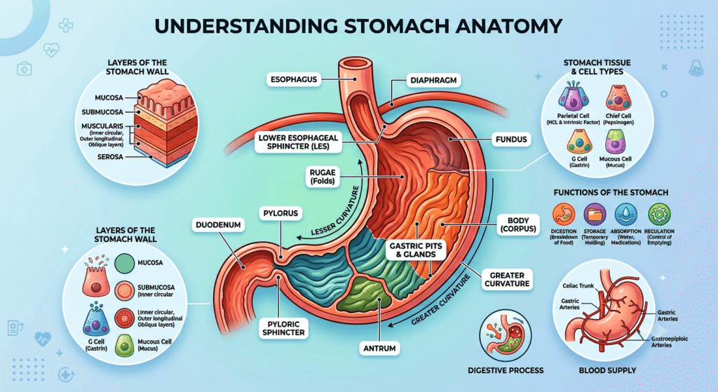
IBS vs. IBD: What Is the Difference?

IBS vs. IBD

IBS vs. IBD: What Is the Difference?

IBS (Irritable Bowel Syndrome) is a functional disorder causing chronic abdominal pain, bloating, and altered bowel habits, but no structural damage. IBD (Inflammatory Bowel Disease), including Crohn’s and ulcerative colitis, is a serious autoimmune condition causing chronic inflammation, tissue ulcers, and visible damage to the digestive tract.

Introduction

Digestive disorders affect millions of people worldwide, but many people are confused by the terms IBS and IBD. Although their names sound similar and both can cause symptoms such as abdominal pain, diarrhea, and changes in bowel habits, they are fundamentally different conditions. Irritable Bowel Syndrome (IBS) is a functional gastrointestinal disorder that impacts how the digestive tract works without causing visible inflammation or permanent damage. Inflammatory Bowel Disease (IBD), on the other hand, is a group of chronic inflammatory conditions that cause ongoing inflammation and structural damage to the gastrointestinal tract.

Understanding the difference between IBS vs. IBD is important because their causes, diagnostic approaches, treatments, and long-term health implications vary significantly. While IBS can greatly impact quality of life, IBD may lead to serious complications if left untreated. This guide explores the key differences between IBS and IBD, including their symptoms, causes, diagnosis, treatment options, and when to seek medical attention.

Understanding IBS and IBD

IBS vs. IBD Anatomy

What Is IBS?

Irritable Bowel Syndrome (IBS) is a functional gastrointestinal disorder. This means that the digestive tract appears structurally normal but does not function properly.

In IBS:

  • The intestines are not damaged.
  • No significant inflammation is present.
  • Symptoms arise from abnormal gut motility, altered gut-brain communication, and increased intestinal sensitivity.

IBS affects millions of people worldwide and is one of the most common reasons for gastroenterology consultations.

Common Types of IBS

  • IBS-D: Predominantly diarrhea
  • IBS-C: Predominantly constipation
  • IBS-M: Mixed diarrhea and constipation
  • IBS-U: Unclassified

What Is IBD?

Inflammatory Bowel Disease (IBD) is a group of chronic inflammatory disorders that cause actual damage to the digestive tract.

The two major forms are:

Crohn’s Disease

Can impact any part of the gastrointestinal tract, from the mouth to the anus.

Characteristics:

  • Patchy areas of inflammation (“skip lesions”)
  • Involves the entire bowel wall thickness
  • May cause fistulas and strictures

Ulcerative Colitis

Affects only the colon and rectum.

Characteristics:

  • Continuous inflammation
  • Limited primarily to the inner lining of the colon
  • Often causes bloody diarrhea (Gastrointestinal bleeding)

Unlike IBS, IBD causes visible inflammation and tissue injury.

IBS vs. IBD: Key Differences at a Glance

FeatureIBSIBD
Type of ConditionFunctional disorderInflammatory disease
Intestinal DamageNoYes
InflammationMinimal or absentSignificant
BleedingRareCommon
Cancer RiskNo increased riskIncreased risk in some patients
Weight LossUncommonCommon
FeverRareMay occur
Blood TestsUsually normalOften abnormal
Colonoscopy FindingsTypically normalInflammation visible
Life-Threatening ComplicationsRarePossible

Causes of IBS

The exact cause of IBS remains incompletely understood.

Most experts believe multiple factors contribute.

Common Causes and Risk Factors

Gut-Brain Axis Dysfunction

The gut and brain communicate continuously.

In IBS:

  • Signals become exaggerated.
  • The brain can interpret the normal, everyday movements of the gut as painful sensations.

Abnormal Intestinal Motility

Food may move:

  • Too quickly (diarrhea)
  • Too slowly (constipation)

Visceral Hypersensitivity

Visceral hypersensitivity is an increased sensitivity to sensations arising from internal organs, especially the gastrointestinal (GI) tract. People with visceral hypersensitivity may experience pain, discomfort, bloating, or urgency from normal levels of intestinal stretching or activity that would not bother most people.

Stress and Psychological Factors

Stress does not cause IBS directly but often worsens symptoms.

Common associations include:

  • Anxiety
  • Depression
  • Chronic stress

Microbiome Changes

Alterations in gut bacteria may contribute to symptoms.

Less Common Contributing Factors

  • Prior gastrointestinal infections
  • Food intolerances
  • Small intestinal bacterial overgrowth (SIBO)
  • Genetic predisposition

Causes of IBD

Unlike IBS, IBD involves a true inflammatory process.

Immune System Dysfunction

The immune system mistakenly attacks the digestive tract.

This leads to:

  • Chronic inflammation
  • Tissue injury
  • Ulcer formation

Genetic Factors

Numerous genetic variants increase susceptibility.

Having a close relative with IBD significantly raises risk.

Environmental Factors

Potential triggers include:

  • Smoking (especially Crohn’s disease)
  • Western dietary patterns
  • Certain infections
  • Urban living environments

Microbiome Alterations

Changes in intestinal bacteria may activate abnormal immune responses.

Symptoms of IBS

IBS symptoms typically fluctuate over time.

Many patients describe periods of improvement followed by flare-ups.

Typical Symptoms

Abdominal Pain

A hallmark symptom.

Pain often:

  • Improves after bowel movements
  • Varies in intensity
  • Recurs frequently

Bloating

Patients frequently describe:

  • Abdominal fullness
  • Distension
  • Excess gas

Diarrhea

Particularly in IBS-D.

May include:

  • Urgency
  • Frequent loose stools

Constipation

Common in IBS-C.

May involve:

  • Hard stools
  • Straining
  • Incomplete evacuation

Mixed Bowel Habits

Alternating constipation and diarrhea.

Symptoms Not Typical of IBS

The presence of these symptoms should prompt further evaluation:

  • Blood in stool
  • Fever
  • Significant weight loss
  • Nighttime symptoms
  • Persistent anemia

Symptoms of IBD

IBD symptoms are often more severe and progressive.

Digestive Symptoms

Chronic Diarrhea

May occur multiple times daily.

Blood in Stool

Particularly common in ulcerative colitis.

Abdominal Pain

Usually caused by active inflammation.

Urgent Bowel Movements

Patients may struggle to delay defecation.

Systemic Symptoms

Because IBD is an inflammatory disease, symptoms may affect the whole body.

Fatigue

One of the most common complaints.

Weight Loss

Results from:

  • Poor absorption
  • Reduced appetite
  • Chronic inflammation

Fever

May occur during active disease.

Extraintestinal Manifestations

IBD can impact organs beyond the gut.

Examples include:

  • Arthritis
  • Eye inflammation
  • Skin disorders
  • Liver disease

For medical students, these extraintestinal manifestations are important diagnostic clues.

How IBS and IBD Are Diagnosed

Diagnosis begins with a thorough clinical evaluation.

Medical History

Doctors assess:

  • Symptom patterns
  • Duration
  • Family history
  • Dietary triggers
  • Associated symptoms

Physical Examination

May reveal:

  • Abdominal tenderness
  • Signs of malnutrition
  • Weight loss
  • Dehydration

IBS examinations are often normal.

IBD may show objective abnormalities.

Blood Tests

Common tests include:

  • Complete Blood Count (CBC)
  • C-reactive Protein (CRP)
  • Erythrocyte Sedimentation Rate (ESR)
  • Electrolytes
  • Liver function tests
  • Lipid profile
  • RBS & HbA1c

Findings

IBS:

  • Usually normal
  • RBS & HbA1c (abnormal finding if patient is diabetic)

IBD:

May show:

  • Elevated inflammatory markers
  • Anemia
  • Nutritional deficiencies

Stool Tests

Doctors may test for:

  • Infection
  • Blood
  • Fecal calprotectin

Elevated fecal calprotectin strongly suggests intestinal inflammation and helps distinguish IBD from IBS.

Endoscopy and Colonoscopy

These are among the most essential investigations for IBD.

Doctors can:

  • Visualize inflammation
  • Obtain biopsies
  • Assess disease severity

IBS typically shows normal findings.

Imaging Studies

Ultrasound

Can evaluate:

  • Bowel wall thickness
  • Abscesses
  • Inflammation

CT Scan

Useful for:

  • Complications
  • Acute flare-ups
  • Abscess detection

MRI

Particularly valuable for:

  • Crohn’s disease
  • Fistulas
  • Pelvic involvement

Biopsy

Tissue samples obtained during colonoscopy confirm the diagnosis.

Potential Complications of IBS

IBS does not damage the intestines, but it can significantly impact quality of life.

Complications include:

  • Chronic discomfort
  • Anxiety
  • Depression
  • Social limitations
  • Work and school absenteeism

Importantly, IBS does not increase the risk of colon cancer.

Potential Complications of IBD

IBD can lead to serious complications.

Intestinal Complications

Crohn’s Disease

  • Fistulas
  • Abscesses
  • Strictures
  • Bowel obstruction

Ulcerative Colitis

  • Severe bleeding
  • Toxic megacolon
  • Colon perforation

Nutritional Problems

Patients may develop:

  • Iron deficiency
  • Vitamin B12 deficiency
  • Protein malnutrition

Cancer Risk

Long-standing IBD increases the risk of colorectal cancer, especially when inflammation is extensive and poorly controlled.

Treatment of IBS

Treatment focuses on symptom management.

Dietary Changes

Many patients benefit from:

  • Low-FODMAP diets
  • Increased fiber (selected patients)
  • Identifying trigger foods

Common triggers include:

  • Caffeine
  • Alcohol
  • Fatty foods

Stress Management

Helpful strategies include:

  • Cognitive behavioral therapy
  • Meditation
  • Regular exercise
  • Mindfulness techniques

Medications

Depending on symptoms:

  • Anti diarrheals
  • Laxatives
  • Antispasmodics
  • Certain antidepressants

Treatment of IBD

Treatment aims to control inflammation and prevent complications.

Anti-Inflammatory Medications

Examples:

  • Mesalamine
  • Sulfasalazine

Corticosteroids

Used for short-term flare control.

Examples:

  • Prednisone
  • Budesonide

Immunomodulators

Help suppress abnormal immune activity.

Examples:

  • Azathioprine
  • Methotrexate

Biologic Therapies

Modern treatments targeting specific inflammatory pathways.

Examples:

  • Anti-TNF agents
  • Integrin inhibitors
  • IL-12/23 inhibitors

Surgery

It may be necessary for:

  • Severe disease
  • Obstruction
  • Fistulas
  • Cancer prevention

Surgery can cure ulcerative colitis by removing the colon but does not cure Crohn’s disease.

Living With IBS and IBD

Receiving a diagnosis can feel overwhelming. Fortunately, both conditions can be managed effectively.

Practical Lifestyle Advice

Keep a Symptom Diary

Track:

  • Foods
  • Stress levels
  • Medications
  • Symptoms

Patterns often emerge.

Follow Medical Advice

Regular follow-up improves outcomes.

Stay Physically Active

Exercise can:

  • Improve bowel function
  • Reduce stress
  • Support overall health

Prioritize Mental Health

Digestive disorders and emotional health are closely linked.

Seeking support is a sign of strength, not weakness.

When to Seek Medical Attention

Contact a healthcare professional if you experience:

  • Blood in stool
  • Persistent weight loss
  • Fever
  • Severe abdominal pain
  • Nighttime diarrhea
  • Symptoms that worsen despite treatment

These may indicate IBD or another serious condition rather than IBS.

Final Thoughts

The differentiation between IBS and IBD remains the most crucial topic in gastroenterology. Although similar in many ways as far as symptoms and severity are concerned, there exist several differences that make each of the diseases biologically unique.

On the patient’s side, learning about the difference between the two disorders will help decrease anxiety levels and get proper medical attention. For a medical student, on the other hand, such differentiation becomes key in assessing the problem.

Most importantly, whether it is IBS or IBD that a person has, adequate management and good relations with his/her healthcare provider can truly change one’s life.

Disclaimer: The information provided in this blog post is for educational and informational purposes only and should not be considered medical advice. It is not intended to replace professional medical consultation, diagnosis, or treatment.
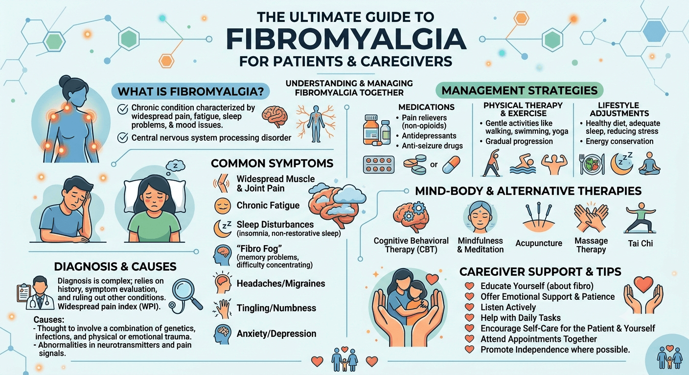
Splenomegaly: A Comprehensive Guide

Splenomegaly

to Causes, Symptoms, Diagnosis, and Treatment

Introduction

Discovering that you have an enlarged spleen or hearing the term splenomegaly for the first time can be unsettling. Patients often wonder whether it is serious, while medical students may be trying to understand the many conditions that can cause it. The good news is that splenomegaly is not a disease itself; rather, it is a sign that something else is affecting the body.

Explore splenomegaly (enlarged spleen) with this comprehensive guide covering causes, symptoms, diagnosis, treatment, complications, and warning signs that require medical attention.

This guide aims to bridge the gap between patient education and medical learning. Whether you are seeking reassurance about your health or building your clinical knowledge, understanding splenomegaly begins with understanding the remarkable organ at the center of it all: the spleen.

What Is Splenomegaly?

Splenomegaly is the medical term for an enlarged spleen.

In healthy adults, the spleen is usually about the size of a clenched fist and is typically not felt during a physical examination. When the spleen becomes larger than normal, it is described as splenomegaly.

From a clinical perspective, splenomegaly is important because it often serves as a clue to an underlying medical condition. These conditions range from temporary infections to chronic liver disease, blood disorders, autoimmune diseases, and cancers.

For patients, it is helpful to remember that an enlarged spleen is a symptom or finding not a diagnosis by itself.

Understanding the Spleen: Anatomy and Function

Where Is the Spleen Located?

The spleen sits in the upper left part of the abdomen, tucked beneath the rib cage and behind the stomach.

Normally, the spleen is protected by the lower ribs and cannot be felt externally.

Spleen Anatomy

What Does the Spleen Do?

Although many people rarely think about the spleen, it plays several critical roles:

1. Blood Filtration

The spleen acts like a sophisticated filter, removing:

  • Old red blood cells
  • Damaged blood cells
  • Cellular debris

2. Immune Defense

The spleen helps the body fight infections by:

  • Producing immune cells
  • Filtering bacteria and viruses from the bloodstream
  • Supporting antibody production

3. Blood Storage

The spleen stores:

  • Platelets
  • White blood cells
  • Small reserves of blood

4. Fetal Blood Production

Before birth, the spleen helps produce blood cells. Certain diseases can reactivate this function later in life, contributing to enlargement.

How Does Splenomegaly Develop?

The spleen enlarges when it becomes overworked, congested, infiltrated by abnormal cells, or stimulated by immune activity.

Common mechanisms include:

  • Increased destruction of blood cells
  • Excess immune activation
  • Blood flow obstruction
  • Cancer cell infiltration
  • Infection-related inflammation

For medical students, splenomegaly is often categorized as:

  1. Congestive
  2. Infectious
  3. Hematologic
  4. Neoplastic
  5. Inflammatory/autoimmune
  6. Infiltrative

This framework helps narrow the differential diagnosis.

Causes of Splenomegaly

The causes are numerous, but they can be organized into major categories.

Infections

Infections are among the most common causes worldwide.

Viral Infections

Examples include:

  • Infectious Mononucleosis
  • COVID-19
  • Viral hepatitis
  • Cytomegalovirus (CMV)
  • HIV infection

Bacterial Infections

Examples:

  • Tuberculosis
  • Infective endocarditis
  • Typhoid fever

Parasitic Infections

Common in certain regions:

  • Malaria
  • Leishmaniasis
  • Schistosomiasis

Liver Disease and Portal Hypertension

One of the most common causes of significant splenomegaly in adults is liver disease.

Conditions include:

  • Cirrhosis
  • Chronic hepatitis
  • Portal vein thrombosis

When blood flow through the liver becomes restricted, pressure increases in the portal venous system. Blood backs up into the spleen, causing congestive splenomegaly.

Blood Disorders

Several hematologic conditions enlarge the spleen.

Hemolytic Anemias

The spleen removes damaged red blood cells. If destruction becomes excessive, the spleen enlarges.

Examples:

  • Sickle Cell Disease
  • Hereditary spherocytosis
  • Autoimmune hemolytic anemia

Myeloproliferative Disorders

Examples:

  • Polycythemia Vera
  • Primary Myelofibrosis
  • Essential thrombocythemia

These conditions often produce marked enlargement.

Cancers

Cancer-related splenomegaly usually occurs when abnormal cells infiltrate the spleen.

Examples include:

  • Leukemia
  • Lymphoma
  • Metastatic cancers

Massive splenomegaly is classically associated with certain leukemias and Myeloproliferative diseases.

Autoimmune and Inflammatory Diseases

The immune system can stimulate splenic enlargement.

Examples:

  • Systemic Lupus Erythematosus
  • Rheumatoid Arthritis
  • Felty syndrome
  • Sarcoidosis

Storage and Infiltrative Disorders

These are less common but important.

Examples:

  • Gaucher Disease
  • Amyloidosis
  • Niemann-Pick disease

Abnormal substances accumulate within the spleen, causing enlargement.

Symptoms of Splenomegaly

Many people with mild splenomegaly have no symptoms at all.

When symptoms occur, they may arise directly from the enlarged spleen or from the underlying disease.

Upper Left Abdominal Pain

Patients may experience:

  • Dull discomfort
  • Pressure sensation
  • Fullness under the ribs

Pain Radiating to the Left Shoulder

Known as Kehr’s sign, this may occur if the spleen irritates the diaphragm.

Early Satiety

A growing spleen can press against the stomach.

Patients often report:

I feel full after eating only a small amount.

Abdominal Distension

A very enlarged spleen may create a visible abdominal bulge.

Symptoms Related to the Underlying Cause

Infection-Related Symptoms

  • Fever
  • Chills
  • Fatigue
  • Night sweats

Blood Disorder Symptoms

  • Weakness
  • Shortness of breath
  • Easy bruising

Cancer-Related Symptoms

  • Weight loss
  • Persistent fatigue
  • Enlarged lymph nodes
  • Night sweats

Liver Disease Symptoms

  • Jaundice
  • Swollen abdomen
  • Leg swelling

How Is Splenomegaly Diagnosed?

Diagnosis involves determining both:

  1. Whether the spleen is enlarged
  2. Why it is enlarged

Medical History

Doctors ask about:

  • Recent infections
  • Travel history
  • Alcohol use
  • Family history
  • Weight loss
  • Fever
  • Bleeding problems

For medical students, history-taking often provides the strongest clues to etiology.

Physical Examination

During examination, the physician feels beneath the left rib cage.

A normal spleen is usually not palpable.

Findings may include:

  • Palpable spleen tip
  • Massive enlargement extending across the abdomen
  • Associated liver enlargement

Blood Tests

Blood work frequently reveals the underlying cause.

Common tests include:

TestPurpose
Complete Blood Count (CBC)Evaluates red cells, white cells, platelets
Peripheral Blood SmearExamines blood cell morphology
Liver Function TestsAssesses liver disease
Viral TestingDetects viral infections
Inflammatory MarkersEvaluates immune disorders
Coagulation StudiesAssesses clotting function

Ultrasound

First-Line Imaging Study

Ultrasound is:

  • Safe
  • Inexpensive
  • Radiation-free

It accurately measures spleen size and evaluates blood flow.

CT Scan

CT scans provide detailed images and help identify:

  • Tumors
  • Abscesses
  • Trauma
  • Portal hypertension

MRI

MRI is particularly useful when:

  • Soft tissue characterization is needed
  • Vascular abnormalities are suspected
  • Radiation exposure should be minimized

Bone Marrow Biopsy

When blood cancers or marrow disorders are suspected, a bone marrow biopsy may be necessary.

This test can diagnose:

  • Leukemia
  • Lymphoma
  • Myelofibrosis
  • Other hematologic diseases

For medical students, this is typically a key step in evaluating unexplained splenomegaly with abnormal blood counts.

Treatment of Splenomegaly

The most important principle is simple:

Treat the underlying cause, not just the enlarged spleen.

Treatment of Infections

Examples include:

  • Antibiotics for bacterial infections
  • Antiviral therapies when indicated
  • Antiparasitic medications for malaria and related diseases

Once the infection resolves, the spleen often returns to normal size.

Treatment of Liver Disease

Management may involve:

  • Avoiding alcohol
  • Treating viral hepatitis
  • Managing portal hypertension
  • Liver transplantation in advanced cases

Treatment of Blood Disorders

Depending on the condition:

  • Corticosteroids
  • Immunosuppressive medications
  • Blood transfusions
  • Targeted therapies

Treatment of Cancer

Cancer treatment may include:

  • Chemotherapy
  • Radiation therapy
  • Immunotherapy
  • Stem cell transplantation

Successful treatment often reduces spleen size.

Splenectomy (Spleen Removal)

In selected situations, removal of the spleen may be necessary.

Common indications:

  • Severe hypersplenism
  • Splenic rupture
  • Certain blood disorders
  • Persistent symptomatic enlargement

However, splenectomy increases susceptibility to certain infections.

Patients require:

  • Vaccinations
  • Preventive medical follow-up
  • Education about infection risks

Prognosis

The outlook for splenomegaly varies considerably.

Generally Favorable Prognosis

Examples:

  • Infectious mononucleosis
  • Temporary viral infections
  • Treatable bacterial infections

Variable Prognosis

Examples:

  • Cirrhosis
  • Autoimmune disorders
  • Chronic hemolytic anemia

More Serious Prognosis

Examples:

  • Advanced leukemia
  • Aggressive lymphoma
  • Severe myelofibrosis

Ultimately, prognosis depends on:

  • Underlying diagnosis
  • Severity of disease
  • Response to treatment
  • Presence of complications

Living With Splenomegaly

For patients, receiving a diagnosis of splenomegaly can feel overwhelming. Fortunately, many people live well while the underlying condition is investigated and treated.

Protect Your Spleen

Avoid activities with a high risk of abdominal trauma, including:

  • Contact sports
  • Martial arts
  • High-impact activities unless approved by your doctor

Attend Follow-Up Appointments

Regular monitoring helps track:

  • Spleen size
  • Blood counts
  • Treatment effectiveness

Know the Warning Signs

Seek urgent medical attention if you develop:

  • Severe left-sided abdominal pain
  • Fainting
  • Sudden dizziness
  • Significant bleeding
  • High fever

These symptoms may indicate a serious complication.

Maintain General Health

Helpful measures include:

  • Eating a balanced diet
  • Staying physically active as advised
  • Avoiding excessive alcohol
  • Keeping vaccinations up to date

Managing chronic illnesses carefully

Splenomegaly FAQ

Key Takeaways

For patients, the most important message is reassurance: many causes of splenomegaly are treatable, and modern diagnostic tools allow physicians to identify the underlying problem effectively. For medical students, splenomegaly remains a classic clinical finding that reflects diverse pathophysiologic processes and demands a systematic diagnostic approach. Understanding why the spleen enlarges is often the key to understanding the disease itself.

Disclaimer: The information provided in this blog post is for educational and informational purposes only and should not be considered medical advice. It is not intended to replace professional medical consultation, diagnosis, or treatment. Always seek the guidance of a qualified healthcare professional regarding any medical condition or health-related concerns.
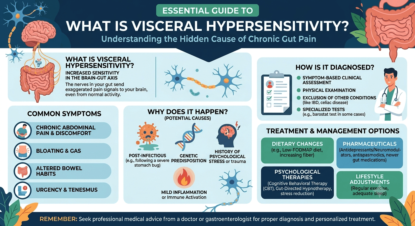
Pain in Epigastric Region

Pain in Epigastric region

What Your Belly Is Trying to Tell You

Pain in Epigastric Region: What Your Belly Is Trying to Tell You. Discover the common causes, symptoms, warning signs, diagnosis, treatment options, and when to seek medical attention for upper abdominal pain.

Introduction

Pain in the upper middle part of your abdomen can be alarming. This area, known as the epigastric region, sits just below the breastbone and above the belly button. While occasional discomfort may result from something as simple as overeating, persistent or severe pain can signal an underlying medical condition that requires attention.

Pain in Epigastric Region: What Your Belly Is Trying to Tell You. Discover the common causes, symptoms, warning signs, diagnosis, treatment options, and when to seek medical attention for upper abdominal pain.

Understanding what your body is trying to tell you can help you seek the right care at the right time. This article explores the causes, symptoms, diagnosis, treatment, and prevention of epigastric pain based on current medical knowledge.

Understanding the Epigastric Region

The abdomen is divided into several regions to help healthcare professionals identify the source of pain. The epigastric region is located in the upper central abdomen.

Several important organs are found in or near this area, including:

  • Stomach
  • Pancreas
  • Liver
  • Gallbladder
  • Duodenum (first part of the small intestine)
  • Lower esophagus
  • Major blood vessels

Because so many organs are located here, pain in this region can have numerous causes.

Why Epigastric Pain Should Not Be Ignored

Epigastric pain can range from mild discomfort to severe, debilitating pain. The sensation may be:

  • Burning
  • Aching
  • Sharp
  • Cramping
  • Gnawing
  • Pressure-like

In some cases, the pain is harmless and temporary. However, it can also indicate conditions such as ulcers, gallstones, pancreatitis, or even heart disease.

The duration, intensity, and associated symptoms often provide clues about the underlying cause.

Common Symptoms That Accompany Epigastric Pain

Pain rarely occurs alone. Many patients experience additional symptoms that help narrow down the diagnosis.

Common accompanying symptoms include:

SymptomPossible Association
HeartburnAcid reflux, GERD
NauseaGastritis, pancreatitis
VomitingUlcers, infections
BloatingDyspepsia, indigestion
Loss of appetiteGastritis, ulcers
FeverInfection, pancreatitis
Chest discomfortGERD, cardiac causes
Weight lossChronic digestive disease

Gastroesophageal Reflux Disease (GERD)

One of the most common causes of pain in the epigastric region is GERD, commonly known as acid reflux.

GERD occurs when stomach acid flows backward into the esophagus, irritating its lining.

How Acid Reflux Causes Pain

Symptoms often include:

  • Burning sensation in the upper abdomen
  • Heartburn
  • Sour taste in the mouth
  • Difficulty swallowing
  • Symptoms worsening after meals

Certain foods can trigger GERD, including:

  • Spicy foods
  • Fatty meals
  • Chocolate
  • Caffeine
  • Alcohol

Frequent reflux should be evaluated because long-term acid exposure can damage the esophagus.

Gastritis and Stomach Inflammation

Gastritis refers to inflammation of the stomach lining. It may develop suddenly or gradually over time.

Common causes include:

  • Helicobacter pylori infection
  • Excessive alcohol consumption
  • Long-term use of NSAIDs such as ibuprofen
  • Stress-related illness
  • Autoimmune conditions

Risk Factors for Gastritis

  • Helicobacter pylori (H. pylori) infection: One of the most common causes of chronic gastritis worldwide. This bacterium weakens the stomach’s protective barrier and promotes inflammation.
  • Regular use of non-steroidal anti-inflammatory drugs (NSAIDs): Medications such as aspirin, ibuprofen, and naproxen can damage the stomach lining, especially when used frequently or in high doses.
  • Excessive alcohol consumption: Alcohol irritates and erodes the stomach mucosa, increasing the risk of inflammation and bleeding.
  • Smoking: Tobacco use impairs the stomach’s natural defense mechanisms and delays healing of the gastric mucosa.
  • Severe physical stress: Major surgery, severe burns, trauma, or critical illness can lead to stress-related gastritis.
  • Autoimmune disorders: In autoimmune gastritis, the immune system mistakenly attacks the stomach lining, leading to chronic inflammation and vitamin B12 deficiency.
  • Advancing age: Older adults are at greater risk due to thinning of the stomach lining and increased likelihood of H. pylori infection or medication use.
  • Bile reflux: Backflow of bile from the small intestine into the stomach can irritate the gastric mucosa.
  • Chronic medical conditions: Diseases such as chronic kidney disease, liver disease, HIV infection, and Crohn’s disease may increase the risk of gastritis.
  • Dietary and lifestyle factors: Frequent consumption of highly spicy foods, excessive caffeine, and poor dietary habits may worsen symptoms in susceptible individuals, although they are not primary causes.

Peptic Ulcers

Peptic ulcers are open sores that develop in the stomach or upper small intestine.

These ulcers commonly result from:

  • H. pylori infection
  • Frequent NSAID use
  • Smoking
  • Excessive alcohol intake

Signs of a Stomach or Duodenal Ulcer

Symptoms may include:

  • Gnawing upper abdominal pain
  • Pain that improves or worsens after eating
  • Bloating
  • Nausea
  • Nighttime discomfort

Complications such as bleeding ulcers require immediate medical attention.

Gallbladder Disorders

The gallbladder stores bile, a digestive fluid produced by the liver.

When gallstones form, they may block bile ducts and trigger pain.

Gallstones and Upper Abdominal Pain

Typical symptoms include:

  • Sudden upper abdominal pain
  • Pain after fatty meals
  • Pain radiating to the back or right shoulder
  • Nausea and vomiting

Gallbladder pain can sometimes be mistaken for stomach-related discomfort because it occurs near the epigastric region.

Pancreatitis

The pancreas plays a vital role in digestion and blood sugar regulation.

Inflammation of the pancreas, known as pancreatitis, can cause severe epigastric pain.

Acute vs. Chronic Pancreatitis

Acute pancreatitis develops suddenly and may require hospitalization.

Chronic pancreatitis develops gradually and can cause long-term digestive problems.

Common symptoms include:

  • Severe upper abdominal pain
  • Pain radiating to the back
  • Nausea
  • Vomiting
  • Fever

Excessive alcohol use and gallstones are among the leading causes.

Heart-Related Causes of Epigastric Pain

Many people assume heart problems always cause chest pain. In reality, heart disease can sometimes present as upper abdominal discomfort.

When It Mimics a Heart Attack

Heart-related warning signs may include:

  • Pressure-like epigastric pain
  • Sweating
  • Shortness of breath
  • Dizziness
  • Pain spreading to the jaw, neck, or arm

Individuals with these symptoms should seek emergency medical care immediately.

Functional Dyspepsia

Functional dyspepsia refers to chronic indigestion without an identifiable structural cause.

Patients often report:

  • Fullness after eating
  • Bloating
  • Burning discomfort
  • Early satiety

Although not life-threatening, it can significantly affect quality of life.

Diagnosing Epigastric Pain

Doctors begin with a detailed medical history and physical examination.

Additional tests may include:

Laboratory Tests

  • Complete blood count
  • Liver function tests
  • Viral marker
  • Lipid profile
  • Pancreatic enzyme tests (S. Amylase, S. Lipase)
  • H. pylori testing
  • RBS, HbA1c

Imaging Studies

  • Ultrasound (whole abdomen)
  • CT scan
  • MRI

Endoscopy

An upper gastrointestinal endoscopy allows doctors to directly examine the esophagus, stomach, and duodenum.

This test is especially useful when ulcers, gastritis, or bleeding are suspected.

Treatment Options

Treatment depends entirely on the underlying cause.

Medications

Common medications include:

  • Antacids
  • Proton pump inhibitors
  • H2 blockers
  • Antibiotics for H. pylori infection
  • Pain management medications

Procedures

Some conditions require intervention:

  • Gallbladder surgery
  • Endoscopic ulcer treatment
  • Pancreatic procedures

Dietary Management

Many patients benefit from:

  • Smaller meals
  • Reduced fatty food intake
  • Limiting caffeine
  • Avoiding alcohol
  • Maintaining hydration

Lifestyle Changes for Prevention

Healthy habits can reduce the risk of many digestive disorders.

Consider the following strategies:

Eat Mindfully

  • Avoid overeating
  • Chew food thoroughly
  • Eat slowly

Maintain a Healthy Weight

Excess body weight increases abdominal pressure and can worsen reflux symptoms.

Avoid Smoking

Smoking damages the digestive tract and increases ulcer risk.

Manage Stress

Chronic stress may aggravate digestive symptoms and worsen functional gastrointestinal disorders.

Stay Physically Active

Regular exercise supports digestive health and overall well-being.

For additional information World Gastroenterology Organisation.

When to Seek Emergency Care

Seek immediate medical attention if epigastric pain is accompanied by:

  • Severe chest pain
  • Difficulty breathing
  • Vomiting blood
  • Black or tarry stools
  • Sudden severe abdominal pain
  • Fainting
  • High fever
  • Persistent vomiting

These symptoms may indicate a serious medical emergency.

Frequently Asked Questions

1. What is the most common cause of pain in the epigastric region?

Acid reflux, gastritis, and indigestion are among the most common causes of epigastric pain.

2. Can stress cause epigastric pain?

Yes. Stress can worsen digestive disorders and contribute to symptoms such as indigestion and stomach discomfort.

3. Is epigastric pain always related to the stomach?

No. The pancreas, gallbladder, liver, heart, and nearby organs can also cause pain in this region.

4. When should I worry about epigastric pain?

You should seek medical evaluation if the pain is severe, persistent, recurrent, or accompanied by alarming symptoms such as weight loss, vomiting blood, or chest pain.

5. Can a heart attack feel like stomach pain?

Yes. Some heart attacks present as upper abdominal discomfort, especially in older adults, women, and people with diabetes.

6. What foods should I avoid if I have recurring epigastric pain?

Common triggers include spicy foods, fatty meals, alcohol, caffeinated beverages, and acidic foods.

7. Can ulcers heal on their own?

Some ulcers may improve temporarily, but proper diagnosis and treatment are important to prevent complications.

8. Does pancreatitis always cause severe pain?

Acute pancreatitis usually causes severe pain, while chronic pancreatitis may produce persistent but less intense discomfort.

Conclusion

Pain in the epigastric region: What Your Belly Is Trying to Tell You is a question worth paying attention to because this seemingly simple symptom can have many different causes. While indigestion and acid reflux are common explanations, conditions such as ulcers, gallbladder disease, pancreatitis, and even heart disease can also be responsible.

The key is to recognize accompanying symptoms, understand potential triggers, and seek medical evaluation when pain is severe, persistent, or associated with warning signs. Early diagnosis often leads to more effective treatment and better long-term outcomes.

Disclaimer: The information provided in this blog post is for educational and informational purposes only and should not be considered medical advice. It is not intended to replace professional medical consultation, diagnosis, or treatment. Always seek the guidance of a qualified healthcare professional regarding any medical condition or health-related concerns. The author and publisher are not responsible for any actions taken based on the information presented in this article.
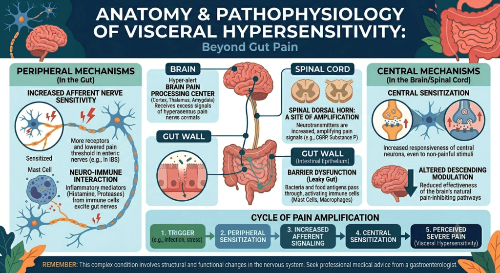
Benign Prostatic Hyperplasia (BPH): Warning Signs You Should Never Ignore

Benign Prostatic Hyperplasia

And Effective Treatments That Can Improve Your Life

Introduction

Benign Prostatic Hyperplasia (BPH), commonly referred to as an enlarged prostate, is among the most frequent urinary disorders seen in older men. The condition occurs when the prostate gland gradually increases in size and compresses the urethra, leading to lower urinary tract symptoms (LUTS). Although BPH is a non-cancerous condition, it can greatly reduce quality of life and may result in serious complications if not properly managed.

Learn everything about Benign Prostatic Hyperplasia (BPH), including causes, symptoms, diagnosis, treatment options, lifestyle changes, and prevention tips in this complete guide for patients and caregivers.

BPH becomes increasingly common after the age of 50. Its progression is usually slow, with symptoms ranging from mild urinary discomfort to severe urinary obstruction. A solid understanding of prostate anatomy, disease mechanisms, clinical features, diagnosis, treatment options, and possible complications is valuable for both patients and medical students.

Anatomy and Role of the Prostate

The prostate is a small gland situated beneath the bladder and anterior to the rectum. It surrounds the first segment of the urethra, called the prostatic urethra.

That’s correct. In healthy young men, the prostate gland typically weighs around 20 grams. Its main role is to produce prostatic secretions that form part of semen and help support sperm survival.

Prostate Anatomy

Prostate Zones

The prostate contains several anatomical regions:

  • Peripheral Zone: Most prostate cancers develop here.
  • Transitional Zone: The primary site where BPH occurs.
  • Central Zone: Encircles the ejaculatory ducts.
  • Anterior Fibromuscular Stroma: Mainly composed of muscle tissue.

With aging, both stromal and epithelial cells in the transitional zone undergo hyperplasia, causing prostate enlargement.

What Is BPH?

https://www.youtube.com/watch?v=WfOTIf8QEm0

Benign Prostatic Hyperplasia refers to the non-malignant increase in the number of smooth muscle and epithelial cells within the prostate gland. This enlargement may partially obstruct urinary flow.

The term includes:

  • Benign: Non-cancerous
  • Hyperplasia: Increase in cell quantity

Although BPH is different from prostate cancer, both diseases can exist together in elderly men.

Epidemiology

BPH is closely linked to advancing age.

Age Distribution

  • Rare before 40 years of age
  • Frequently develops after age 50
  • Seen in nearly half of men by age 60
  • Histological evidence is present in 80–90% of men by age 80

Not every man with prostate enlargement develops symptoms.

Risk Factors

Several factors contribute to the development of BPH:

1. Aging

The most significant contributing factor.

2. Hormonal Influences

Testosterone is converted into dihydrotestosterone (DHT) by the enzyme 5-alpha reductase. DHT promotes prostate growth.

3. Family History

Men with affected family members may experience earlier onset.

4. Obesity and Metabolic Syndrome

Conditions such as diabetes, hypertension, obesity, and abnormal lipid levels are associated with higher risk.

5. Lack of Physical Activity

Sedentary habits may worsen disease progression.

6. Cardiovascular Disease

Impaired vascular health may contribute to prostate enlargement.

Pathophysiology

BPH develops through both mechanical and functional mechanisms.

Static Component

An enlarged prostate physically compresses the urethra, increasing resistance to urinary flow.

Dynamic Component

Increased smooth muscle tone mediated by alpha-adrenergic receptors further narrows the urethra.

Bladder Adaptations

Initially, the bladder compensates by increasing muscular contractions. Over time, chronic obstruction may lead to:

  • Bladder wall thickening
  • Reduced compliance
  • Urinary retention
  • Detrusor dysfunction

Clinical Symptoms of BPH

The symptoms are collectively known as Lower Urinary Tract Symptoms (LUTS) and are categorized as obstructive or irritative.

Obstructive Symptoms

  • Weak urinary stream
  • Difficulty initiating urination
  • Intermittent urine flow
  • Straining during urination
  • Feeling of incomplete bladder emptying
  • Post-void dribbling
  • Urinary retention

Irritative Symptoms

  • Increased urinary frequency
  • Urgency
  • Nocturia
  • Urge incontinence

International Prostate Symptom Score (IPSS)

The IPSS is commonly used to evaluate symptom severity.

Severity Levels

  • Mild: 0–7
  • Moderate: 8–19
  • Severe: 20–35

This scoring system helps determine appropriate management.

Complications of Untreated BPH

If left untreated, BPH can result in significant complications:

  1. Acute urinary retention
  2. Chronic urinary retention
  3. Recurrent urinary tract infections
  4. Bladder stone formation
  5. Hematuria
  6. Hydronephrosis
  7. Renal impairment or failure

Diagnosis

Diagnosis combines history-taking, physical examination, laboratory testing, and imaging.

Clinical History

Important information includes:

  • Severity and duration of symptoms
  • Frequency of Nocturia
  • Episodes of urinary retention
  • Presence of hematuria
  • Medication use
  • Neurological symptoms

Physical Examination

General Examination

Assessment of hydration, neurological status, and bladder distension.

Abdominal Examination

A palpable bladder may indicate retention.

Digital Rectal Examination (DRE)

Typical BPH findings include:

  • Enlarged gland
  • Smooth surface
  • Firm consistency
  • Non-tender prostate

Hard nodules may raise suspicion for prostate cancer.

Laboratory and Imaging Studies

Urinalysis

CBC,

Lipid Profile

RBS, HbA1c

Serum Creatinine

Assesses kidney function Test (KFT)

Prostate-Specific Antigen (PSA)

PSA levels may rise in:

  • BPH
  • Prostatitis
  • Prostate cancer

Imaging and Additional Tests

  • Ultrasound for prostate size and residual urine
  • Post-void residual volume measurement
  • Uroflowmetry to assess urinary flow
  • Cystoscopy in selected patients

Differential Diagnosis

Conditions resembling BPH include:

  • Prostate cancer
  • Urinary tract infection
  • Bladder cancer
  • Urethral stricture
  • Neurogenic bladder
  • Overactive bladder
  • Chronic prostatitis

Management of BPH

Conservative Measures

Mild symptoms may improve with lifestyle adjustments.

Lifestyle Recommendations

  • Reduce evening fluid intake
  • Limit caffeine and alcohol
  • Practice scheduled voiding
  • Avoid decongestants
  • Maintain healthy weight and exercise regularly

Medical Therapy

Alpha-1 Adrenergic Blockers

Examples:

  • Tamsulosin
  • Alfuzosin
  • Silodosin
  • Doxazosin

These medications relax smooth muscle in the prostate and bladder neck, improving urinary flow.

Common Side Effects

  • Dizziness
  • Orthostatic hypotension
  • Retrograde ejaculation

5-Alpha Reductase Inhibitors

Examples:

  • Finasteride
  • Dutasteride

These drugs reduce DHT production and gradually shrink prostate size.

Benefits

  • Lower risk of retention
  • Reduced need for surgery

Side Effects

  • Reduced libido
  • Erectile dysfunction
  • Lower ejaculate volume

Most effective in men with larger prostates.

Combination Therapy

Alpha blockers and 5-alpha reductase inhibitors may be combined for moderate to severe symptoms.

Phosphodiesterase-5 Inhibitors

Tadalafil may help men with both BPH and erectile dysfunction.

Surgical Treatment

Surgery is considered when:

  • Medications fail
  • Recurrent urinary retention occurs
  • Kidney function declines
  • Recurrent infections develop
  • Bladder stones are present

Common Procedures

Transurethral Resection of the Prostate (TURP)

Considered the traditional gold-standard surgery.

Advantages

  • Excellent symptom relief
  • Improved urinary flow

Possible Complications

  • Bleeding
  • Infection
  • Retrograde ejaculation
  • Urethral stricture
  • TUR syndrome

Laser Procedures

Examples:

  • HoLEP
  • GreenLight laser therapy

Benefits include less bleeding and shorter hospitalization.

Open or Simple Prostatectomy

Reserved for extremely enlarged prostates.

Minimally Invasive Therapies

Examples:

  • UroLift
  • Rezūm therapy
  • Prostatic artery embolization

These may better preserve sexual function in selected patients.

Acute Urinary Retention

This is a medical emergency.

Symptoms

  • Severe lower abdominal pain
  • Inability to urinate
  • Distended bladder

Treatment

  • Immediate catheterization
  • Alpha blocker therapy
  • Further evaluation for definitive management

BPH Versus Prostate Cancer

FeatureBPHProstate Cancer
NatureBenignMalignant
Main LocationTransitional zonePeripheral zone
DRE FindingsSmooth enlargementHard nodules
MetastasisAbsentPossible

Prevention and Risk Reduction

Although BPH cannot always be prevented, healthy habits may slow progression.

Helpful Measures

  • Maintain ideal body weight
  • Exercise routinely
  • Control diabetes and hypertension
  • Eat fruits and vegetables
  • Avoid smoking
  • Limit alcohol use

Prognosis

The outlook for BPH is generally excellent with appropriate treatment. Many men experience major improvement in urinary symptoms and quality of life through medications or surgery. Delayed care, however, can increase the risk of bladder and kidney complications.

Important Facts

  • BPH mainly affects the transitional zone.
  • DHT has a central role in disease development.
  • Symptoms are divided into obstructive and irritative LUTS.
  • DRE and IPSS are important assessment tools.
  • PSA is useful but not specific for cancer.
  • Alpha blockers provide rapid relief.
  • 5-alpha reductase inhibitors shrink prostate tissue.
  • TURP remains a major surgical option.
  • Acute urinary retention is an emergency.

When to Seek Medical Care

Patients should seek evaluation if they experience:

  • Difficulty urinating
  • Weak stream
  • Blood in urine
  • Frequent nighttime urination
  • Painful urination
  • Sudden inability to urinate
  • Recurrent urinary infections

Early treatment helps prevent complications and improves long-term outcomes.

Conclusion

Benign Prostatic Hyperplasia is a very common condition in aging men and a major cause of lower urinary tract symptoms. While non-cancerous, it can substantially affect daily living and may lead to urinary retention, infections, bladder dysfunction, and kidney damage if untreated.

Modern advances in diagnosis and therapy have significantly improved outcomes. Treatment options range from lifestyle modifications and medications to minimally invasive procedures and surgery. A thorough understanding of BPH is important for both patients and medical trainees.

With proper medical management and regular follow-up, most men with BPH can achieve effective symptom control and maintain a high quality of life.

Disclaimer: The information provided in this blog post is for educational and informational purposes only and should not be considered medical advice. It is not intended to replace professional medical consultation, diagnosis, or treatment. Always seek the guidance of a qualified healthcare professional regarding any medical condition or health-related concerns. The author and publisher are not responsible for any actions taken based on the information presented in this article.
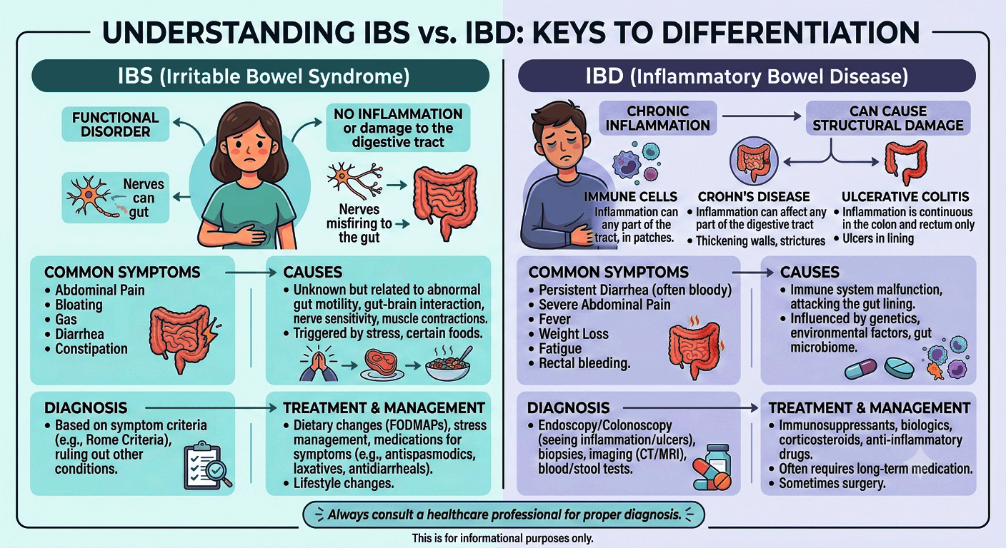
Hidden Causes of Chronic Abdominal Pain

Hidden Causes of Chronic Abdominal Pain

And Digestive Problems

Introduction

Chronic abdominal pain and ongoing digestive problems are among the leading causes of medical consultations worldwide. Many people experience symptoms such as bloating, stomach discomfort, constipation, diarrhea, nausea, indigestion, or unexplained weight loss for months or even years without identifying the true cause. While some conditions are mild and manageable, others may indicate serious disorders involving the digestive tract, liver, pancreas, kidneys, immune system, hormones, or even cancer.

Discover the hidden causes of chronic abdominal pain and digestive problems, including IBS, ulcers, gallbladder disease, food intolerances, pancreatitis, and more. Learn warning signs, diagnosis, and treatment options.

For patients, learning about the hidden causes of persistent digestive symptoms can encourage earlier medical evaluation and treatment. For medical students, chronic abdominal pain is an important clinical topic that requires to be detailed history-taking, physical examination, and a structured diagnostic approach.

This article explores the lesser-known causes of chronic abdominal pain and digestive disorders, along with their symptoms, diagnostic methods, treatment options, and warning signs that should not be overlooked.

What Is Chronic Abdominal Pain?

Chronic abdominal pain generally refers to pain that continues for at least three months. It may occur constantly or come and go, affecting different regions of the abdomen.

The abdominal cavity contains several important organs, including:

  • Stomach
  • Small intestine
  • Large intestine (colon)
  • Liver
  • Gallbladder
  • Pancreas
  • Kidneys
  • Urinary tract
  • Blood vessels
  • Reproductive organs

Because these organs share nerve pathways, pinpointing the exact source of abdominal pain can sometimes be difficult.

Common digestive symptoms associated with chronic abdominal pain include:

  • Bloating
  • Excessive gas
  • Nausea
  • Vomiting
  • Heartburn
  • Diarrhea
  • Constipation
  • Reduced appetite
  • Weight loss
  • Feeling full quickly
  • Fatigue
  • Blood in stool
  • Difficulty swallowing

Persistent digestive complaints should always be properly evaluated, as they may indicate structural, inflammatory, infectious, metabolic, or functional disorders.

Functional Gastrointestinal Disorders

One of the most frequent hidden causes of chronic abdominal pain is functional gastrointestinal disorders, where symptoms are present even though no clear structural abnormality is found.

Irritable Bowel Syndrome (IBS)

Irritable Bowel Syndrome is a common condition marked by:

  • Recurrent abdominal discomfort
  • Bloating
  • Changes in bowel habits
  • Constipation
  • Diarrhea
  • Mucus in stool

Symptoms often become worse with stress, specific foods, or hormonal fluctuations.

Types of IBS

  • IBS with constipation (IBS-C)
  • IBS with diarrhea (IBS-D)
  • Mixed IBS (IBS-M)

Possible Causes

  • Abnormal intestinal movement
  • Increased gut sensitivity
  • Dysfunction of the gut-brain connection
  • Altered gut bacteria
  • Psychological stress

Diagnosis and Management

IBS is usually diagnosed after ruling out more serious conditions. Treatment may involve:

  • Dietary adjustments
  • Stress management
  • Fiber supplements
  • Probiotics
  • Antispasmodic medications
  • Psychological therapy when appropriate

Food Intolerances and Hidden Dietary Triggers

Many individuals with chronic digestive symptoms unknowingly suffer from food intolerances.

Lactose Intolerance

Lactose intolerance develops when the body does not produce enough lactase enzyme to digest dairy products.

Symptoms

  • Bloating
  • Gas
  • Abdominal cramping
  • Diarrhea
  • Nausea

Symptoms typically occur after consuming milk or dairy products.

Gluten Sensitivity and Celiac Disease

Celiac disease is an autoimmune disorder triggered by gluten consumption.

Symptoms

  • Persistent diarrhea
  • Weight loss
  • Iron deficiency anemia
  • Abdominal pain
  • Bloating
  • Fatigue
  • Malnutrition

If untreated, celiac disease can damage the small intestine and lead to long-term complications.

Diagnosis

  • Tissue transglutaminase antibody testing
  • Endoscopy with intestinal biopsy

Treatment

A lifelong gluten-free diet is essential.

Fructose and FODMAP Intolerance

Certain carbohydrates can ferment in the intestines and trigger:

  • Bloating
  • Gas
  • Diarrhea
  • Cramping

Low-FODMAP diets may benefit selected patients.

Inflammatory Bowel Disease (IBD)

Inflammatory Bowel Disease includes:

  • Crohn’s disease
  • Ulcerative colitis

These conditions involve chronic inflammation of the gastrointestinal tract.

Common Symptoms

  • Persistent abdominal pain
  • Chronic diarrhea
  • Blood in stool
  • Weight loss
  • Fatigue
  • Fever
  • Malnutrition

Crohn’s Disease

Crohn’s disease can affect any portion of the digestive tract from mouth to anus.

Possible Complications

  • Intestinal narrowing
  • Fistulas
  • Abscesses
  • Malabsorption

Ulcerative Colitis

Ulcerative colitis mainly affects the colon and rectum.

Symptoms

  • Bloody diarrhea
  • Urgent bowel movements
  • Rectal bleeding
  • Abdominal cramps

Diagnosis and Treatment

Diagnosis may include colonoscopy, biopsy, imaging, and stool tests. Treatment options include:

  • Anti-inflammatory medications
  • Immunosuppressants
  • Biologic therapies
  • Surgery in severe cases

Chronic Gastritis and Peptic Ulcers

Chronic Gastritis

Gastritis refers to inflammation of the stomach lining and may result from:

  • Helicobacter pylori infection
  • Long-term NSAID use
  • Alcohol consumption
  • Autoimmune disease
  • Stress-related injury

Symptoms

  • Upper abdominal discomfort
  • Nausea
  • Bloating
  • Indigestion
  • Early fullness

Peptic Ulcer Disease

Peptic ulcers develop in the stomach or duodenum.

Warning Signs

  • Burning stomach pain
  • Vomiting blood
  • Black stools
  • Anemia
  • Weight loss

Diagnosis and Treatment

Diagnosis often involves endoscopy and H. pylori testing. Treatment may include:

  • Proton pump inhibitors
  • Antibiotics
  • Avoiding NSAIDs

Gallbladder Disorders

Gallbladder problems are often mistaken for indigestion or acidity.

Gallstones

Gallstones can temporarily block bile flow.

Symptoms

  • Right upper abdominal pain
  • Pain after fatty meals
  • Nausea
  • Vomiting
  • Bloating

Pain may spread to the back or right shoulder.

Chronic Cholecystitis

Repeated gallbladder inflammation can lead to ongoing digestive discomfort.

Diagnosis and Treatment

Doctors may use ultrasound, liver function tests, or CT scans. Treatment often involves dietary modifications or gallbladder removal surgery.

Pancreatic Disorders

The pancreas plays an essential role in digestion and blood sugar regulation.

Chronic Pancreatitis

This condition causes long-term pancreatic inflammation and scarring.

Causes

  • Alcohol use
  • Gallstones
  • Genetic disorders
  • Autoimmune diseases
  • High triglyceride levels

Symptoms

  • Upper abdominal pain radiating to the back
  • Fatty stools
  • Weight loss
  • Malabsorption
  • Diabetes

Complications

  • Pancreatic insufficiency
  • Nutritional deficiencies
  • Chronic pain

Pancreatic Cancer

Pancreatic cancer may initially present with subtle digestive symptoms.

Warning Signs

  • Persistent abdominal pain
  • Unexplained weight loss
  • Jaundice
  • Appetite loss
  • Newly diagnosed diabetes

Liver Diseases and Digestive Symptoms

Several chronic liver disorders can contribute to digestive complaints.

Fatty Liver Disease

Nonalcoholic fatty liver disease is increasingly common due to obesity and diabetes.

Symptoms

  • Right upper abdominal discomfort
  • Fatigue
  • Weakness
  • Mild digestive symptoms

Chronic Hepatitis

Chronic liver inflammation may cause:

  • Abdominal pain
  • Nausea
  • Appetite loss
  • Jaundice
  • Enlarged liver

Cirrhosis

Advanced liver disease may lead to:

  • Fluid accumulation in the abdomen
  • Digestive problems
  • Gastrointestinal bleeding
  • Malnutrition

Small Intestinal Bacterial Overgrowth (SIBO)

SIBO occurs when excessive bacteria grow in the small intestine.

Symptoms

  • Bloating
  • Gas
  • Diarrhea
  • Abdominal discomfort
  • Malabsorption
  • Vitamin deficiencies

Risk Factors

  • Diabetes
  • Intestinal surgery
  • Motility disorders
  • Chronic pancreatitis

Treatment

Management may include antibiotics, nutritional support, and treatment of underlying conditions.

Chronic Infections

Some infections can produce long-term digestive symptoms.

Parasitic Infections

Possible symptoms include:

  • Chronic diarrhea
  • Weight loss
  • Abdominal cramps
  • Malnutrition

Abdominal Tuberculosis

Tuberculosis may affect the intestines or abdominal lymph nodes.

Symptoms

  • Chronic abdominal pain
  • Fever
  • Weight loss
  • Altered bowel habits

Gynecological Causes in Women

Certain reproductive disorders may mimic gastrointestinal disease.

Endometriosis

This condition occurs when tissue similar to the uterine lining grows outside the uterus.

Symptoms

  • Chronic pelvic pain
  • Painful periods
  • Pain during intercourse
  • Bloating
  • Bowel-related symptoms

Ovarian Disorders

Ovarian cysts or tumors may cause:

  • Abdominal fullness
  • Pelvic pain
  • Digestive discomfort
  • Early satiety

Kidney and Urinary Tract Causes

Kidney disorders may also present as abdominal discomfort.

Kidney Stones

Symptoms may include:

  • Flank pain
  • Lower abdominal pain
  • Nausea
  • Vomiting
  • Blood in urine

Chronic Kidney Disease

Chronic kidney disease may lead to:

  • Nausea
  • Poor appetite
  • Vomiting
  • Fatigue
  • Abdominal discomfort

Hormonal and Metabolic Causes

Diabetes Mellitus

Diabetes can damage the nerves controlling digestion.

Diabetic Gastroparesis

This condition slows stomach emptying and may cause:

  • Nausea
  • Vomiting
  • Bloating
  • Early fullness
  • Abdominal discomfort

Thyroid Disorders

  • Hypothyroidism may lead to constipation and bloating
  • Hyperthyroidism may cause diarrhea

Psychological Factors and the Gut-Brain Connection

Mental health significantly affects digestive function.

Conditions linked to digestive symptoms include:

  • Anxiety
  • Depression
  • Chronic stress
  • Panic disorders

Stress may worsen:

  • IBS
  • Acid reflux
  • Functional dyspepsia
  • Appetite changes

The gut-brain axis plays a major role in chronic digestive symptoms.

Gastrointestinal Cancers

Persistent digestive complaints should never be ignored because some cancers develop gradually.

Colorectal Cancer

Possible symptoms include:

  • Changes in bowel habits
  • Blood in stool
  • Weight loss
  • Anemia
  • Chronic abdominal pain

Stomach Cancer

Early symptoms may include:

  • Indigestion
  • Feeling full quickly
  • Weight loss
  • Nausea

Ovarian Cancer

Ovarian cancer may also present with bloating and abdominal discomfort.

Warning Signs Requiring Immediate Medical Attention

The following symptoms may indicate serious disease:

  • Blood in stool
  • Vomiting blood
  • Black tarry stools
  • Significant weight loss
  • Persistent vomiting
  • Difficulty swallowing
  • Ongoing fever
  • Night sweats
  • Severe anemia
  • Jaundice
  • New digestive symptoms in older adults
  • Family history of gastrointestinal cancer

Prompt medical evaluation is essential when these symptoms occur.

Diagnostic Evaluation

A thorough and systematic assessment is necessary.

Medical History

Doctors commonly ask about:

  • Pain location and duration
  • Relationship to meals
  • Bowel habits
  • Weight changes
  • Medication use
  • Diet
  • Family history
  • Psychological stress

Physical Examination

The abdomen is checked for:

  • Tenderness
  • Masses
  • Enlarged organs
  • Fluid buildup
  • Signs of malnutrition

Laboratory Tests

Common investigations include:

  • Complete blood count
  • Liver function tests
  • Kidney function tests
  • Blood sugar testing
  • Inflammatory markers
  • Thyroid function tests
  • Stool analysis
  • Urine analysis

Imaging and Procedures

Depending on symptoms, tests may include:

  • Ultrasound
  • CT scan
  • MRI
  • Endoscopy
  • Colonoscopy
  • Breath testing
  • Biopsy

Treatment Approaches

Treatment depends on the underlying condition.

Lifestyle and Dietary Changes

  • Avoid trigger foods
  • Eat balanced meals
  • Stay hydrated
  • Reduce alcohol intake
  • Quit smoking
  • Exercise regularly
  • Manage stress

Medications

Treatment may involve:

  • Acid-reducing drugs
  • Antibiotics
  • Anti-inflammatory medications
  • Probiotics
  • Antispasmodics
  • Digestive enzyme supplements
  • Immunosuppressants

Surgery

Some conditions may require surgery, including:

  • Gallstones
  • Certain cancers
  • Severe inflammatory bowel disease
  • Intestinal obstruction

Prevention and Long-Term Digestive Health

Healthy habits can reduce the risk of chronic digestive disorders.

Preventive Measures

  • Eat fiber-rich foods
  • Limit processed foods
  • Maintain a healthy weight
  • Exercise consistently
  • Avoid unnecessary NSAID use
  • Limit alcohol intake
  • Practice food safety
  • Undergo recommended cancer screenings

Seeking medical care early often helps prevent complications.

Conclusion

Chronic abdominal pain and digestive problems can arise from many hidden causes, including functional disorders, food intolerances, inflammatory diseases, infections, metabolic conditions, and cancers. Since symptoms often overlap, accurate diagnosis requires careful evaluation and systematic clinical reasoning.

Persistent digestive symptoms should never be ignored, especially when accompanied by weight loss, bleeding, anemia, fever, or severe pain. Early diagnosis and treatment improve outcomes and help prevent complications.

For medical students, chronic abdominal pain demonstrates the importance of detailed history-taking, broad clinical thinking, and understanding the complex interactions between digestive, metabolic, infectious, hormonal, and psychological factors.

Timely medical evaluation, healthy lifestyle choices, and evidence-based care remain essential for maintaining digestive health and improving quality of life.

Hidden Signs of Gastrointestinal Bleeding

Gastrointestinal bleeding

You Should Never Ignore

Introduction

Gastrointestinal (GI) bleeding refers to bleeding that occurs anywhere within the digestive system, extending from the mouth to the rectum. The severity can vary widely, ranging from mild, slow blood loss to severe and life-threatening hemorrhage. While some individuals experience obvious symptoms such as vomiting blood or black stools, others develop subtle signs that often go unnoticed until serious complications arise.

This article discusses the concealed symptoms, causes, risk factors, diagnostic approaches, treatment methods, and prevention of gastrointestinal bleeding.

Identifying these less obvious symptoms early is essential for patients and healthcare students alike. Prompt diagnosis can reduce the risk of complications including severe anemia, shock, organ damage, and even death.

Understanding Gastrointestinal Bleeding

GI bleeding is not considered a disease on its own; instead, it is a symptom of another disorder affecting the digestive tract.

The digestive system includes:

  • Mouth
  • Esophagus
  • Stomach
  • Small intestine
  • Large intestine (colon)
  • Rectum
  • Anus

Types of GI Bleeding

Upper Gastrointestinal Bleeding

Occurs above the ligament of Treitz and commonly affects:

  • Esophagus
  • Stomach
  • Duodenum

Lower Gastrointestinal Bleeding

Occurs below the ligament of Treitz and may involve:

  • Small intestine
  • Colon
  • Rectum
  • Anus

Bleeding may also be categorized as:

  • Acute: sudden and severe
  • Chronic: slow and ongoing
  • Occult: hidden blood not visible in stool

Why GI Bleeding Is Serious

Many people believe gastrointestinal bleeding always produces dramatic symptoms. However, slow chronic bleeding can continue unnoticed for weeks or even months.

Unrecognized blood loss may lead to:

  • Iron deficiency anemia
  • Persistent fatigue
  • Reduced oxygen supply to tissues
  • Increased strain on the heart
  • Dizziness and falls
  • Circulatory instability
  • Hypovolemic shock in severe cases

In some situations, GI bleeding may be the earliest sign of conditions such as stomach ulcers, inflammatory bowel disease, or gastrointestinal cancers.

Hidden Signs of Gastrointestinal Bleeding

1. Persistent Fatigue and Weakness

One of the most common but overlooked signs of chronic GI bleeding is ongoing fatigue.

Gradual blood loss lowers hemoglobin levels, reducing oxygen delivery throughout the body. Patients may experience:

  • Constant tiredness
  • Lack of energy
  • Difficulty focusing
  • Reduced ability to exercise
  • General weakness

Iron deficiency anemia in adults without a clear explanation should raise concern for occult GI bleeding.

2. Pale Skin and Pale Eyelids

Long-term blood loss may cause anemia, resulting in:

  • Pale skin
  • Pale nail beds
  • Pale inner eyelids (conjunctival pallor)

Because this develops slowly, many patients fail to notice it.

3. Shortness of Breath With Mild Activity

Lower hemoglobin levels reduce the blood’s oxygen-carrying ability.

Symptoms may include:

  • Breathlessness while walking
  • Difficulty climbing stairs
  • Fast heartbeat during exertion
  • Poor stamina

Severe anemia may even trigger chest discomfort in elderly patients or those with heart disease.

4. Dizziness or Fainting

Blood loss decreases circulating blood volume and oxygen supply.

Patients may develop:

  • Lightheadedness
  • Dizziness when standing
  • Near-fainting episodes
  • Syncope (fainting)

Dizziness accompanied by black stools or vomiting blood requires urgent medical attention.

5. Black, Tarry Stools (Melena)

Melena is a classic symptom of upper GI bleeding.

The stool may appear:

  • Black
  • Sticky
  • Tarry
  • Foul-smelling

This occurs because blood becomes digested while traveling through the digestive tract.

6. Hidden Blood in Stool

Occult bleeding involves blood that is not visible to the naked eye.

Patients may have:

  • Normal-looking stool
  • Chronic anemia
  • Fatigue without obvious bleeding

Tests such as FOBT or FIT can identify hidden blood in stool.

Possible causes include:

  • Colon polyps
  • Colorectal cancer
  • Peptic ulcers
  • Inflammatory bowel disease
  • Angiodysplasia

7. Vomiting Blood (Hematemesis)

Hematemesis is a dangerous sign of upper GI bleeding.

Vomited material may appear:

  • Bright red
  • Dark red
  • Similar to coffee grounds

Coffee-ground vomit usually indicates partially digested blood.

Common causes include:

  • Peptic ulcer disease
  • Esophageal varices
  • Gastritis
  • Mallory-Weiss tears

Immediate medical care is necessary.

8. Abdominal Pain or Discomfort

Some bleeding disorders also cause abdominal symptoms such as:

  • Burning upper abdominal pain
  • Cramping
  • Bloating
  • Tenderness
  • Persistent discomfort after eating

These symptoms may suggest:

9. Unexplained Weight Loss

Weight loss combined with GI bleeding may indicate a serious underlying illness.

Potential causes include:

  • Stomach cancer
  • Colorectal cancer
  • Chronic inflammatory bowel disease
  • Malabsorption disorders

10. Changes in Bowel Habits

Lower GI bleeding may occur alongside altered bowel patterns.

Warning signs include:

  • Persistent constipation
  • Chronic diarrhea
  • Narrow stools
  • Increased urgency
  • Feeling of incomplete bowel emptying

These symptoms may occur in:

  • Colorectal cancer
  • Inflammatory bowel disease
  • Diverticular disease
  • Hemorrhoids

11. Rapid Heartbeat or Palpitations

To compensate for blood loss, the body increases heart rate.

Patients may notice:

  • Heart palpitations
  • Racing pulse
  • Anxiety-like feelings
  • Weak pulse

Severe blood loss may progress to shock.

12. Rectal Bleeding

Visible rectal bleeding may appear:

  • Bright red
  • Maroon-colored
  • Mixed with stool
  • On toilet paper

Although hemorrhoids are common, rectal bleeding should always be evaluated.

Common Causes of GI Bleeding

Frequent causes include:

  • Peptic ulcer disease
  • Gastritis
  • Esophageal varices
  • Hemorrhoids
  • Diverticular disease
  • Inflammatory bowel disease
  • Gastrointestinal cancers
  • Angiodysplasia

Risk Factors

Factors that increase risk include:

  • NSAID use
  • Aspirin and blood thinners
  • Liver disease
  • Heavy alcohol use
  • Smoking
  • Older age
  • Previous GI disorders

Diagnosis

Doctors may use:

  • Medical history and physical examination
  • Complete blood count (CBC)
  • Stool testing (FOBT/FIT)
  • Liver function tests
  • Upper endoscopy (EGD)
  • Colonoscopy
  • Capsule endoscopy
  • CT angiography

Treatment

Treatment depends on the source and severity of bleeding.

Emergency Care

Severe cases may require:

  • IV fluids
  • Blood transfusion
  • Oxygen therapy
  • Monitoring of vital signs

Medications

Common treatments include:

  • Proton pump inhibitors (PPIs)
  • Antibiotics for H. pylori
  • Vasoconstrictor therapy such as octreotide

Endoscopic Treatment

Endoscopy may control bleeding through:

  • Injection therapy
  • Thermal coagulation
  • Hemoclip placement
  • Variceal band ligation

Surgery

Surgery may be necessary if:

  • Bleeding cannot be controlled
  • Cancer is present
  • Perforation develops
  • Recurrent severe bleeding occurs

Complications of Untreated GI Bleeding

Ignoring GI bleeding may lead to:

  • Severe anemia
  • Hypovolemic shock
  • Organ failure
  • Cardiac complications
  • Malnutrition
  • Death

When Immediate Medical Attention Is Needed

Seek emergency care for:

  • Vomiting blood
  • Black tarry stools
  • Severe rectal bleeding
  • Fainting
  • Rapid heartbeat
  • Chest pain
  • Severe weakness
  • Shortness of breath
  • Confusion

Prevention Strategies

Helpful preventive measures include:

  • Avoiding excessive NSAID use
  • Limiting alcohol intake
  • Quitting smoking
  • Eating a balanced diet rich in fiber and iron
  • Undergoing recommended cancer screening
  • Investigating unexplained anemia promptly

Important Learning Points

  • GI bleeding can remain hidden and symptom-free
  • Iron deficiency anemia in adults requires investigation
  • Melena usually indicates upper GI bleeding
  • Hematochezia commonly suggests lower GI bleeding
  • Hemodynamic instability signals major blood loss
  • Endoscopy is both diagnostic and therapeutic
  • NSAIDs are a major cause of peptic ulcer bleeding

Conclusion

Hidden gastrointestinal bleeding can gradually harm health before severe symptoms become obvious. Fatigue, anemia, dizziness, black stools, unexplained weight loss, and bowel habit changes should never be ignored.

Early medical assessment can prevent serious complications and improve outcomes. For medical students, understanding the many presentations of GI bleeding is essential for timely diagnosis and treatment.

Recognizing these warning signs early may save lives, particularly when bleeding is linked to ulcers, inflammatory bowel disease, or gastrointestinal cancers. Anyone experiencing symptoms suggestive of GI bleeding should seek prompt medical evaluation.

Disclaimer: The information provided in this blog post is for educational and informational purposes only and should not be considered medical advice. It is not intended to replace professional medical consultation, diagnosis, or treatment. Always seek the guidance of a qualified healthcare professional regarding any medical condition or health-related concerns. The author and publisher are not responsible for any actions taken based on the information presented in this article.
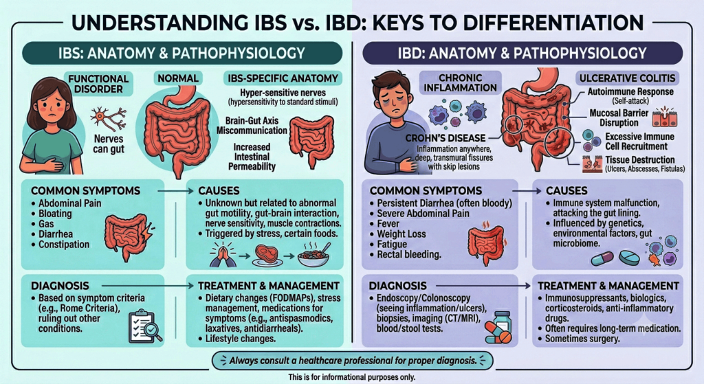
Right Upper Quadrant Pain:

right upper quadrant pain

Causes, Symptoms, and When to Seek Medical Help

Introduction

Right upper quadrant (RUQ) pain refers to discomfort felt on the upper right side of the abdomen beneath the ribs. This area contains several vital organs responsible for digestion, metabolism, filtration, and circulation. The pain can vary from mild, short-term discomfort to intense pain that may require immediate medical care.

Learn about right upper quadrant pain, including its causes, symptoms, diagnosis, treatment options, and when to seek medical attention for RUQ pain.

The type, intensity, and duration of the pain often help identify its underlying cause. Some conditions, such as indigestion or muscle strain, are relatively minor, while others including gallbladder disorders, liver disease, kidney infections, or appendicitis may be serious. Recognizing related symptoms and possible causes is important for prompt diagnosis and treatment.

Organs Associated with Right Upper Quadrant Pain

Several organs are located within or near the RUQ, and pain may arise directly from them or from nearby structures.

1. Liver

The liver is the body’s largest internal organ and plays a major role in detoxification, metabolism, bile formation, and nutrient storage. Conditions such as liver inflammation, enlargement, infection, or tumors may trigger RUQ pain.

2. Gallbladder

The gallbladder stores bile produced by the liver and releases it during digestion. Gallstones, inflammation, or blockage of bile flow commonly lead to severe RUQ discomfort.

3. Bile Ducts

These channels carry bile from the liver and gallbladder to the intestine. Infections or blockages in these ducts may result in pain, fever, and jaundice.

4. Right Kidney

Part of the right kidney lies near the RUQ. Kidney infections, stones, or inflammation can cause pain that spreads toward the flank or back.

5. Pancreas

Although mainly central in location, the head of the pancreas extends toward the right side. Pancreatic inflammation may produce pain in the upper abdomen and RUQ.

6. Intestines

The upper portions of the intestines can also contribute to RUQ pain due to inflammation, infection, gas buildup, or digestive disorders.

7. Right Lung and Pleura

Conditions affecting the lower right lung or pleural lining, such as pneumonia or pleuritis, may create pain felt in the RUQ.

8. Muscles and Ribs

Injuries or strain involving muscles, ribs, or the chest wall can mimic pain originating from internal organs.

Mechanisms Behind Right Upper Quadrant Pain

The development of RUQ pain depends on the affected organ and the disease process involved.

Inflammation

Inflammatory conditions stimulate pain receptors and cause tissue swelling. Disorders such as hepatitis, pancreatitis, cholecystitis, and kidney infections commonly produce pain through inflammation.

Obstruction

Blockage within ducts or hollow organs raises internal pressure and stretches tissues, resulting in discomfort. Gallstones obstructing the bile duct are a common example.

Infection

Viral, bacterial, or parasitic infections trigger immune responses that cause tissue injury, swelling, fever, and pain.

Ischemia

Reduced blood supply deprives tissues of oxygen and nutrients, causing damage and pain.

Muscle Spasm

Spasms in smooth muscles of the intestines or bile ducts may cause intermittent cramping sensations.

Capsule Stretching

Organs such as the liver and kidneys are enclosed in fibrous coverings. Enlargement or swelling stretches these capsules and leads to discomfort.

Common Causes of Right Upper Quadrant Pain

1. Gallstones (Cholelithiasis)

Gallstones are hardened deposits inside the gallbladder that may block bile flow and cause sudden, severe pain known as biliary colic.

Symptoms

  • Sharp RUQ pain after eating fatty foods
  • Nausea and vomiting
  • Pain spreading to the right shoulder or back

2. Acute Cholecystitis

This refers to inflammation of the gallbladder, usually caused by gallstone blockage.

Symptoms

  • Continuous severe RUQ pain
  • Fever
  • Tenderness over the gallbladder
  • Nausea and vomiting

3. Hepatitis

Hepatitis is liver inflammation caused by viruses, alcohol, medications, or autoimmune conditions.

Symptoms

  • RUQ discomfort
  • Fatigue
  • Reduced appetite
  • Jaundice
  • Dark urine

4. Fatty Liver Disease

Excess fat buildup in the liver may enlarge the organ and produce discomfort.

Risk Factors

  • Obesity
  • Diabetes
  • High cholesterol
  • Heavy alcohol use

5. Liver Abscess

A liver abscess is a pus-filled infection within the liver.

Symptoms

  • Fever and chills
  • Severe RUQ pain
  • Weight loss
  • Weakness

6. Pancreatitis

Inflammation of the pancreas may cause upper abdominal pain extending toward the RUQ.

Causes

  • Gallstones
  • Alcohol misuse
  • Elevated triglycerides

7. Peptic Ulcer Disease

Ulcers in the stomach or duodenum can produce pain that resembles RUQ discomfort.

8. Kidney Stones

Stones in the right kidney may cause flank pain radiating to the RUQ.

Symptoms

  • Severe colicky pain
  • Blood in urine
  • Pain during urination

9. Kidney Infection (Pyelonephritis)

A bacterial kidney infection can lead to fever, flank pain, and RUQ discomfort.

10. Pneumonia

Infection of the lower right lung may irritate the diaphragm and cause referred abdominal pain.

11. Appendicitis

Although usually associated with lower abdominal pain, appendicitis may initially present with upper abdominal discomfort.

12. Musculoskeletal Causes

Muscle strain, rib fractures, or chest wall inflammation can also produce localized RUQ pain.

Symptoms Associated with Right Upper Quadrant Pain

Symptoms differ depending on the underlying condition.

Common Symptoms

  • Sharp or dull pain in the RUQ
  • Burning or cramping sensation
  • Pain radiating to the shoulder or back
  • Nausea and vomiting
  • Fever
  • Bloating
  • Loss of appetite
  • Fatigue
  • Abdominal tenderness

Warning Signs Requiring Immediate Attention

  • Jaundice
  • Persistent vomiting
  • High fever
  • Breathing difficulty
  • Black stools
  • Severe abdominal stiffness
  • Fainting or dizziness
  • Confusion
  • Low BP (Hypotension)
  • Rebound tenderness.

These symptoms may indicate a serious medical emergency.

Diagnosis of Right Upper Quadrant Pain

Proper diagnosis requires a detailed medical history and physical examination.

Medical History

Doctors typically ask about:

  • Duration and nature of pain
  • Relationship to meals
  • Associated symptoms
  • Alcohol use
  • Medication history
  • Previous illnesses

Physical Examination

The physician may assess:

  • Tenderness
  • Enlarged organs
  • Fever
  • Jaundice
  • Abdominal guarding

Laboratory Tests

Blood Tests

  • Complete blood count (CBC)
  • Liver function tests (LFTs)
  • Kidney function tests
  • Lipid Profile
  • Pancreatic enzyme levels (S Amylase, S Lipase)
  • Inflammatory markers
  • Blood sugar (RBS)

Urine Analysis

Helps identify kidney infections or blood in the urine.

Imaging Studies

Ultrasound

Often the first imaging test used to evaluate gallstones, liver conditions, and bile duct disorders.

CT Scan

Provides detailed images of abdominal organs.

MRI or MRCP

Useful for examining bile ducts and liver structures.

Chest X-ray

May reveal pneumonia or lung-related causes.

Endoscopy

Used to detect ulcers or gastrointestinal diseases.

When to Seek Medical Care

Medical attention is recommended if RUQ pain:

  • Persists for several hours
  • Becomes severe or progressively worse
  • Occurs with fever
  • Causes jaundice
  • Is associated with vomiting
  • Occurs alongside chest pain or breathing problems
  • Leads to weakness or fainting
  • Involves blood in vomit or stool

Sudden, intense abdominal pain requires immediate emergency evaluation.

Treatment of Right Upper Quadrant Pain

Treatment depends on the underlying cause.

1. Medications

Pain Relievers

Used to control mild to moderate pain.

Antibiotics

Prescribed for bacterial infections such as kidney infections or cholecystitis.

Antiviral Medications

Helpful in certain viral hepatitis infections.

Antacids and Proton Pump Inhibitors

Used for ulcers and acid reflux conditions.

Antispasmodics

Help reduce spasms in the bile ducts or intestines.

2. Dietary Changes

  • Avoid fried and fatty foods
  • Limit alcohol consumption
  • Eat smaller, frequent meals
  • Stay well hydrated
  • Increase fruits and vegetables intake

3. Surgical Treatment

Cholecystectomy

Removal of the gallbladder for gallstones or gallbladder inflammation.

Drainage of Liver Abscess

Performed when pus accumulates inside the liver.

Surgery for Appendicitis or Obstruction

Needed in emergency situations.

4. Management of Liver Disorders

  • Weight loss for fatty liver disease
  • Avoiding alcohol
  • Treating hepatitis
  • Regular liver monitoring

5. Treatment of Kidney Conditions

  • Adequate hydration
  • Antibiotics for infections
  • Removal of kidney stones if required

Prevention and Precautions

Many causes of RUQ pain can be reduced through healthy lifestyle habits.

Healthy Diet

  • Limit oily and processed foods
  • Eat balanced, fiber-rich meals
  • Reduce excess sugar intake

Maintain a Healthy Weight

Obesity increases the likelihood of gallstones and fatty liver disease.

Avoid Excess Alcohol

Alcohol can damage both the liver and pancreas.

Stay Hydrated

Proper hydration supports digestive and kidney health.

Exercise Regularly

Physical activity improves metabolism and overall organ function.

Vaccination

Vaccines for hepatitis A and B help prevent viral hepatitis.

Safe Medication Practices

Avoid unnecessary medications that may harm the liver.

Routine Medical Checkups

Early detection of liver disease, gallstones, or metabolic disorders lowers the risk of complications.

Conclusion

Right upper quadrant pain is a common symptom with many possible causes involving the liver, gallbladder, kidneys, pancreas, lungs, intestines, or musculoskeletal structures. While some conditions are mild, others can become life-threatening if not treated promptly.

Recognizing associated symptoms, warning signs, and risk factors is essential for early diagnosis and proper treatment. Healthy lifestyle choices, balanced nutrition, preventive care, and timely medical evaluation all play an important role in minimizing complications and improving overall health outcomes.

Anyone experiencing persistent or severe RUQ pain should seek professional medical care for accurate diagnosis and treatment.

Disclaimer: The information provided in this blog post is for educational and informational purposes only and should not be considered medical advice. It is not intended to replace professional medical consultation, diagnosis, or treatment.
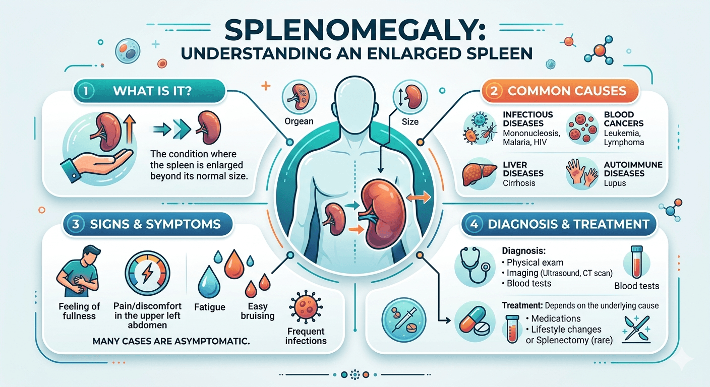
Pancreatitis: A Complete Medical Guide For Patients And Caregivers

Understanding Pancreatitis acute and chronic

Introduction

Pancreatitis is a condition marked by inflammation of the pancreas, which may occur suddenly as acute pancreatitis or develop gradually as chronic pancreatitis. The pancreas, located behind the stomach, plays a vital role in both digestion and blood sugar regulation. It produces enzymes that aid in digesting fats, proteins, and carbohydrates, while also releasing hormones like insulin and glucagon to control glucose levels.

This guide explains pancreatitis in detail, including its causes, symptoms, diagnostic methods, treatment approaches, possible complications, and recovery recommendations.

Pancreatitis develops when digestive enzymes become activated within the pancreas rather than in the small intestine. As a result, the pancreas begins to digest itself, leading to inflammation and tissue damage. The severity may vary from mild inflammation that improves with supportive care to severe disease involving organ failure and systemic complications.

Acute pancreatitis appears suddenly and often resolves with proper treatment, while chronic pancreatitis causes ongoing inflammation that permanently damages pancreatic tissue and gradually impairs pancreatic function.

Understanding the disease process, warning signs, causes, and treatment strategies is essential for early diagnosis and prevention of serious complications.

How Pancreatitis Develops

The primary problem in pancreatitis is the early activation of digestive enzymes inside pancreatic tissue. Normally, these enzymes remain inactive until they reach the duodenum. In pancreatitis, this protective system fails.

Enzyme Activation and Self-Digestion

Activation of trypsin within pancreatic acinar cells triggers a chain reaction of enzyme activation. These enzymes begin damaging pancreatic tissue, leading to:

  • Cellular destruction
  • Fat necrosis
  • Bleeding
  • Swelling
  • Inflammation

This destructive process is known as pancreatic autodigestion.

Inflammatory Reaction

When pancreatic injury occurs, inflammatory chemicals such as cytokines and chemokines are released. These attract immune cells and worsen tissue damage. Severe inflammation can spread throughout the body and lead to systemic inflammatory response syndrome (SIRS).

Impaired Blood Circulation

Inflammation increases vascular permeability and swelling, reducing blood flow within the pancreas. Poor circulation may eventually cause tissue death and pancreatic necrosis.

Systemic Complications

In severe pancreatitis, inflammatory substances may enter the bloodstream and affect other organs, potentially causing:

  • Acute respiratory distress syndrome (ARDS)
  • Acute kidney injury
  • Shock
  • Sepsis
  • Multi-organ failure

Chronic Pancreatitis

Repeated inflammatory episodes can lead to fibrosis, calcification, and permanent destruction of pancreatic tissue. Over time, both digestive and hormonal functions decline, resulting in malabsorption and diabetes.

Causes of Pancreatitis

Pancreatitis may develop from several underlying conditions. The causes often differ between acute and chronic disease.

Common Causes of Acute Pancreatitis

Gallstones

Gallstones are one of the most frequent causes. Small stones can block the pancreatic duct or ampulla of Vater, triggering enzyme activation and inflammation.

Alcohol Use

Heavy alcohol consumption damages pancreatic cells and thickens pancreatic secretions, contributing to duct blockage and inflammation.

Hypertriglyceridemia

Very high triglyceride levels may trigger pancreatitis due to the production of toxic free fatty acids.

Medications

Some medications linked to pancreatitis include:

  • Azathioprine
  • Valproic acid
  • Thiazide diuretics
  • Corticosteroids
  • Didanosine

Infections

Certain viral infections, including mumps, hepatitis viruses, and cytomegalovirus, can inflame the pancreas.

Trauma

Abdominal injuries or surgical procedures may damage pancreatic tissue.

ERCP Procedures

Endoscopic Retrograde Cholangiopancreatography (ERCP) is a recognized cause of procedure-related pancreatitis.

Autoimmune Disorders

Autoimmune pancreatitis occurs when the immune system attacks pancreatic tissue.

Metabolic Conditions

Additional causes include:

  • Hypercalcemia
  • Genetic abnormalities
  • Obesity

Idiopathic Cases

In some patients, no specific cause can be identified.

Causes of Chronic Pancreatitis

Common causes include:

  • Long-term alcohol abuse
  • Smoking
  • Genetic disorders
  • Autoimmune pancreatitis
  • Repeated acute pancreatitis attacks
  • Obstructive pancreatic duct disease

Symptoms and Signs of Pancreatitis

Clinical manifestations vary depending on the severity and duration of inflammation.

Symptoms of Acute Pancreatitis

Abdominal Pain

Severe upper abdominal pain is the most characteristic symptom. The pain often:

  • Radiates to the back
  • Starts suddenly
  • Becomes worse after meals
  • Improves slightly when leaning forward

Nausea and Vomiting

Persistent nausea and repeated vomiting are frequently present.

Fever

Inflammation or infection may lead to elevated body temperature.

Abdominal Tenderness

The abdomen may become swollen and painful on examination.

Loss of Appetite

Eating often worsens pain, causing patients to avoid food.

Tachycardia and Low Blood Pressure

Severe disease may cause dehydration and circulatory collapse.

Severe Features

Serious pancreatitis may present with:

  • Breathing difficulty
  • Cyanosis
  • Confusion
  • Jaundice
  • Multi-organ dysfunction

Features of Chronic Pancreatitis

Persistent or Recurrent Pain

Pain may continue for long periods or occur in repeated episodes.

Malabsorption

Reduced digestive enzyme production may cause:

  • Steatorrhea
  • Weight loss
  • Nutritional deficiencies

Diabetes Mellitus

Damage to insulin-producing cells can lead to secondary diabetes.

Pancreatic Calcification

Calcifications may appear on imaging studies.

Diagnosis of Pancreatitis

Diagnosis combines clinical symptoms, laboratory testing, and imaging studies.

Diagnostic Criteria for Acute Pancreatitis

A diagnosis is typically confirmed when at least two of the following are present:

  1. Characteristic abdominal pain
  2. Elevated pancreatic enzyme levels
  3. Imaging findings consistent with pancreatitis

Laboratory Tests

Important investigations include:

  • Serum amylase and lipase
  • Complete blood count (CBC)
  • Liver function tests
  • Kidney function tests
  • Serum calcium
  • Lipid profile
  • Blood glucose, RBS, and HbA1c
  • C-reactive protein (CRP)
  • ESR

Lipase is generally more specific and remains elevated longer than amylase.

Imaging Studies

Abdominal Ultrasound

Useful for identifying gallstones and biliary obstruction.

Contrast-Enhanced CT Scan

CT imaging helps evaluate:

  • Pancreatic inflammation
  • Necrosis
  • Fluid collections
  • Complications

MRI and MRCP

Helpful for examining pancreatic ducts and surrounding soft tissue.

Endoscopic Ultrasound (EUS)

Effective for detecting small gallstones and structural abnormalities.

Severity Assessment

Several scoring systems help predict disease severity, including:

  • Ranson criteria
  • APACHE II score
  • BISAP score
  • Glasgow score

Treatment of Pancreatitis

Management depends on the severity, cause, and associated complications.

Initial Treatment of Acute Pancreatitis

Fluid Replacement

Aggressive intravenous hydration is critical to maintain circulation and prevent complications.

Pain Relief

Strong analgesics are commonly needed to control severe abdominal pain.

Nutritional Support

Early enteral nutrition is preferred over prolonged fasting. Severe cases may require feeding through a nasojejunal tube.

Bowel Rest

Temporary restriction of oral intake may be necessary during severe episodes.

Oxygen Therapy

Supplemental oxygen is given if respiratory problems develop.

Treating Underlying Causes

Gallstone Pancreatitis

  • ERCP may be performed to relieve biliary obstruction
  • Cholecystectomy helps prevent recurrence

Alcohol-Induced Pancreatitis

Complete alcohol avoidance is essential.

Hypertriglyceridemia

Management may include:

  • Insulin therapy
  • Lipid-lowering medications
  • Plasmapheresis in severe cases

Antibiotics

Preventive antibiotics are not routinely recommended and are reserved for infected necrosis or confirmed infections.

Managing Complications

Pancreatic Necrosis

Severe necrosis may require drainage procedures or surgery.

Pancreatic Pseudocysts

Large or symptomatic pseudocysts may need endoscopic or surgical drainage.

Organ Failure

Patients with respiratory, renal, or circulatory failure often require intensive care support.

Treatment of Chronic Pancreatitis

Lifestyle Changes

Recommended measures include:

  • Stopping alcohol consumption
  • Quitting smoking
  • Following dietary modifications

Pancreatic Enzyme Replacement

Enzyme supplements help improve digestion and reduce steatorrhea.

Diabetes Treatment

Insulin therapy may become necessary.

Pain Management

Pain control strategies may involve:

  • Analgesic medications
  • Nerve blocks
  • Endoscopic procedures
  • Surgery in selected patients

Surgical Treatment

Surgery may be considered for:

  • Duct obstruction
  • Severe chronic pain
  • Complications
  • Suspicion of cancer

Prognosis

The outlook depends on disease severity, complications, and underlying causes.

Acute Pancreatitis

Most cases are mild and improve completely with supportive care. However, severe disease carries significant risk because of:

  • Pancreatic necrosis
  • Sepsis
  • Multi-organ failure

Early diagnosis and prompt treatment improve survival and outcomes.

Chronic Pancreatitis

Chronic pancreatitis is progressive and irreversible. Long-term complications may include:

  • Persistent pain
  • Malnutrition
  • Diabetes mellitus
  • Pancreatic insufficiency
  • Increased pancreatic cancer risk

Lifestyle modifications and regular follow-up can improve quality of life.

Prevention

Several strategies may reduce the likelihood of pancreatitis.

Healthy Lifestyle Measures

  • Avoid excessive alcohol use
  • Stop smoking
  • Maintain a healthy weight
  • Follow a balanced low-fat diet

Managing Metabolic Disorders

  • Control triglyceride levels
  • Treat hypercalcemia
  • Manage diabetes effectively

Preventing Gallstones

Early treatment of gallstones can lower the risk of biliary pancreatitis.

Monitoring Medications

Patients taking medications associated with pancreatitis should be carefully monitored.

Conclusion

Pancreatitis is a potentially serious inflammatory condition affecting the pancreas in either acute or chronic forms. Gallstones and alcohol use remain the most common causes, although infections, metabolic disorders, medications, and autoimmune diseases may also contribute.

Patients often experience severe abdominal pain, nausea, vomiting, and systemic symptoms. Early diagnosis using laboratory tests and imaging studies is essential for effective treatment and prevention of complications. Supportive care, fluid therapy, nutritional support, and addressing the underlying cause remain the foundation of management.

Greater awareness, timely intervention, and long-term lifestyle changes can significantly improve outcomes and quality of life for individuals living with pancreatitis.

Disclaimer: The information provided in this blog post is for educational and informational purposes only and should not be considered medical advice. It is not intended to replace professional medical consultation, diagnosis, or treatment.
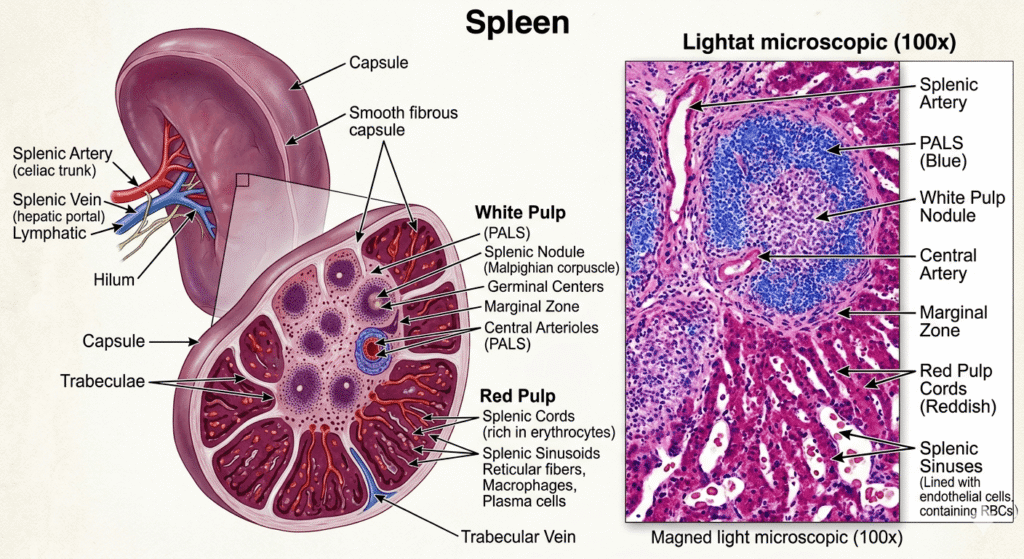
Inflammatory Bowel Disease A Complete Medical Guide

Understanding inflammatory bowl disease

Introduction

Inflammatory Bowel Disease (IBD) refers to a group of long-term disorders that cause inflammation within the digestive system. The two primary types are Crohn’s disease and ulcerative colitis. These conditions can greatly impact daily life by leading to symptoms such as abdominal discomfort, diarrhea, tiredness, weight reduction, and poor nutrient absorption.

Learn about Inflammatory Bowel Disease, including its causes, symptoms, diagnosis, complications, and treatment options in this complete medical guide.

IBD should not be confused with Irritable Bowel Syndrome (IBS). IBS is a functional digestive disorder without intestinal inflammation, whereas IBD involves chronic inflammation that may damage the bowel lining and result in severe complications.

Although the exact cause remains uncertain, experts believe IBD develops from an abnormal immune reaction influenced by genetics, environmental exposures, and gut microorganisms. The disease commonly alternates between active flare-ups and periods of remission.

Types of Inflammatory Bowel Disease

1. Crohn’s Disease

Crohn’s disease may affect any section of the gastrointestinal tract, from the mouth to the anus. However, it most often occurs in the lower small intestine and the beginning of the large intestine. Inflammation can involve all layers of the bowel wall.

Main Characteristics

  • Patchy inflamed areas known as “skip lesions”
  • Deep ulcer formation and thickened bowel walls
  • Risk of fistulas, abscesses, and intestinal narrowing
  • Possible effects on the skin, eyes, and joints

2. Ulcerative Colitis

Ulcerative colitis primarily affects the colon and rectum. The inflammation usually spreads continuously from the rectum and mainly involves the inner lining of the intestine.

Main Characteristics

  • Continuous inflammation starting in the rectum
  • Frequent bloody diarrhea
  • Higher long-term risk of colon cancer
  • More superficial inflammation than Crohn’s disease
Understanding inflammatory bowl disease IBD its Anatomy and Pathology

Pathophysiology of IBD

IBD develops through a complicated interaction involving immune dysfunction, intestinal bacteria, inherited factors, and environmental influences.

1. Immune System Abnormalities

Normally, the immune system protects against harmful organisms while tolerating healthy gut bacteria. In IBD, the immune system mistakenly attacks intestinal tissue.

This can result in:

  • Persistent inflammation
  • Increased inflammatory chemicals
  • Ulceration and tissue injury
  • Reduced healing ability

2. Genetic Predisposition

Research has identified multiple genes linked to IBD, especially Crohn’s disease. Individuals with affected family members are more likely to develop the condition.

Genetic changes may:

  • Disrupt immune regulation
  • Damage intestinal barrier function
  • Alter gut bacteria interactions

3. Gut Microbiome Disturbance

The intestines contain beneficial microorganisms important for digestion and immunity. In IBD, imbalance in these bacteria may trigger excessive immune activity.

4. Environmental Influences

Environmental exposures may activate the disease in genetically susceptible individuals.

Potential triggers include:

  • Smoking
  • Certain medications
  • Diets high in processed foods
  • Stress
  • Infections
  • Pollution

5. Intestinal Injury

Ongoing inflammation may damage the bowel lining and lead to:

  • Ulcers
  • Bleeding
  • Scar tissue
  • Narrowed intestines
  • Poor nutrient absorption
  • Fistulas in Crohn’s disease

Causes of Inflammatory Bowel Disease

The precise cause of IBD is unknown, but several contributing factors have been identified.

1. Genetic Factors

  • Family history of IBD
  • Inherited gene mutations
  • Increased risk among close relatives

2. Autoimmune Reactions

The immune system may wrongly attack healthy intestinal tissue, causing chronic inflammation.

3. Environmental Factors

  • Smoking raises Crohn’s disease risk
  • Urban lifestyles may contribute
  • Diets rich in unhealthy fats and processed foods
  • Pollutant exposure

4. Microbial Changes

Alterations in gut bacteria may stimulate abnormal immune responses.

5. Psychological Stress

Stress does not directly cause IBD, but it may worsen symptoms and provoke flare-ups.

Symptoms of Inflammatory Bowel Disease

Symptoms differ depending on the location and severity of inflammation.

Digestive Symptoms

  • Chronic diarrhea
  • Abdominal cramps and pain
  • Blood in stool
  • Urgent bowel movements
  • Rectal bleeding
  • Weight loss
  • Reduced appetite
  • Bloating
  • Nausea

General Symptoms

  • Fatigue
  • Fever
  • Weakness
  • Malnutrition
  • Delayed growth in children

Effects Outside the Intestines

Skin

  • Erythema nodosum
  • Pyoderma gangrenosum

Joints

  • Arthritis
  • Joint swelling and pain

Eyes

  • Uveitis
  • Episcleritis

Liver and Bile Ducts

Diagnosis of IBD

Diagnosis involves medical evaluation, laboratory testing, imaging, and endoscopic procedures.

1. Medical History and Examination

Doctors evaluate:

  • Symptom duration and intensity
  • Family history
  • Weight changes
  • Nutritional condition

2. Blood Tests

Blood work may reveal:

  • Anemia
  • Elevated inflammatory markers such as ESR and CRP
  • Electrolyte disturbances
  • Nutritional deficiencies

3. Stool Testing

Stool tests help exclude infections and identify signs of intestinal inflammation.

4. Colonoscopy

Colonoscopy is a major diagnostic procedure that allows physicians to:

  • Examine intestinal inflammation
  • Detect ulcers and bleeding
  • Collect biopsy samples

5. Imaging Techniques

  • CT scans
  • MRI enterography
  • Abdominal ultrasound

These tests help identify complications such as fistulas, abscesses, and narrowed bowel segments.

6. Biopsy

Tissue samples taken during endoscopy help confirm the diagnosis and differentiate Crohn’s disease from ulcerative colitis.

Risk Factors for IBD

1. Family History

Having a close relative with IBD significantly increases the likelihood of developing the condition.

2. Age

IBD often appears between ages 15 and 35, although it can occur at any stage of life.

3. Smoking

Smoking is strongly linked to Crohn’s disease and may worsen its severity.

4. Diet

Highly processed foods and unhealthy fats may contribute to inflammation.

5. Environmental Factors

  • Urban living
  • Pollution
  • Westernized lifestyle

6. Ethnicity

Certain ethnic populations experience higher rates of IBD.

7. Medications

Some medications associated with increased risk include:

  • Nonsteroidal anti-inflammatory drugs (NSAIDs)
  • Antibiotics
  • Oral contraceptives

When to Seek Medical Attention

A doctor should be consulted if any of the following occur:

  • Diarrhea lasting several weeks
  • Blood in stool
  • Severe abdominal pain
  • Unexplained weight loss
  • Fever with digestive symptoms
  • Extreme fatigue
  • Persistent rectal bleeding
  • Difficulty eating due to nausea or pain

Immediate medical care is necessary for severe dehydration, intestinal blockage, or heavy bleeding.

Prognosis of IBD

IBD is a chronic lifelong illness, but many individuals can successfully manage symptoms with proper treatment and healthy lifestyle changes.

Factors Influencing Prognosis

  • Severity of inflammation
  • Frequency of flare-ups
  • Treatment response
  • Presence of complications
  • Smoking habits

Potential Complications

Crohn’s Disease

  • Fistulas
  • Intestinal strictures
  • Abscesses
  • Malnutrition

Ulcerative Colitis

  • Severe bleeding
  • Toxic megacolon
  • Higher colorectal cancer risk

Long-Term Outlook

With early diagnosis and effective treatment:

  • Many patients achieve remission
  • Quality of life improves
  • Complications are reduced
  • Nutritional status can be maintained

Ongoing medical follow-up and treatment adherence are important for long-term disease management.

Lifestyle and Dietary Management

Dietary Tips

  • Eat smaller, frequent meals
  • Maintain good hydration
  • Avoid foods that trigger symptoms
  • Reduce processed food intake
  • Limit dairy if intolerant
  • Choose nutrient-rich foods

Stress Reduction

  • Meditation
  • Yoga
  • Counseling
  • Adequate sleep

Smoking Cessation

Stopping smoking is particularly important for individuals with Crohn’s disease.

Conclusion

Inflammatory Bowel Disease is a chronic condition involving inflammation of the digestive tract, mainly in the form of Crohn’s disease and ulcerative colitis. The disorder develops due to a combination of immune abnormalities, genetic factors, environmental triggers, and disturbances in gut bacteria.

IBD can produce significant digestive symptoms and may also affect other body systems, including the skin, joints, and eyes. Early diagnosis and proper treatment are essential to minimize complications and improve quality of life.

While there is currently no permanent cure, modern therapies, dietary strategies, and lifestyle modifications allow many people with IBD to maintain long-term remission and live productive lives. Consistent medical care, medication adherence, and healthy daily habits remain essential for successful management.

Disclaimer: The information provided in this blog post is for educational and informational purposes only and should not be considered medical advice. It is not intended to replace professional medical consultation, diagnosis, or treatment.
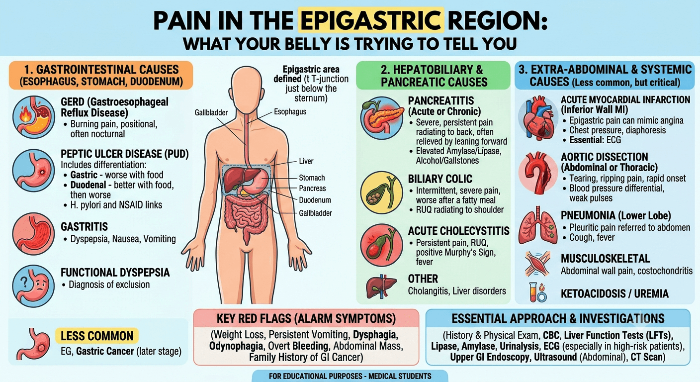
Pain in the Left Lower Quadrant (LLQ)

Pain in the left lower quadrant

Causes, Symptoms & Treatment

Pathophysiology

Pain in the left lower quadrant (LLQ) arises from irritation, inflammation, obstruction, ischemia, or distension of structures located in or referred to the LLQ region. The sensation of pain in this region is mediated by visceralsomatic, or referred nerve pathways.

Mechanisms of Pain

  • Visceral pain originates from the internal organs (e.g., sigmoid colon, ureter, ovary). These structures are innervated by autonomic afferent fibers, and the pain is typically dull, poorly localized, and colicky in nature. It results from distension, ischemia, or inflammation.
  • Somatic pain arises when parietal peritoneum or abdominal wall structures are irritated. This pain is sharp, well localized, and often accompanied by guarding or rebound tenderness.
  • Referred pain occurs when sensory fibers from distant organs share the same spinal segment. For example, Ureteric colic can radiate to the groin due to shared innervation (T10–L2 segments).

Nerve Pathways

  • The lower thoracic (T10–T12) and lumbar (L1–L2) spinal nerves transmit afferent signals from abdominal viscera.
  • Pain signals from the sigmoid colon and left ureter often converge in these spinal segments, making localization challenging.

Physiological Contributors

  • Inflammation: Releases prostaglandins, bradykinin, and cytokines that sensitize nociceptors.
  • Ischemia: Causes lactic acidosis and stimulation of chemoreceptors.
  • Distension or obstruction: Activates stretch receptors, causing cramping pain.

Organs Involved

The left lower quadrant of the abdomen contains several visceral and vascular structures. Pain in this region can originate from one or more of the following:

Organ/SystemRelevant ConditionsClinical Notes
Sigmoid ColonDiverticulitis, volvulus, colitis, ischemiaMost common source of LLQ pain in adults; typically presents with fever, altered bowel habits, localized tenderness
Left Ovary & Fallopian Tube (Females)Ovarian cyst, torsion, ectopic pregnancy, pelvic inflammatory disease (PID)Gynecological sources often mimic intestinal pathology
Left UreterUreteric colic due to renal stones or infectionPain radiates to groin, may be associated with hematuria
Left Kidney (Lower Pole)Pyelonephritis, hydronephrosisFlank pain that may extend to LLQ
Descending ColonColitis, inflammatory bowel disease (IBD), malignancyChronic pain, often with altered bowel habits or bleeding
Left Iliac VesselsThrombosis, aneurysm (rare)May cause dull, aching pain
Musculoskeletal StructuresAbdominal wall strain, herniaPain localized to movement or palpation

Causes

Acute Causes

SystemConditionDescription / Key Features
GastrointestinalDiverticulitisInflammation of sigmoid diverticula; LLQ pain, fever, leukocytosis, and altered bowel movements
Infectious colitisCaused by bacterial or viral infection; associated with diarrhea and systemic symptoms
Bowel obstructionColicky pain, distension, vomiting; may result from adhesions, hernia, or tumor
Ischemic colitisSudden onset pain with rectal bleeding in elderly or vascular-compromised patients
GenitourinaryUreteric colicSharp, radiating pain to groin; hematuria; caused by stone impaction
PyelonephritisDull, constant pain with fever, chills, urinary frequency, and costovertebral angle tenderness
Gynecological (Females)Ovarian torsionSudden, severe unilateral pain; may follow cyst rupture; surgical emergency
Ectopic pregnancyLower abdominal pain, amenorrhea, and vaginal bleeding; positive pregnancy test
Pelvic inflammatory disease (PID)Bilateral lower pain, fever, discharge; often sexually transmitted
MusculoskeletalRectus sheath hematomaFollowing trauma or anticoagulation; localized pain and bruising

Chronic Causes

SystemConditionDescription / Key Features
GastrointestinalIrritable Bowel Syndrome (IBS)Recurrent pain with bowel habit changes; relieved by defecation
Chronic diverticular diseaseRecurrent LLQ discomfort post-diverticulitis; bloating, constipation
Colorectal cancerProgressive, dull pain; change in bowel habits, blood in stool
GenitourinaryChronic PyelonephritisRecurrent flank pain, low-grade fever, dysuria
GynecologicalEndometriosisCyclical LLQ pain, Dysmenorrhea, infertility
Ovarian cyst (benign)Dull, intermittent pain or fullness sensation
MusculoskeletalHernia, muscle strainExacerbated by activity; palpable tenderness

Line of Management

Initial Assessment

History

·         Onset, duration, character, and radiation of pain.

·         Associated symptoms: nausea, bowel changes, urinary complaints, menstrual history (in females).

Physical Examination

·         Inspection, palpation (guarding, rebound), percussion, and auscultation.

·         Digital rectal and pelvic exams if indicated.

Diagnostic Evaluation

TestPurpose / Findings
Complete blood count (CBC)Leukocytosis in infection/inflammation
UrinalysisDetects hematuria, infection (for ureteric causes)
Serum electrolytes, renal function testsEvaluate dehydration, renal impairment
β-hCG (in females)Exclude ectopic pregnancy
Abdominal ultrasoundFirst-line for gynecological or urinary causes
CT abdomen and pelvis (contrast-enhanced)Gold standard for diverticulitis, obstruction, abscess
ColonoscopyFor chronic pain, IBD, or malignancy suspicion
Pelvic MRIFor detailed gynecological or soft tissue pathology

Line of Management

Medical Management

Diverticulitis: Broad-spectrum antibiotics (e.g., ciprofloxacin + metronidazole), bowel rest, fluids.

·         Ureteric colic: NSAIDs, hydration, alpha-blockers (tamsulosin), lithotripsy if indicated.

·         PID / Endometriosis: Antibiotics, hormonal therapy, or laparoscopic management.

·         IBS: Dietary modification, probiotics, antispasmodics.

·         Colitis: Antibiotics (if bacterial), corticosteroids or aminosalicylates for IBD.

Surgical Management

·         Perforated diverticulitis / abscess: Hartmann’s procedure or drainage.

·         Ovarian torsion / ectopic pregnancy: Emergency surgery.

·         Obstruction or malignancy: Resection, stenting, or bypass.

Follow-up Care

  • Lifestyle modification: High-fiber diet, adequate hydration, regular exercise.
  • Surveillance colonoscopy for chronic diverticular disease or post-malignancy.
  • Regular imaging for renal stones or cyst recurrence.
  • Patient education on warning signs (fever, severe pain, rectal bleeding).

Summary

Pain in the left lower quadrant is a multifactorial clinical presentation requiring a methodical diagnostic approach. The most common cause in adults is sigmoid diverticulitis, while in women of reproductive age, gynecological causes such as ovarian torsion or ectopic pregnancy must be urgently ruled out. A combination of thorough history-taking, focused physical examination, and targeted imaging ensures timely diagnosis and appropriate management, minimizing complications.

Disclaimer: The information provided in this blog post is for educational and informational purposes only and should not be considered medical advice. It is not intended to replace professional medical consultation, diagnosis, or treatment. Always seek the guidance of a qualified healthcare professional regarding any medical condition or health-related concerns. The author and publisher are not responsible for any actions taken based on the information presented in this article.
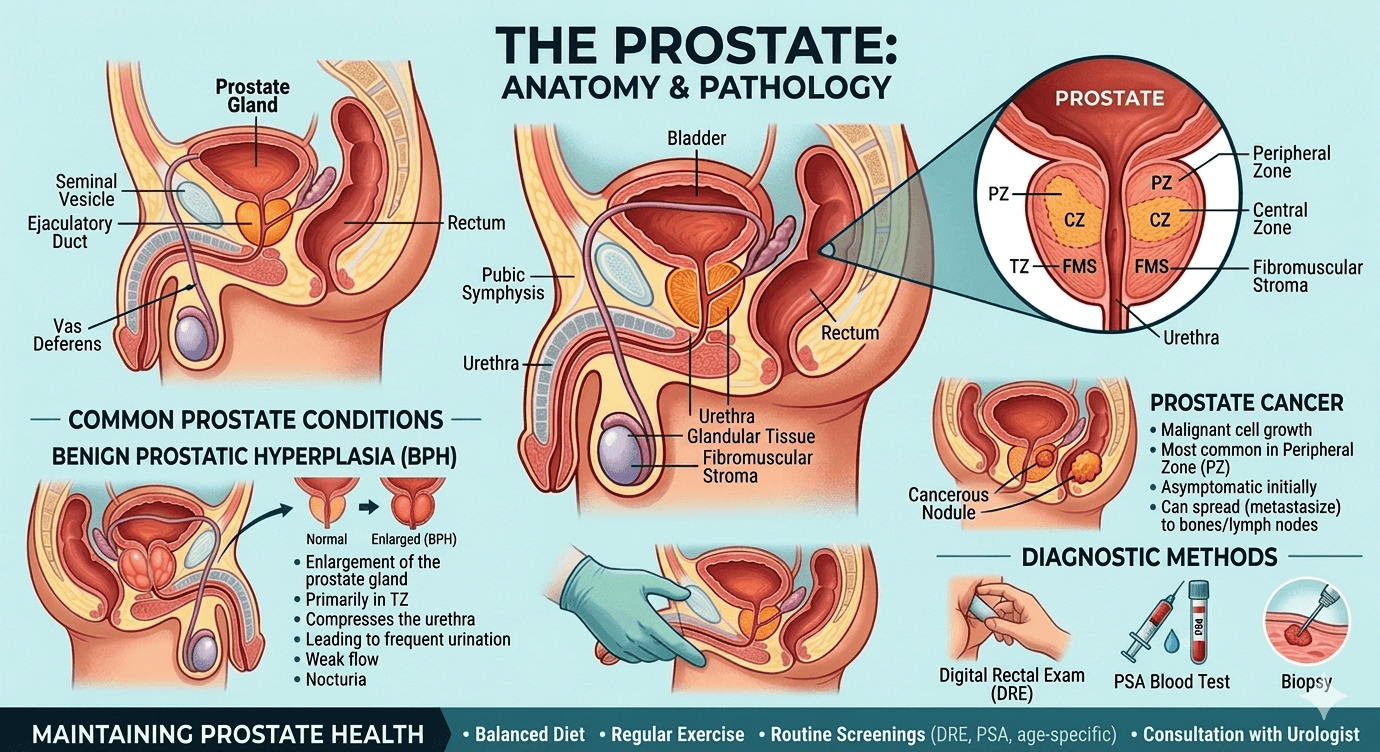
Viral Hepatitis: A Concise Clinical Overview

Viral hepatitis its types, causes, complications, and symptoms

Viral hepatitis refers to inflammation of the liver caused by specific viruses that target liver cells. The main culprits are hepatitis A, B, C, D, and E viruses. These infections vary in how they spread, how long they last, and how severely they affect the liver.

Pathophysiology

The liver damage in viral hepatitis results from two main processes: the direct impact of the virus on liver cells and the body’s immune response to infection.

  • Inside the liver cells:
    The viruses enter hepatocytes and replicate using the cell’s machinery. Some, like hepatitis B, can integrate parts of their genetic material into the host’s DNA, allowing them to persist for years. Others, such as hepatitis A and E, are cleared after a short infection. Viral replication can interfere with normal cell functions like protein synthesis and metabolism.
  • Immune response:
    The immune system detects infected liver cells and attacks them. Cytotoxic T cells and inflammatory cytokines cause cell death and inflammation, leading to elevated liver enzymes and jaundice. When the immune attack continues over time, it triggers fibrosis, cirrhosis, and can eventually lead to liver cancer.
  • Potential complications:
    These include acute liver failure from massive cell death, chronic hepatitis that progresses to cirrhosis, and hepatocellular carcinoma. Coinfection (for example, HBV with HDV) or a weakened immune system can make the disease more severe.

Types of Viral Hepatitis

Hepatitis A (HAV)

  • Type: RNA virus (Picornavirus).
  • Transmission: Fecal–oral route via contaminated food, water, or contact with infected individuals.
  • Course: Usually an acute, short-term illness. Rarely causes severe liver failure. No chronic infection. An effective vaccine is available.

Hepatitis B (HBV)

  • Type: DNA virus (Hepadnavirus).
  • Transmission: Blood and body fluids (birth, sex, needles, transfusions).
  • Course: May be acute or chronic. Infants infected at birth have a higher risk of developing chronic disease. Chronic HBV can lead to cirrhosis and liver cancer. A highly effective vaccine and antiviral treatments (tenofovir, entecavir) are available.

Hepatitis C (HCV)

  • Type: RNA virus (Flavivirus).
  • Transmission: Primarily through blood exposure (needles, transfusions before screening, unsafe medical practices).
  • Course: Often becomes chronic and can slowly cause fibrosis, cirrhosis, and liver cancer. Modern antiviral drugs (DAAs) cure over 95% of cases. No vaccine yet.

Hepatitis D (HDV)

  • Type: Defective RNA virus that requires HBV to replicate.
  • Transmission: Same as HBV.
  • Course: Occurs as a Coinfection with HBV or as a super infection in HBV carriers. Super infection tends to cause more severe disease. Prevention relies on HBV vaccination.

Hepatitis E (HEV)

  • Type: RNA virus (Hepevirus).
  • Transmission: Fecal–oral, often through contaminated water; can also spread from animals.
  • Course: Usually acute and self-limited but can be severe in pregnant women. Chronic HEV may occur in immunocompromised individuals. A vaccine exists in some countries but is not widely used.

Causes and Risk Factors

Viral hepatitis is mainly caused by infection with hepatitis viruses such as Hepatitis A, Hepatitis B, Hepatitis C, Hepatitis D, and Hepatitis E. These viruses attack the liver and cause inflammation, which can lead to liver damage if not treated properly.

Several risk factors increase the chance of developing viral hepatitis. These include poor hygiene, unsafe drinking water, blood transfusions without proper screening, intravenous drug use, multiple sexual partners, healthcare exposure to infected blood, tattooing or piercing with unsterilized equipment, and weakened immunity. Lack of vaccination, especially for hepatitis A and B, also increases the risk of infection.

Clinical Features

The symptoms of viral hepatitis may vary from mild illness to serious liver complications. During the initial stage, patients often report tiredness, weakness, fever, reduced appetite, nausea, vomiting, body aches, and pain or discomfort in the upper right side of the abdomen. As the condition advances, jaundice can appear, leading to yellowing of the skin and eyes, dark urine, and pale-colored stools.

Certain individuals may also develop itching, liver enlargement, and mild pain in the joints. In chronic infections, particularly Hepatitis B and Hepatitis C, symptoms can remain unnoticed for many years before progressing to complications such as liver fibrosis, cirrhosis, or liver failure. Advanced disease may cause mental confusion, increased bleeding tendency, and abdominal swelling as a result of severe liver damage.

Line of Management

Prevention:

  • Vaccines: Available for HAV and HBV (which also prevents HDV). Universal infant HBV vaccination is key to prevention.
  • Public health measures: Safe food and water, blood screening, sterile injections, safe sex, and harm reduction programs for people who inject drugs.
  • Expanding vaccination and treatment access supports WHO’s global elimination goals.

Diagnosis and Screening:

  • HAV: IgM anti-HAV confirms acute infection.
  • HBV: HBsAg, anti-HBs, and HBV DNA for diagnosis and monitoring.
  • HCV: Anti-HCV for screening; HCV RNA to confirm active infection.
  • HDV: Test in HBsAg-positive individuals using anti-HDV or HDV RNA.
  • HEV: IgM anti-HEV or HEV RNA testing for acute cases.
    Screening recommendations vary, but many countries now advocate one-time HBV and HCV testing for adults.

Treatment

  • HAV and HEV: Supportive care only; rest, hydration, and avoiding alcohol or liver-toxic drugs.
  • HBV: Long-term antivirals (tenofovir, entecavir) to control viral replication and prevent progression to cirrhosis or liver cancer.
  • HCV: Short courses of oral DAAs that cure most infections. Common oral DAAs include sofosbuvir, ledipasvir, daclatasvir, velpatasvir, and glecaprevir/pibrentasvir. Treatment choice depends on the HCV genotype, liver condition, and patient-related factors.
  • HDV: Difficult to treat; pegylated interferon and newer therapies are under study. HBV vaccination remains the best prevention.

Lifestyle and follow-up

Avoid alcohol, manage metabolic conditions, and ensure vaccination of household contacts. Long-term follow-up includes liver enzyme testing, fibrosis assessment, and cancer screening (ultrasound, and AFP every six months for cirrhotic patients).

Patient education

Education should include the importance of completing prescribed treatment, attending regular follow-up visits, and monitoring liver function. Patients should avoid alcohol, smoking, and unnecessary medications that may further damage the liver. A balanced diet, adequate rest, and healthy lifestyle habits can help support liver recovery.

Family members and close contacts may also require screening or vaccination to reduce transmission. Proper counseling helps improve treatment adherence, reduce stigma, and enhance the overall quality of life of patients with viral hepatitis.

Disclaimer: The information provided in this blog post is for educational and informational purposes only and should not be considered medical advice. It is not intended to replace professional medical consultation, diagnosis, or treatment. Always seek the guidance of a qualified healthcare professional regarding any medical condition or health-related concerns. The author and publisher are not responsible for any actions taken based on the information presented in this article.
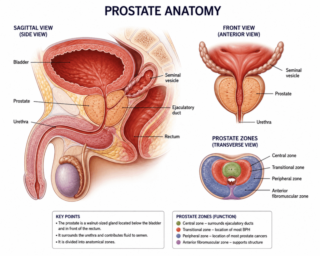
Hepatorenal Syndrome (HRS)

Understanding Hepatorenal Syndrome

Causes, Pathophysiology, and Management

Definition and Overview

Hepatorenal syndrome is defined as the development of renal failure in individuals with severe liver disease, most often cirrhosis, in the absence of any identifiable cause of kidney dysfunction. The syndrome results from alterations in blood flow and vascular regulation, leading to intense constriction of blood vessels in the kidneys and failure of the renal filtration process. HRS typically occurs in the context of worsening liver function, often triggered by infections, gastrointestinal bleeding, or excessive use of diuretics.

Pathophysiology

The main mechanism underlying HRS is an imbalance between vasodilation and vasoconstriction, resulting in reduced blood flow to the kidneys.

  1. Splanchnic Vasodilation

Portal hypertension in liver disease triggers the release of vasodilators such as nitric oxide.

This widens blood vessels in the Splanchnic circulation (gut and abdominal organs), lowering effective blood volume.

Activation of Vasoconstrictor Systems

To compensate, the body activates the RAAS (rennin–angiotensin–aldosterone system), sympathetic nervous system, and antidiuretic hormone.

These responses cause sodium and water retention, Ascites, and progressive renal vasoconstriction.

  1. Systemic Inflammation

Bacterial translocation from the gut and endotoxin release aggravate vascular dysfunction and accelerate renal impairment.

Causes and Risk Factors

HRS does not develop spontaneously but is usually precipitated by specific events in patients with advanced cirrhosis or acute liver failure.

Primary liver conditions:

  • Decompensated cirrhosis (most common) is the stage of chronic liver disease where the liver can no longer perform its essential functions adequately, and serious complications begin to appear.
  • Severe alcoholic hepatitis
  • Acute fulminant hepatic failure

Precipitating factors:

  • Spontaneous bacterial peritonitis (SBP)
  • Large-volume paracentesis without albumin replacement
  • Gastrointestinal bleeding
  • Overuse of diuretics leading to hypovolemia.
  • Severe infections or sepsis

Types of Hepatorenal Syndrome

Hepatorenal syndrome (HRS) is a severe complication of advanced liver disease in which kidney function deteriorates quickly even though the kidneys remain structurally intact. It most often develops in individuals with decompensated cirrhosis and significant portal hypertension.

Type 1 Hepatorenal Syndrome (HRS-AKI)

This is the rapidly worsening form of HRS. Kidney failure appears over a period of days to weeks and is marked by a significant increase in serum creatinine levels. Common triggers include infections such as spontaneous bacterial peritonitis, gastrointestinal hemorrhage, or profound dehydration. Patients may experience decreased urine output, increasing ascites, confusion, and marked fatigue. Type 1 HRS has a poor outlook and demands immediate management with vasoconstrictor medications, albumin administration, and consideration of liver transplantation.

Type 2 Hepatorenal Syndrome

This form is more gradual and chronic in nature. Kidney impairment develops slowly and is frequently linked with refractory Ascites that responds poorly to diuretic treatment. Early symptoms are usually milder, although the disorder may worsen progressively. Treatment focuses on managing Ascites, providing albumin therapy, and assessing suitability for liver transplantation.

Updated Classification

Current guidelines divide HRS into:

  • HRS-AKI (Acute Kidney Injury) → equivalent to the previous Type 1 HRS
  • HRS-NAKI (Non-Acute Kidney Injury) → includes chronic or subacute kidney impairment, similar to the former Type 2 HRS

Prompt recognition and treatment are essential because Hepatorenal syndrome is linked to high mortality in patients with advanced liver disease.

HRS Type 1: Rapidly progressive, with doubling of serum creatinine (>2.5 mg/dL) or halving of creatinine clearance within 2 weeks. Poor prognosis, median survival ~2 weeks if untreated.

HRS Type 2: More slowly progressive, commonly associated with refractory Ascites. Median survival ~6 months.

Diagnosis

HRS is a diagnosis of exclusion, made after ruling out structural kidney injury or other causes of renal dysfunction. Key criteria include:

  • Advanced liver disease with portal hypertension and Ascites
  • Low GFR (elevated serum creatinine or low urine output)
  • No shock, nephrotoxic drug use, or structural kidney disease
  • Lack of improvement after diuretic withdrawal and plasma volume expansion with albumin

Lab Tests

  • CBC
  • LFT (Liver function test)
  • KFT (kidney function test) with electrolytes.
  • Lipid Profile
  • RBS with HBA1C
  • Urinalysis

Imaging

  • USG (whole abdomen)
  • CT Scan or MRI (whole abdomen)

Management

1. General Principles

  • Remove or correct precipitating factors (infections, bleeding, over-diuresis).
  • Discontinue nephrotoxic drugs (NSAIDs, Aminoglycosides, contrast agents).
  • Avoid hypovolemia and correct electrolyte imbalances.

2. Pharmacological Therapy

Vasoconstrictors + Albumin are the mainstay:

Terlipressin + Albumin (gold standard where available).

Alternatives: Norepinephrine, Midodrine + Octreotide + Albumin (especially where Terlipressin is not available).

Albumin: Expands plasma volume and counteracts circulatory dysfunction.

3. Supportive Measures

  • Careful fluid balance.
  • Treat infections aggressively (antibiotics, albumin infusion for SBP).
  • Avoid further renal insults.

4. Renal Replacement Therapy

  • Dialysis may be required as a bridge to liver transplantation.
  • Not a definitive therapy but supports patients with severe renal dysfunction.

5. Definitive Treatment

  • Liver Transplantation: The only curative treatment, as it corrects the underlying liver failure and restores renal function in most cases.
  • Simultaneous liver–kidney transplantation may be considered in selected patients with prolonged renal dysfunction.

Prognosis

  • Untreated HRS has extremely poor survival (weeks to months).
  • Early recognition and initiation of vasoconstrictor therapy with albumin improve short-term outcomes.
  • Liver transplantation remains the only intervention that significantly improves long-term survival.

Conclusion

Hepatorenal syndrome is a severe complication of advanced liver disease, characterized by functional renal failure due to systemic and Splanchnic circulatory disturbances. Prompt recognition, correction of precipitating factors, vasoconstrictor plus albumin therapy, and timely referral for liver transplantation are critical for improving patient outcomes.

Disclaimer: The information provided in this blog post is for educational and informational purposes only and should not be considered medical advice. It is not intended to replace professional medical consultation, diagnosis, or treatment. Always seek the guidance of a qualified healthcare professional regarding any medical condition or health-related concerns. The author and publisher are not responsible for any actions taken based on the information presented in this article.
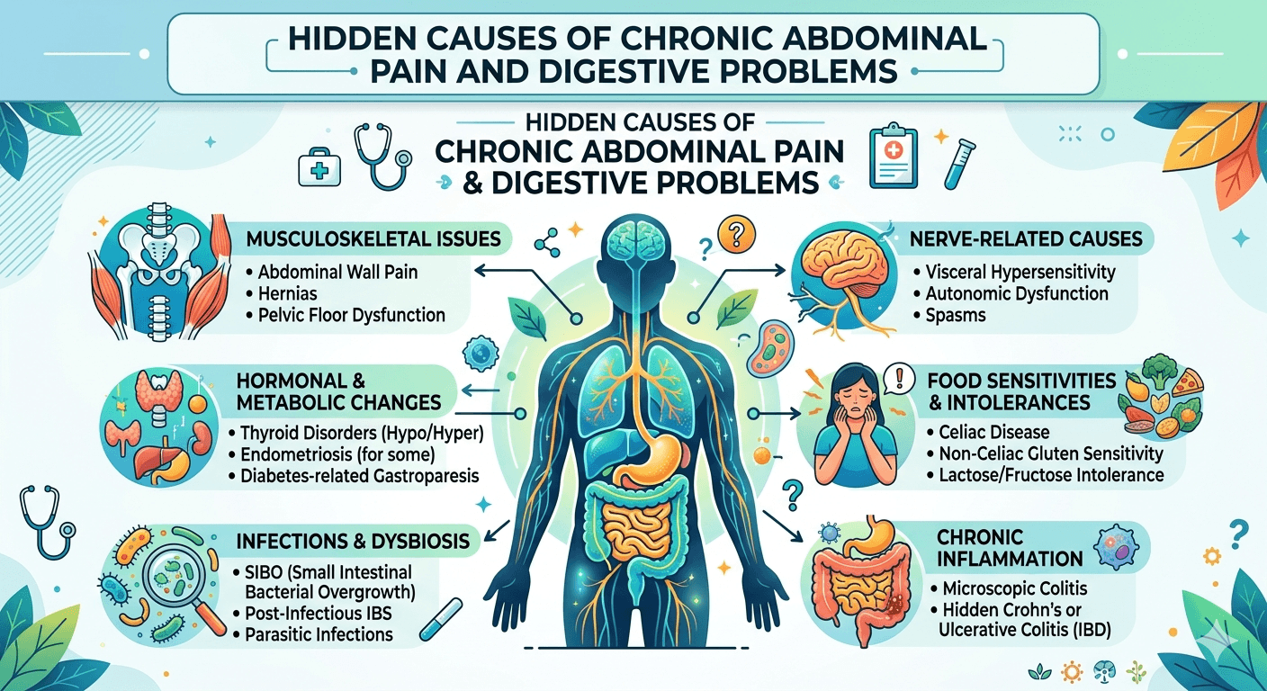
Read more about diabetes and organ damage

Read More

Fatty Liver Disease: 25 FAQs You Must Know

Fatty Liver Disease: 25 FAQs You Must Know

Introduction

Fatty liver disease is becoming one of the most common health issues today, especially in India. What makes it dangerous is how quietly it develops.

Most people don’t realize they have it until it progresses.

This guide answers the most important questions about fatty liver in a simple, practical way so you can take action early.

What is Fatty Liver Disease?

Fatty Liver Disease refers to excessive fat accumulation in liver cells. It is broadly classified into:

  • Non-alcoholic fatty liver disease (NAFLD) – not related to alcohol use
  • Alcoholic fatty liver disease (AFLD) – caused by excessive alcohol consumption

When fat exceeds 5–10% of liver weight, it is considered abnormal.

What causes Fatty Liver Disease?

Common causes include:

  • Obesity and metabolic syndrome
  • Insulin resistance and type 2 diabetes
  • High cholesterol and triglycerides
  • Excess alcohol intake
  • Poor diet (high sugar, refined carbs, processed food)
  • Sedentary lifestyle
  • Certain medications (e.g., steroids, chemotherapy drugs)

What are the early symptoms of Fatty Liver Disease?

Early stages are often silent. When symptoms occur, they may include:

  • Fatigue
  • Mild discomfort in the upper right abdomen
  • Bloating
  • Loss of appetite

What are the warning signs of advanced disease?

As the disease progresses (to NASH or cirrhosis), symptoms may include:

  • Persistent abdominal pain
  • Jaundice (yellowing of skin and eyes)
  • Swelling in legs or abdomen (ascites)
  • Easy bruising
  • Confusion (hepatic encephalopathy)

What is the difference between NAFLD and NASH?

Nonalcoholic Fatty Liver Disease (NAFLD) is a condition in which excess fat builds up in the liver in people who drink little or no alcohol.

In many people, NAFLD causes no major symptoms and may remain stable for years. It is often linked with:

  • Obesity
  • Type 2 diabetes
  • Insulin resistance
  • High cholesterol
  • Metabolic dysfunction

Nonalcoholic Steatohepatitis (NASH) is a more serious form of NAFLD.

In NASH, the liver not only contains excess fat but also develops:

  • Inflammation
  • Liver cell injury
  • Scarring (fibrosis)

Over time, NASH can progress to:

  • Advanced fibrosis
  • Cirrhosis
  • Liver failure
  • Liver cancer

Is Fatty Liver Disease reversible?

Yes, especially in early stages. Lifestyle changes can significantly reduce liver fat and even reverse damage:

  • Weight loss (5–10% body weight)
  • Regular exercise
  • Healthy diet

Advanced stages like cirrhosis are usually irreversible.

How is Fatty Liver Disease diagnosed?

Diagnosis typically involves:

  • Blood tests (liver enzymes: ALT, AST)
  • Imaging (ultrasound, CT scan, MRI)
  • FibroScan (to assess liver stiffness)
  • Liver biopsy (gold standard, used in selected cases)

What is the best diet for Fatty Liver Disease?

A liver-friendly diet includes:

  • Fruits and vegetables
  • Whole grains
  • Lean protein (fish, chicken, legumes)
  • Healthy fats (olive oil, nuts)

Avoid:

  • Sugary drinks
  • Processed foods
  • Trans fats
  • Excess salt and alcohol

Which foods should be strictly avoided?

  • Soft drinks and high-fructose corn syrup
  • Fried foods
  • Packaged snacks
  • Red and processed meat
  • Alcohol

Can exercise help in Fatty Liver Disease?

Yes. Exercise improves insulin sensitivity and reduces liver fat.

  • Aim for 150–300 minutes/week of moderate activity
  • Include both aerobic exercise and strength training

Is Fatty Liver Disease dangerous?

It can be. While early stages are harmless, untreated disease may progress to:

  • NASH
  • Fibrosis
  • Cirrhosis
  • Liver cancer

Can non-obese people get Fatty Liver Disease?

Yes. This is called lean NAFLD and is common in Asian populations. Causes include:

  • Genetic predisposition
  • Poor diet
  • Metabolic abnormalities

Does Fatty Liver affect other organs?

Yes. It is closely linked with:

  • Heart disease
  • Type 2 diabetes
  • Kidney disease

Cardiovascular disease is the leading cause of death in these patients.

Are there any medications for Fatty Liver Disease?

Currently, no specific FDA-approved drugs for NAFLD exist. However:

  • Vitamin E (in selected patients)
  • Pioglitazone (for insulin resistance)
  • New drugs are under research
  • L-Ornithine L-Aspartate (LOLA) is a stable salt of two amino acids used primarily to treat liver diseases.
  • Ursodeoxycholic Acid is a hepatoprotective medication.

Treatment mainly focuses on lifestyle changes.

How long does it take to reverse Fatty Liver?

Improvement can be seen within:

  • 4–12 weeks with lifestyle changes
  • Significant reversal may take 3–6 months or longer
  • Consistency is key.

Is alcohol completely banned in Fatty Liver Disease?

  • In NAFLD: Best to avoid or strictly limit alcohol
  • In AFLD: Complete abstinence is necessary

Can Fatty Liver Disease lead to liver cancer?

Yes. Long-standing NASH and cirrhosis increase the risk of hepatocellular carcinoma (HCC).

Is Fatty Liver common in India?

Yes, prevalence is rising rapidly due to:

  • Sedentary lifestyle
  • High carbohydrate diet
  • Increasing obesity and diabetes rates

Studies estimate 25–30% of urban Indians may have NAFLD.

Can Fatty Liver be prevented?

Yes, prevention strategies include:

  • Maintaining a healthy weight
  • Regular physical activity
  • Balanced diet
  • Avoiding alcohol and processed foods
  • Managing diabetes and cholesterol

When should you see a doctor?

Consult a doctor if you have:

  • Persistent fatigue or abdominal discomfort
  • Abnormal liver function tests
  • Risk factors like obesity, diabetes, or alcohol use

Is Fatty Liver the same as liver failure?

No. Fatty liver is an early, often reversible stage. Liver failure occurs in advanced disease like cirrhosis.

Can herbal or natural remedies cure Fatty Liver?

Some may help support liver health (e.g., coffee, omega-3s), but:

  • No herbal remedy alone can cure fatty liver
  • Always consult a doctor before using supplements

Does drinking coffee help Fatty Liver?

Yes, moderate coffee consumption (2–3 cups/day) may:

  • Reduce liver inflammation
  • Slow disease progression

Can children develop Fatty Liver Disease?

Yes, especially with:

  • Childhood obesity
  • Poor diet and lack of exercise

This is becoming increasingly common worldwide.

What is the prognosis of Fatty Liver Disease?

  • Early stage: Excellent prognosis with lifestyle changes
  • Advanced stage (cirrhosis): Serious, may require liver transplant

Disclaimer: The information provided in this blog post is for educational and informational purposes only and should not be considered medical advice. It is not intended to replace professional medical consultation, diagnosis, or treatment. Always seek the guidance of a qualified healthcare professional regarding any medical condition or health-related concerns.