Insulin Resistance: Causes, Symptoms, Diagnosis & Treatment

Understanding insulin resistance, its causes, impact on the body, sign & symptoms

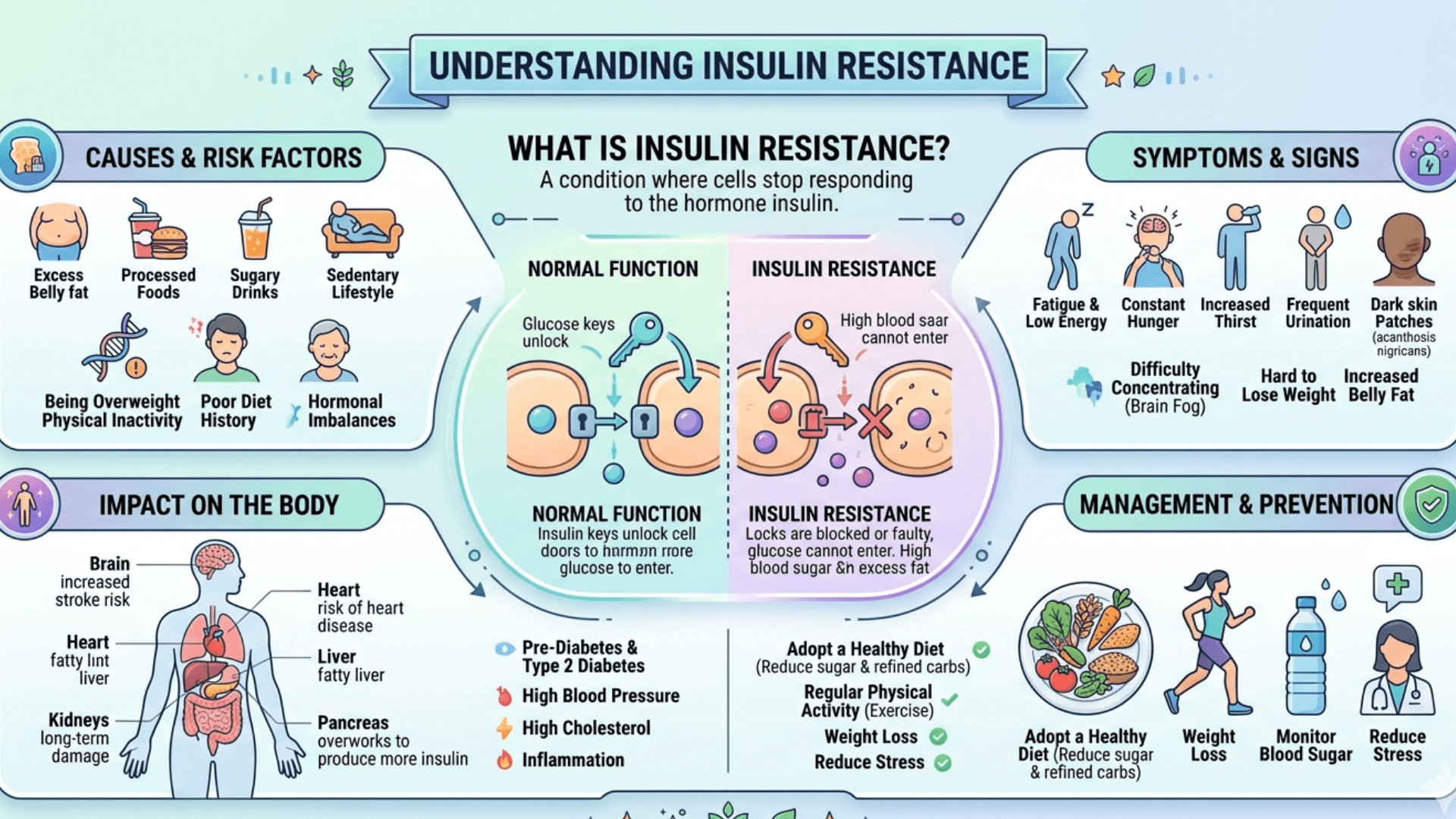
Insulin resistance is a key factor behind many metabolic problems, including type 2 diabetes, obesity, and fatty liver disease. Here’s a clear breakdown of what happens in the body, what causes it, and how it’s managed.

Pathophysiology What’s Happening Inside the Body

Insulin resistance means your body’s cells especially in your muscles, liver, and fat don’t respond as well to insulin as they should. Because of that, your body needs to make more insulin to keep blood sugar levels normal.

Normally, insulin helps move glucose (sugar) into cells for energy and tells the liver to slow down glucose production. When this process breaks down, muscles take up less glucose, fat tissue releases more fatty acids, and the liver keeps pumping out sugar all leading to higher blood glucose and insulin levels.

What causes this breakdown in signaling?

  • Damaged insulin signaling pathways. The key proteins involved in the insulin pathway stop working efficiently, blocking normal glucose uptake.
  • Fat buildup in the wrong places. When fat accumulates in the liver or muscles (not just under the skin), it interferes with insulin’s action a process called Lipotoxicity.
  • Mitochondrial issues. These “power plants” of the cell may not produce energy effectively, further impairing how the body handles glucose and fat.
  • Chronic inflammation. Stressed fat tissue releases inflammatory chemicals (like TNF-α and IL-6) and attracts immune cells, which worsen insulin resistance throughout the body.

How it affects different organs

  • Muscles: They use most of the glucose after a meal. With insulin resistance, they can’t take up enough sugar, so blood glucose rises.
  • Liver: Insulin normally stops the liver from making glucose. When the liver becomes resistant, it overproduces glucose and stores more fat, creating a cycle of worsening insulin resistance.
  • Fat tissue: Unhealthy fat cells leak fatty acids into the bloodstream and send inflammatory signals that disrupt insulin’s work in other tissues.

What Causes Insulin Resistance

There’s rarely just one cause. It’s usually a mix of lifestyle, genetics, and hormonal factors.

  1. Excess belly fat
    Visceral (deep abdominal) fat is the biggest modifiable risk factor. It’s metabolically active and releases inflammatory compounds that interfere with insulin action.
  2. Inactivity and low muscle mass
    Exercise boosts insulin sensitivity by improving how muscles use glucose. A sedentary lifestyle does the opposite.
  3. Unhealthy diet
    Diets high in refined carbs, added sugars, and saturated fats promote weight gain and fat buildup in the liver and muscles.
  4. Genetics
    Some people are more prone to insulin resistance, especially when combined with weight gain or poor diet.
  5. Hormonal or medical conditions
    Disorders like PCOS, Cushing’s syndrome, hypothyroidism, or acromegaly can worsen insulin resistance. Certain medications such as steroids or some antipsychotics can also play a role.
  6. Aging
    With age, muscle mass tends to decline while belly fat increases. Both reduce insulin sensitivity.
  7. Sleep problems and stress
    Chronic stress and poor sleep raise cortisol and other hormones that make the body less responsive to insulin.
  8. Other metabolic issues
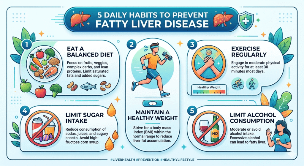
    Conditions like fatty liver disease and abnormal cholesterol often go hand-in-hand with insulin resistance and make it worse.

How It’s Managed

The goal is to improve how the body responds to insulin, lower blood sugar levels, and prevent diabetes or related complications. Management involves lifestyle changes first, then medication if needed.

Lifestyle Changes (the Foundation)

  1. Weight loss
    Losing even 5–10% of your body weight can significantly improve insulin sensitivity.
  2. Exercise
    Combine cardio (like brisk walking, swimming, or cycling) with strength training. Aim for at least 150 minutes of moderate activity a week, plus resistance exercises twice a week.
  3. Healthy eating
    Focus on whole, unprocessed foods. A Mediterranean-style diet — rich in vegetables, lean proteins, whole grains, and healthy fats has strong evidence for improving metabolic health.
  4. Sleep and habits
    Get enough quality sleep, limit alcohol, and avoid smoking. All three affect how the body regulates glucose and insulin.

Medications (When Needed)

If lifestyle changes aren’t enough, medications can help improve insulin sensitivity or control blood sugar.

  1. Metformin
    Often the first choice. It reduces sugar production in the liver and slightly improves insulin sensitivity.
  2. Thiazolidinediones (e.g., pioglitazone)
    These drugs help fat cells work better and shift fat storage away from the liver and muscles. They’re effective but can cause weight gain or fluid retention.
  3. GLP-1 receptor agonists (e.g., semaglutide, liraglutide)
    These medications help with blood sugar control and appetite, often leading to significant weight loss which further improves insulin resistance.
  4. Other medications
  5. SGLT2 inhibitors help the body excrete extra glucose through urine and improve heart and kidney health.
  6. DPP-4 inhibitors modestly lower blood sugar without weight gain.

Medication choice depends on the person’s blood sugar, weight goals, other health issues, and preferences.

When to Start Medication

For people with prediabetes, lifestyle change comes first. Metformin is considered if those changes aren’t enough especially in people with a high BMI, younger age, or a history of gestational diabetes.

Monitoring Progress

Doctors usually track weight, waist size, blood pressure, fasting glucose, HbA1c, and cholesterol. Follow-ups are done every 3–12 months depending on the situation. Regular feedback and structured programs make lifestyle changes more sustainable.

Summary

  • Insulin resistance means the body’s cells don’t respond properly to insulin, mainly due to fat buildup, inflammation, and energy imbalance.
  • Belly fat and inactivity are the biggest modifiable causes.
  • Weight loss and regular exercise remain the most powerful ways to reverse insulin resistance.
  • Medications like Metformin or GLP-1 agonists can help when lifestyle measures alone aren’t enough.

Disclaimer: The information provided in this blog post is for educational and informational purposes only and should not be considered medical advice. It is not intended to replace professional medical consultation, diagnosis, or treatment. Always seek the guidance of a qualified healthcare professional regarding any medical condition or health-related concerns. The author and publisher are not responsible for any actions taken based on the information presented in this article.

Viral Hepatitis: A Concise Clinical Overview

Viral hepatitis its types, causes, complications, and symptoms

Viral hepatitis refers to inflammation of the liver caused by specific viruses that target liver cells. The main culprits are hepatitis A, B, C, D, and E viruses. These infections vary in how they spread, how long they last, and how severely they affect the liver.

Pathophysiology

The liver damage in viral hepatitis results from two main processes: the direct impact of the virus on liver cells and the body’s immune response to infection.

  • Inside the liver cells:
    The viruses enter hepatocytes and replicate using the cell’s machinery. Some, like hepatitis B, can integrate parts of their genetic material into the host’s DNA, allowing them to persist for years. Others, such as hepatitis A and E, are cleared after a short infection. Viral replication can interfere with normal cell functions like protein synthesis and metabolism.
  • Immune response:
    The immune system detects infected liver cells and attacks them. Cytotoxic T cells and inflammatory cytokines cause cell death and inflammation, leading to elevated liver enzymes and jaundice. When the immune attack continues over time, it triggers fibrosis, cirrhosis, and can eventually lead to liver cancer.
  • Potential complications:
    These include acute liver failure from massive cell death, chronic hepatitis that progresses to cirrhosis, and hepatocellular carcinoma. Coinfection (for example, HBV with HDV) or a weakened immune system can make the disease more severe.

Types of Viral Hepatitis

Hepatitis A (HAV)

  • Type: RNA virus (Picornavirus).
  • Transmission: Fecal–oral route via contaminated food, water, or contact with infected individuals.
  • Course: Usually an acute, short-term illness. Rarely causes severe liver failure. No chronic infection. An effective vaccine is available.

Hepatitis B (HBV)

  • Type: DNA virus (Hepadnavirus).
  • Transmission: Blood and body fluids (birth, sex, needles, transfusions).
  • Course: May be acute or chronic. Infants infected at birth have a higher risk of developing chronic disease. Chronic HBV can lead to cirrhosis and liver cancer. A highly effective vaccine and antiviral treatments (tenofovir, entecavir) are available.

Hepatitis C (HCV)

  • Type: RNA virus (Flavivirus).
  • Transmission: Primarily through blood exposure (needles, transfusions before screening, unsafe medical practices).
  • Course: Often becomes chronic and can slowly cause fibrosis, cirrhosis, and liver cancer. Modern antiviral drugs (DAAs) cure over 95% of cases. No vaccine yet.

Hepatitis D (HDV)

  • Type: Defective RNA virus that requires HBV to replicate.
  • Transmission: Same as HBV.
  • Course: Occurs as a Coinfection with HBV or as a super infection in HBV carriers. Super infection tends to cause more severe disease. Prevention relies on HBV vaccination.

Hepatitis E (HEV)

  • Type: RNA virus (Hepevirus).
  • Transmission: Fecal–oral, often through contaminated water; can also spread from animals.
  • Course: Usually acute and self-limited but can be severe in pregnant women. Chronic HEV may occur in immunocompromised individuals. A vaccine exists in some countries but is not widely used.

Causes and Risk Factors

Viral hepatitis is mainly caused by infection with hepatitis viruses such as Hepatitis A, Hepatitis B, Hepatitis C, Hepatitis D, and Hepatitis E. These viruses attack the liver and cause inflammation, which can lead to liver damage if not treated properly.

Several risk factors increase the chance of developing viral hepatitis. These include poor hygiene, unsafe drinking water, blood transfusions without proper screening, intravenous drug use, multiple sexual partners, healthcare exposure to infected blood, tattooing or piercing with unsterilized equipment, and weakened immunity. Lack of vaccination, especially for hepatitis A and B, also increases the risk of infection.

Clinical Features

The symptoms of viral hepatitis may vary from mild illness to serious liver complications. During the initial stage, patients often report tiredness, weakness, fever, reduced appetite, nausea, vomiting, body aches, and pain or discomfort in the upper right side of the abdomen. As the condition advances, jaundice can appear, leading to yellowing of the skin and eyes, dark urine, and pale-colored stools.

Certain individuals may also develop itching, liver enlargement, and mild pain in the joints. In chronic infections, particularly Hepatitis B and Hepatitis C, symptoms can remain unnoticed for many years before progressing to complications such as liver fibrosis, cirrhosis, or liver failure. Advanced disease may cause mental confusion, increased bleeding tendency, and abdominal swelling as a result of severe liver damage.

Line of Management

Prevention:

  • Vaccines: Available for HAV and HBV (which also prevents HDV). Universal infant HBV vaccination is key to prevention.
  • Public health measures: Safe food and water, blood screening, sterile injections, safe sex, and harm reduction programs for people who inject drugs.
  • Expanding vaccination and treatment access supports WHO’s global elimination goals.

Diagnosis and Screening:

  • HAV: IgM anti-HAV confirms acute infection.
  • HBV: HBsAg, anti-HBs, and HBV DNA for diagnosis and monitoring.
  • HCV: Anti-HCV for screening; HCV RNA to confirm active infection.
  • HDV: Test in HBsAg-positive individuals using anti-HDV or HDV RNA.
  • HEV: IgM anti-HEV or HEV RNA testing for acute cases.
    Screening recommendations vary, but many countries now advocate one-time HBV and HCV testing for adults.

Treatment

  • HAV and HEV: Supportive care only; rest, hydration, and avoiding alcohol or liver-toxic drugs.
  • HBV: Long-term antivirals (tenofovir, entecavir) to control viral replication and prevent progression to cirrhosis or liver cancer.
  • HCV: Short courses of oral DAAs that cure most infections. Common oral DAAs include sofosbuvir, ledipasvir, daclatasvir, velpatasvir, and glecaprevir/pibrentasvir. Treatment choice depends on the HCV genotype, liver condition, and patient-related factors.
  • HDV: Difficult to treat; pegylated interferon and newer therapies are under study. HBV vaccination remains the best prevention.

Lifestyle and follow-up

Avoid alcohol, manage metabolic conditions, and ensure vaccination of household contacts. Long-term follow-up includes liver enzyme testing, fibrosis assessment, and cancer screening (ultrasound, and AFP every six months for cirrhotic patients).

Patient education

Education should include the importance of completing prescribed treatment, attending regular follow-up visits, and monitoring liver function. Patients should avoid alcohol, smoking, and unnecessary medications that may further damage the liver. A balanced diet, adequate rest, and healthy lifestyle habits can help support liver recovery.

Family members and close contacts may also require screening or vaccination to reduce transmission. Proper counseling helps improve treatment adherence, reduce stigma, and enhance the overall quality of life of patients with viral hepatitis.

Disclaimer: The information provided in this blog post is for educational and informational purposes only and should not be considered medical advice. It is not intended to replace professional medical consultation, diagnosis, or treatment. Always seek the guidance of a qualified healthcare professional regarding any medical condition or health-related concerns. The author and publisher are not responsible for any actions taken based on the information presented in this article.

Hepatorenal Syndrome (HRS)

Understanding Hepatorenal Syndrome

Causes, Pathophysiology, and Management

Definition and Overview

Hepatorenal syndrome is defined as the development of renal failure in individuals with severe liver disease, most often cirrhosis, in the absence of any identifiable cause of kidney dysfunction. The syndrome results from alterations in blood flow and vascular regulation, leading to intense constriction of blood vessels in the kidneys and failure of the renal filtration process. HRS typically occurs in the context of worsening liver function, often triggered by infections, gastrointestinal bleeding, or excessive use of diuretics.

Pathophysiology

The main mechanism behind HRS is an imbalance between vasodilation and vasoconstriction, leading to reduced blood flow to the kidneys.

  1. Splanchnic Vasodilation

Portal hypertension in liver disease triggers the release of vasodilators such as nitric oxide.

This widens blood vessels in the Splanchnic circulation (gut and abdominal organs), lowering effective blood volume.

Activation of Vasoconstrictor Systems

To compensate, the body activates the RAAS (rennin–angiotensin–aldosterone system), sympathetic nervous system, and antidiuretic hormone.

These responses cause sodium and water retention, Ascites, and progressive renal vasoconstriction.

  1. Systemic Inflammation

Bacterial translocation from the gut and endotoxin release aggravate vascular dysfunction and accelerate renal impairment.

Causes and Risk Factors

HRS does not develop spontaneously but is usually precipitated by specific events in patients with advanced cirrhosis or acute liver failure.

Primary liver conditions:

  • Decompensated cirrhosis (most common) is the stage of chronic liver disease where the liver can no longer perform its essential functions adequately, and serious complications begin to appear.
  • Severe alcoholic hepatitis
  • Acute fulminant hepatic failure

Precipitating factors:

  • Spontaneous bacterial peritonitis (SBP)
  • Large-volume paracentesis without albumin replacement
  • Gastrointestinal bleeding
  • Over use of diuretics leading to hypovolemia.
  • Severe infections or sepsis

Types of Hepatorenal Syndrome

Hepatorenal syndrome (HRS) is a severe complication of advanced liver disease in which kidney function deteriorates quickly even though the kidneys remain structurally intact. It most often develops in individuals with decompensated cirrhosis and significant portal hypertension.

Type 1 Hepatorenal Syndrome (HRS-AKI)

This is the rapidly worsening form of HRS. Kidney failure appears over a period of days to weeks and is marked by a significant increase in serum creatinine levels. Common triggers include infections such as spontaneous bacterial peritonitis, gastrointestinal hemorrhage, or profound dehydration. Patients may experience decreased urine output, increasing ascites, confusion, and marked fatigue. Type 1 HRS has a poor outlook and demands immediate management with vasoconstrictor medications, albumin administration, and consideration of liver transplantation.

Type 2 Hepatorenal Syndrome

This form is more gradual and chronic in nature. Kidney impairment develops slowly and is frequently linked with refractory Ascites that responds poorly to diuretic treatment. Early symptoms are usually milder, although the disorder may worsen progressively. Treatment focuses on managing Ascites, providing albumin therapy, and assessing suitability for liver transplantation.

Updated Classification

Current guidelines divide HRS into:

  • HRS-AKI (Acute Kidney Injury) → equivalent to the previous Type 1 HRS
  • HRS-NAKI (Non-Acute Kidney Injury) → includes chronic or sub acute kidney impairment, similar to the former Type 2 HRS

Prompt recognition and treatment are essential because Hepatorenal syndrome is linked to high mortality in patients with advanced liver disease.

HRS Type 1: Rapidly progressive, with doubling of serum creatinine (>2.5 mg/dL) or halving of creatinine clearance within 2 weeks. Poor prognosis, median survival ~2 weeks if untreated.

HRS Type 2: More slowly progressive, commonly associated with refractory Ascites. Median survival ~6 months.

Diagnosis

HRS is a diagnosis of exclusion, made after ruling out structural kidney injury or other causes of renal dysfunction. Key criteria include:

  • Advanced liver disease with portal hypertension and Ascites
  • Low GFR (elevated serum creatinine or low urine output)
  • No shock, nephrotoxic drug use, or structural kidney disease
  • Lack of improvement after diuretic withdrawal and plasma volume expansion with albumin

Lab Tests

  • CBC
  • LFT (Liver function test)
  • KFT (kidney function test) with electrolytes.
  • Lipid Profile
  • RBS with HBA1C
  • Urinalysis

Imaging

  • USG (whole abdomen)
  • CT Scan or MRI (whole abdomen)

Management

1. General Principles

  • Remove or correct precipitating factors (infections, bleeding, over-diuresis).
  • Discontinue nephrotoxic drugs (NSAIDs, Aminoglycosides, contrast agents).
  • Avoid hypovolemia and correct electrolyte imbalances.

2. Pharmacological Therapy

Vasoconstrictors + Albumin are the mainstay:

Terlipressin + Albumin (gold standard where available).

Alternatives: Norepinephrine, Midodrine + Octreotide + Albumin (especially where Terlipressin is not available).

Albumin: Expands plasma volume and counteracts circulatory dysfunction.

3. Supportive Measures

  • Careful fluid balance.
  • Treat infections aggressively (antibiotics, albumin infusion for SBP).
  • Avoid further renal insults.

4. Renal Replacement Therapy

  • Dialysis may be required as a bridge to liver transplantation.
  • Not a definitive therapy but supports patients with severe renal dysfunction.

5. Definitive Treatment

  • Liver Transplantation: The only curative treatment, as it corrects the underlying liver failure and restores renal function in most cases.
  • Simultaneous liver–kidney transplantation may be considered in selected patients with prolonged renal dysfunction.

Prognosis

  • Untreated HRS has extremely poor survival (weeks to months).
  • Early recognition and initiation of vasoconstrictor therapy with albumin improve short-term outcomes.
  • Liver transplantation remains the only intervention that significantly improves long-term survival.

Conclusion

Hepatorenal syndrome is a severe complication of advanced liver disease, characterized by functional renal failure due to systemic and Splanchnic circulatory disturbances. Prompt recognition, correction of precipitating factors, vasoconstrictor plus albumin therapy, and timely referral for liver transplantation are critical for improving patient outcomes.

Disclaimer: The information provided in this blog post is for educational and informational purposes only and should not be considered medical advice. It is not intended to replace professional medical consultation, diagnosis, or treatment. Always seek the guidance of a qualified healthcare professional regarding any medical condition or health-related concerns. The author and publisher are not responsible for any actions taken based on the information presented in this article.

Fatty Liver Disease: 25 FAQs You Must Know

Introduction

Fatty liver disease is becoming one of the most common health issues today, especially in India. What makes it dangerous is how quietly it develops.

Most people don’t realize they have it until it progresses.

This guide answers the most important questions about fatty liver in a simple, practical way so you can take action early.

What is Fatty Liver Disease?

Fatty Liver Disease refers to excessive fat accumulation in liver cells. It is broadly classified into:

  • Non-alcoholic fatty liver disease (NAFLD) – not related to alcohol use
  • Alcoholic fatty liver disease (AFLD) – caused by excessive alcohol consumption

When fat exceeds 5–10% of liver weight, it is considered abnormal.

What causes Fatty Liver Disease?

Common causes include:

  • Obesity and metabolic syndrome
  • Insulin resistance and type 2 diabetes
  • High cholesterol and triglycerides
  • Excess alcohol intake
  • Poor diet (high sugar, refined carbs, processed food)
  • Sedentary lifestyle
  • Certain medications (e.g., steroids, chemotherapy drugs)

What are the early symptoms of Fatty Liver Disease?

Early stages are often silent. When symptoms occur, they may include:

  • Fatigue
  • Mild discomfort in the upper right abdomen
  • Bloating
  • Loss of appetite

What are the warning signs of advanced disease?

As the disease progresses (to NASH or cirrhosis), symptoms may include:

  • Persistent abdominal pain
  • Jaundice (yellowing of skin and eyes)
  • Swelling in legs or abdomen (ascites)
  • Easy bruising
  • Confusion (hepatic encephalopathy)

What is the difference between NAFLD and NASH?

Nonalcoholic Fatty Liver Disease (NAFLD) is a condition in which excess fat builds up in the liver in people who drink little or no alcohol.

In many people, NAFLD causes no major symptoms and may remain stable for years. It is often linked with:

  • Obesity
  • Type 2 diabetes
  • Insulin resistance
  • High cholesterol
  • Metabolic dysfunction

Nonalcoholic Steatohepatitis (NASH) is a more serious form of NAFLD.

In NASH, the liver not only contains excess fat but also develops:

  • Inflammation
  • Liver cell injury
  • Scarring (fibrosis)

Over time, NASH can progress to:

  • Advanced fibrosis
  • Cirrhosis
  • Liver failure
  • Liver cancer

Is Fatty Liver Disease reversible?

Yes, especially in early stages. Lifestyle changes can significantly reduce liver fat and even reverse damage:

  • Weight loss (5–10% body weight)
  • Regular exercise
  • Healthy diet

Advanced stages like cirrhosis are usually irreversible.

How is Fatty Liver Disease diagnosed?

Diagnosis typically involves:

  • Blood tests (liver enzymes: ALT, AST)
  • Imaging (ultrasound, CT scan, MRI)
  • FibroScan (to assess liver stiffness)
  • Liver biopsy (gold standard, used in selected cases)

What is the best diet for Fatty Liver Disease?

A liver-friendly diet includes:

  • Fruits and vegetables
  • Whole grains
  • Lean protein (fish, chicken, legumes)
  • Healthy fats (olive oil, nuts)

Avoid:

  • Sugary drinks
  • Processed foods
  • Trans fats
  • Excess salt and alcohol

Which foods should be strictly avoided?

  • Soft drinks and high-fructose corn syrup
  • Fried foods
  • Packaged snacks
  • Red and processed meat
  • Alcohol

Can exercise help in Fatty Liver Disease?

Yes. Exercise improves insulin sensitivity and reduces liver fat.

  • Aim for 150–300 minutes/week of moderate activity
  • Include both aerobic exercise and strength training

Is Fatty Liver Disease dangerous?

It can be. While early stages are harmless, untreated disease may progress to:

  • NASH
  • Fibrosis
  • Cirrhosis
  • Liver cancer

Can non-obese people get Fatty Liver Disease?

Yes. This is called lean NAFLD and is common in Asian populations. Causes include:

  • Genetic predisposition
  • Poor diet
  • Metabolic abnormalities

Does Fatty Liver affect other organs?

Yes. It is closely linked with:

  • Heart disease
  • Type 2 diabetes
  • Kidney disease

Cardiovascular disease is the leading cause of death in these patients.

Are there any medications for Fatty Liver Disease?

Currently, no specific FDA-approved drugs for NAFLD exist. However:

  • vitamin E (in selected patients)
  • Pioglitazone (for insulin resistance)
  • New drugs are under research
  • L-Ornithine L-Aspartate (LOLA) is a stable salt of two amino acids used primarily to treat liver diseases.
  • Ursodeoxycholic Acid is a hepatoprotective medication.

Treatment mainly focuses on lifestyle changes.

How long does it take to reverse Fatty Liver?

Improvement can be seen within:

  • 4–12 weeks with lifestyle changes
  • Significant reversal may take 3–6 months or longer
  • Consistency is key.

Is alcohol completely banned in Fatty Liver Disease?

  • In NAFLD: Best to avoid or strictly limit alcohol
  • In AFLD: Complete abstinence is necessary

Can Fatty Liver Disease lead to liver cancer?

Yes. Long-standing NASH and cirrhosis increase the risk of hepatocellular carcinoma (HCC).

Is Fatty Liver common in India?

Yes, prevalence is rising rapidly due to:

  • Sedentary lifestyle
  • High carbohydrate diet
  • Increasing obesity and diabetes rates

Studies estimate 25–30% of urban Indians may have NAFLD.

Can Fatty Liver be prevented?

Yes, prevention strategies include:

  • Maintaining healthy weight
  • Regular physical activity
  • Balanced diet
  • Avoiding alcohol and processed foods
  • Managing diabetes and cholesterol

When should you see a doctor?

Consult a doctor if you have:

  • Persistent fatigue or abdominal discomfort
  • Abnormal liver function tests
  • Risk factors like obesity, diabetes, or alcohol use

Is Fatty Liver the same as liver failure?

No. Fatty liver is an early, often reversible stage. Liver failure occurs in advanced disease like cirrhosis.

Can herbal or natural remedies cure Fatty Liver?

Some may help support liver health (e.g., coffee, omega-3s), but:

  • No herbal remedy alone can cure fatty liver
  • Always consult a doctor before using supplements

Does drinking coffee help Fatty Liver?

Yes, moderate coffee consumption (2–3 cups/day) may:

  • Reduce liver inflammation
  • Slow disease progression

Can children develop Fatty Liver Disease?

Yes, especially with:

  • Childhood obesity
  • Poor diet and lack of exercise

This is becoming increasingly common worldwide.

What is the prognosis of Fatty Liver Disease?

  • Early stage: Excellent prognosis with lifestyle changes
  • Advanced stage (cirrhosis): Serious, may require liver transplant

Disclaimer: The information provided in this blog post is for educational and informational purposes only and should not be considered medical advice. It is not intended to replace professional medical consultation, diagnosis, or treatment. Always seek the guidance of a qualified healthcare professional regarding any medical condition or health-related concerns.

Diabetic Neuropathy: Causes, Symptoms and Treatments

Understanding diabetic neuropathy its causes, symptoms, types, and management

Diabetic neuropathy refers to nerve damage that occurs as a complication of diabetes, often due to long-term high blood sugar levels. It commonly affects peripheral nerves, resulting in symptoms like pain, tingling, numbness, and weakness, starting in the feet and hands. If untreated, it can lead to serious complications, such as infections, ulcers, or amputations. However, with proper care, the condition can be managed effectively.

Types of Diabetic Neuropathy

There are four primary forms of diabetic neuropathy:

Peripheral Neuropathy: This is the most common type, affecting the feet, legs, hands, and arms. It causes symptoms like tingling, numbness, and pain.

Autonomic Neuropathy: This impacts the nerves controlling involuntary body functions, such as heart rate and digestion. It can result in digestive issues, dizziness, and urinary problems.

Proximal Neuropathy: Also known as diabetic amyotrophy, this type causes pain and muscle weakness in the thighs, hips, or buttocks, usually on one side.

Focal Neuropathy: This involves sudden pain or weakness in specific nerves, often in the eyes or face, and tends to improve over time.

Causes of Diabetic Neuropathy

The primary factor behind diabetic neuropathy is high blood glucose levels, which damage blood vessels that nourish the nerves, leading to nerve dysfunction. Several contributing factors include:

Chronic High Blood Sugar: Consistently elevated blood sugar levels cause harmful substances to accumulate, damaging nerve fibers.

Inflammation: Ongoing inflammation due to diabetes can contribute to nerve damage.

Poor Circulation: Reduced blood flow due to high blood sugar levels deprives nerves of essential nutrients and oxygen.

Genetics: Some individuals may be more prone to developing neuropathy due to genetic factors.

Duration of Diabetes: The longer someone has diabetes, the higher their risk of developing neuropathy, especially if their blood sugar levels have been poorly managed.

Symptoms of Diabetic Neuropathy

The symptoms of diabetic neuropathy vary, but they commonly include:

Tingling or numbness in the hands, feet, or legs

Sharp or burning pain

Increased sensitivity to touch

Muscle weakness and poor coordination, especially in the feet and hands

Digestive problems such as nausea, constipation, and diarrhea (in autonomic neuropathy)

Urinary issues like incontinence or difficulty emptying the bladder

Dizziness or fainting due to low blood pressure upon standing

Managing Diabetic Neuropathy

Managing diabetic neuropathy involves controlling blood sugar levels and addressing symptoms to prevent further nerve damage. Key strategies include:

Blood Sugar Control: Keeping blood sugar within the target range through diet, exercise, and medication is crucial in preventing further nerve damage.

Pain Management: Medications like anti-seizure drugs (gabapentin) or antidepressants (amitriptyline) can help control pain and discomfort caused by nerve damage.

Physical Therapy: Physical therapy can improve strength and balance, particularly in cases of muscle weakness or coordination issues.

Foot Care: Regular foot inspections are vital, as nerve damage can prevent pain sensation, leading to unnoticed injuries and infections.

Lifestyle Adjustments: A balanced diet, regular physical activity, quitting smoking, and managing blood pressure and cholesterol are all essential for controlling the condition.

Surgical Options: In some cases, surgery may be needed to address specific issues caused by neuropathy, such as a compressed nerve.

Routine Monitoring: Regular doctor visits, blood tests, and foot exams are important for tracking the progression of the condition.

Precautions in Diabetic Neuropathy

Diabetic neuropathy is a nerve damage complication caused by prolonged high blood sugar levels. Proper precautions can help slow progression, reduce symptoms, and prevent serious complications.

Important Precautions

  • Maintain good blood sugar control through regular monitoring and proper medication use.
  • Inspect feet daily for cuts, blisters, redness, swelling, or ulcers.
  • Wear comfortable, well-fitted footwear to avoid foot injuries.
  • Avoid walking barefoot, even inside the house.
  • Keep feet clean and moisturized, but avoid excess moisture between toes.
  • Trim toenails carefully to prevent infections.
  • Stop smoking and limit alcohol intake, as both worsen nerve damage and circulation.
  • Exercise regularly to improve blood flow and nerve health.
  • Maintain a healthy body weight.
  • Attend regular medical checkups for blood sugar, kidney function, eye health, and nerve assessment.
  • Avoid exposure to extreme heat or cold because reduced sensation may increase the risk of burns or injuries.

Diet Plan for Diabetic Neuropathy

A balanced diet helps control blood glucose levels and supports nerve function.

Foods to Include

  • Whole grains such as oats, brown rice, quinoa, and whole wheat
  • High-fiber vegetables like spinach, broccoli, carrots, and beans
  • Fresh fruits in moderate portions, especially berries, apples, and guava
  • Lean proteins including fish, eggs, chicken, tofu, and legumes
  • Healthy fats from nuts, seeds, olive oil, and avocado
  • Low-fat dairy products
  • Foods rich in vitamin B12, vitamin D, magnesium, and omega-3 fatty acids

Foods to Avoid

  • Sugary drinks and sweets
  • Refined carbohydrates such as white bread and pastries
  • Fried and processed foods
  • Excess salt and packaged snacks
  • Alcohol and smoking
  • High-fat fast foods

Conclusion

Diabetic neuropathy is a serious condition that can lead to significant complications if not managed properly. Early detection and good blood sugar control are crucial for slowing its progression. With the right treatment plan and regular monitoring, those affected can manage their symptoms and maintain a better quality of life. A healthy diet combined with regular exercise, stress management, and proper diabetes control can significantly reduce the risk of worsening diabetic neuropathy.

Disclaimer: The information provided in this blog post is for educational and informational purposes only and should not be considered medical advice. It is not intended to replace professional medical consultation, diagnosis, or treatment. 

Splenomegaly FAQ: Everything You Need to Know

Understanding splenomegaly its pathogenesis, causes, and consequences

Splenomegaly refers to an abnormal enlargement of the spleen, an organ located in the upper left part of the abdomen that plays a key role in filtering blood and supporting the immune system. It is not a disease itself but a sign of underlying conditions such as infections (like malaria or mononucleosis), liver diseases (such as cirrhosis), blood disorders, or cancers like leukemia and lymphoma. Symptoms may include a feeling of fullness in the left upper abdomen, fatigue, anemia, or frequent infections. Diagnosis typically involves physical examination and imaging tests, while treatment focuses on managing the underlying cause.

1. What is splenomegaly?

Splenomegaly means enlargement of the spleen, an organ located in the upper left side of the abdomen, just below the ribs. Normally, the spleen is about the size of a fist, but in certain diseases it can become significantly larger. An enlarged spleen is not a disease itself  it’s usually a sign of another underlying condition.

2. What does the spleen do in the body?

The spleen has several vital functions:

  • Filters old or damaged blood cells from circulation.
  • Stores blood components, especially platelets and white blood cells.
  • Supports the immune system by producing antibodies and fighting infections.
  • Helps recycle iron from red blood cells.
    Because it is highly vascular (contains lots of blood), the spleen can enlarge when these functions are overactive or disrupted.

3. What are the common causes of splenomegaly?

Splenomegaly can result from a wide range of conditions. The main categories include:

CategoryExamples
InfectionsViral (EBV/mononucleosis, CMV, HIV), Bacterial (endocarditis, brucellosis), Parasitic (malaria, leishmaniasis)
Blood disordersHemolytic anemias, thalassemia, sickle cell disease (early stages)
CancersLymphomas, leukemias, myelofibrosis, chronic myeloid leukemia
Liver and vascular diseasesCirrhosis with portal hypertension, splenic vein thrombosis
Autoimmune or inflammatory diseasesSystemic lupus erythematosus, rheumatoid arthritis (Felty’s syndrome)
Metabolic or storage disordersGaucher disease, Niemann–Pick, Amyloidosis

4. What symptoms can splenomegaly cause?

Many people have no symptoms until the spleen becomes very large. Common symptoms include:

  • A feeling of fullness or discomfort in the upper left abdomen.
  • Early satiety (feeling full quickly) due to stomach compression.
  • Pain in the left shoulder or upper abdomen.
  • Fatigue, pallor, or frequent infections (from anemia or low white cell counts).
  • Easy bruising or bleeding (from low platelets).

5. How is splenomegaly diagnosed?

Diagnosis usually starts with a clinical examination and is confirmed with tests such as:

  • Physical exam: Palpation of the spleen below the left rib cage.
  • Ultrasound or CT scan: Confirms size and appearance of the spleen.
  • Blood tests (CBC, liver tests): To check for anemia, infection, or blood cell abnormalities.
  • Bone marrow or serologic tests: If cancer, infection, or autoimmune causes are suspected.

6. What is hypersplenism, and how is it related?

Hypersplenism refers to an overactive spleen that destroys blood cells too rapidly. It often occurs in chronic splenomegaly. The result can be:

  • Anemia (low red cells)
  • Leucopenia (low white cells)
  • Thrombocytopenia (low platelets)
    Treatment focuses on managing the underlying cause, and in severe cases, partial or total splenectomy may be required.

7. Is splenomegaly dangerous?

An enlarged spleen can be dangerous because:

  • It can rupture easily, especially after trauma, causing internal bleeding.
  • It may trap and destroy blood cells, leading to low counts and complications.
  • The underlying disease (infection, cancer, liver disease) can be serious.
    Patients with splenomegaly should avoid contact sports or heavy trauma until the cause is identified and treated.

8. How is splenomegaly treated?

Treatment depends on the underlying cause, not the size of the spleen itself. Examples include:

  • Infections: Appropriate antibiotics, antivirals, or antiparasitic therapy.
  • Autoimmune diseases: Corticosteroids or immunosuppressants.
  • Blood cancers: Chemotherapy, targeted therapy, or JAK inhibitors (for myelofibrosis).
  • Portal hypertension: Management of liver disease and control of varices.
  • Surgical option (splenectomy): For trauma, severe hypersplenism, or specific hematologic disorders (e.g., hereditary spherocytosis, refractory immune thrombocytopenia).

9. When is splenectomy (spleen removal) needed?

Splenectomy is recommended when:

  • The spleen is causing severe pain, early satiety, or compression symptoms.
  • Cytopenias (low blood counts) due to hypersplenism are severe or unresponsive to medical therapy.
  • Certain diseases (e.g., hereditary spherocytosis, ITP, lymphoma) require removal for cure or control.
    However, splenectomy increases infection risk, so it’s only done when clearly indicated.

10. What are the risks after splenectomy?

Without a spleen, the body becomes more susceptible to infections by encapsulated bacteria such as:

  • Streptococcus pneumoniae
  • Haemophilus influenzae type b
  • Neisseria meningitidis
    This can lead to overwhelming post-splenectomy infection (OPSI), which is rare but life-threatening. Preventive strategies include vaccination and prompt treatment of any fever.

11. What vaccines are needed after spleen removal or dysfunction?

Vaccinations should be given before or shortly after splenectomy, and repeated as needed:

  1. Pneumococcal vaccines  PCV13/15/20 and PPSV23 boosters.
  2. Meningococcal vaccines  both ACWY and B types.
  3. Haemophilus influenzae type b (Hib) vaccine.
  4. Annual influenza vaccine.
    These vaccines significantly reduce infection risk in asplenic or hyposplenic patients.

12. What precautions should I take if I have an enlarged spleen?

  • Avoid contact sports or heavy lifting to prevent rupture.
  • Seek medical advice for any fever, sore throat, or infection symptoms.
  • Stay up to date with vaccinations.
  • Inform healthcare providers about your splenic condition before surgeries or medical treatments.
  • Wear a medical alert bracelet if you have no spleen or functional asplenia.

13. Can the spleen return to normal size?

Yes   if the underlying condition is successfully treated (e.g., infection cleared, inflammation resolved, blood disorder managed), the spleen may shrink back to its normal size. However, in chronic or infiltrative diseases (like myelofibrosis or storage disorders), enlargement may persist.

14. What happens if splenomegaly is left untreated?

Ignoring splenomegaly can lead to serious complications:

  • Splenic rupture (medical emergency with internal bleeding).
  • Severe anemia or thrombocytopenia (risk of bleeding or infection).
  • Progression of the underlying disease (cancer, infection, liver failure).
    Early diagnosis and management are essential to prevent long-term complications.

15. Which doctor should I see for splenomegaly?

Initial evaluation can be done by a primary care physician, but depending on the cause, you may be referred to:

  • Hematologist – for blood disorders or malignancies.
  • Gastroenterologist / Hepatologist  for liver disease and portal hypertension.
  • Infectious disease specialist  for parasitic or chronic infections.
  • Surgeon  if splenectomy or biopsy is required.

16. What is the outlook for people with splenomegaly?

The prognosis depends entirely on the underlying cause:

  • Infectious causes (like mononucleosis or malaria) often resolve completely.
  • Chronic hematologic or neoplastic causes require ongoing management.
  • Patients without a spleen can live normal, healthy lives with proper vaccinations and preventive care.

Key Takeaway

Splenomegaly is a symptom of an underlying condition, not a standalone disease.
Early evaluation, accurate diagnosis, and preventive care  especially against infections are essential for long-term health and safety.

Disclaimer: The information provided in this blog post is for educational and informational purposes only and should not be considered medical advice.

Diabetes and Organ Damage

diabetes and organ damage1

Causes, Complications, and Management

Diabetes mellitus is a chronic metabolic disorder with systemic implications. Beyond abnormal blood glucose regulation, long-standing diabetes can progressively damage multiple organs. Persistent hyperglycemia disrupts vascular integrity, impairs nerve function, and accelerates inflammatory and oxidative processes, leading to both microvascular and macrovascular complications. Recognizing these mechanisms and implementing preventive strategies are essential in reducing morbidity and mortality among patients with diabetes.

Pathophysiology of Organ Damage

The principal mechanism underlying diabetic complications is sustained hyperglycemia, which exerts harmful effects in several ways:

Vascular Dysfunction

Microvascular injury: Affects small blood vessels, particularly in the retina, kidneys, and peripheral nerves.

Macrovascular injury: Involves large arteries, increasing risk of coronary artery disease, cerebrovascular disease, and peripheral arterial disease.

Neuropathy
Hyperglycemia impairs nerve blood supply and function, resulting in sensory, motor, and autonomic deficits.

Inflammation and Oxidative Stress
Excess glucose promotes free radical formation and low-grade inflammation, accelerating cellular and tissue injury.

Major Complications of Diabetes

Ophthalmic (Diabetic Retinopathy): Microvascular leakage and vessel proliferation may cause visual impairment or blindness.

Types:

Non-Proliferative Diabetic Retinopathy (NPDR):

  • Early stage.
  • Features: microaneurysms, dot/blot hemorrhages, hard exudates, macular edema.

Proliferative Diabetic Retinopathy (PDR):

  • Advanced stage.
  • Features: neovascularization, vitreous hemorrhage, tractional retinal detachment.

Symptoms:

  • Often asymptomatic in early stages.
  • Blurred or fluctuating vision.
  • Dark spots (floaters).
  • Vision loss in advanced disease.

Renal (Diabetic Nephropathy)

Progressive glomerular damage can lead to proteinuria, chronic kidney disease, and end-stage renal failure.

Key Features:

  • Early sign: Microalbuminuria (small amounts of protein in urine).
  • Progression: Can advance to proteinuria, reduced kidney function, and eventually chronic kidney disease (CKD) or end-stage renal disease (ESRD).
  • Symptoms: Often silent in the early stages; later may include swelling of feet/ankles, fatigue, high blood pressure, and foamy urine.
  • Risk factors: Poorly controlled blood sugar, long duration of diabetes, hypertension, smoking, and genetic predisposition.

Cardiovascular

 Increased risk of myocardial infarction, heart failure, hypertension, and atherosclerosis.

Key Effects:

  • Atherosclerosis: Faster buildup of fatty plaques in arteries, causing narrowing and stiffness.
  • Coronary Artery Disease (CAD): Higher risk of angina, heart attacks, and ischemic heart disease.
  • Hypertension: Common in diabetics, further straining the heart and vessels.
  • Cardiomyopathy: Structural and functional changes in the heart muscle, leading to heart failure.
  • Peripheral Artery Disease (PAD): Reduced blood flow to limbs, causing pain, ulcers, and risk of gangrene.
  • Stroke: Increased likelihood of cerebrovascular accidents due to vascular damage and clot formation.

Neurological (Peripheral and Autonomic Neuropathy)

Manifestations include pain, numbness, gastrointestinal dysmotility, and erectile dysfunction.

 Peripheral Neuropathy

  • Affects nerves in the hands, feet, and legs.
  • Symptoms: Numbness, tingling, burning or stabbing pain, reduced ability to feel temperature or touch.
  • Complications: Increased risk of foot ulcers, infections, and amputations due to unnoticed injuries.

 Autonomic Neuropathy

Involves nerves controlling involuntary body functions.

Possible effects:

  • Heart: Resting tachycardia, silent heart attacks, abnormal blood pressure regulation.
  • Digestive system: Gastroparesis, constipation, or diarrhea.
  • Urinary system: Bladder dysfunction, urinary retention or incontinence.
  • Sexual function: Erectile dysfunction in men, vaginal dryness in women.
  • Sweating and temperature control abnormalities.

Foot Complications

Neuropathy and ischemia predispose to ulcers, infections, and amputations.

Dermatological and Oral Health Issues

Higher susceptibility to infections, periodontal disease, and delayed wound healing.

Neurological (Central Nervous System)

Elevated risk of cognitive decline, vascular dementia, and Alzheimer’s disease.

Management and Preventive Strategies

Early intervention and sustained management are critical to mitigating complications.

 Glycemic Control

  • Self-monitoring of blood glucose and HbA1c testing.
  • Adherence to pharmacological treatment (oral hypoglycemics, insulin, or combination therapy).
  • Dietary modifications emphasizing low glycemic index foods, fiber, lean protein, and limited refined carbohydrates.

 Cardiovascular Risk Management

  • Blood pressure target: <130/80 mmHg.
  • Lipid control with statins when indicated.
  • Sodium restriction and avoidance of processed foods.

 Lifestyle Modifications

  • At least 150 minutes of moderate-intensity physical activity per week.
  • Weight reduction in overweight/obese individuals.
  • Smoking cessation and moderation of alcohol intake.

 Routine Monitoring and Screening

  • Annual dilated eye examination.
  • Regular urine albumin-to-creatinine ratio and serum creatinine testing for nephropathy.
  • Comprehensive foot examination for neuropathy and ulcer prevention.
  • Cardiovascular assessment based on risk profile.

 Psychosocial and Behavioral Interventions

  • Stress management to reduce cortisol-mediated glucose fluctuations.
  • Sleep hygiene to support metabolic stability.

Conclusion

Diabetes is a multisystem disease with the potential to cause severe organ damage if inadequately controlled. The burden of complications such as retinopathy, nephropathy, cardiovascular disease, and neuropathy underscores the importance of comprehensive care. Through tight glycemic control, cardiovascular risk reduction, lifestyle interventions, and regular screening, the progression of diabetic complications can be significantly delayed or prevented.

Effective diabetes management today is the foundation for long-term organ protection and improved patient outcomes.

Why Does Splenomegaly Occur in Cirrhosis?

Enlarged spleen in liver disease

(Pathophysiology, Causes, Symptoms, and Management Explained)

Introduction

Splenomegaly, or enlargement of the spleen, is a common clinical finding in patients with cirrhosis. It is not just an incidental feature but an important marker of disease progression and portal hypertension. Understanding why splenomegaly occurs in cirrhosis helps clinicians and patients better manage complications and improve outcomes.

What is Splenomegaly?

Splenomegaly is a medical term for an enlarged spleen, an organ located in the upper left side of the abdomen. The spleen plays a key role in filtering blood, removing old or damaged blood cells, and supporting the immune system. When it becomes enlarged, it is usually a sign of an underlying condition such as infections, liver disease (like cirrhosis), blood disorders, or inflammatory diseases. Splenomegaly may be asymptomatic in early stages, but larger enlargement can cause abdominal discomfort, early satiety, and low blood cell counts due to increased sequestration within the spleen.

What is Cirrhosis?

Cirrhosis is a chronic liver disease in which normal liver tissue is gradually replaced by scar tissue (fibrosis), leading to loss of liver function. This scarring disrupts blood flow through the liver and impairs its ability to perform vital tasks such as detoxification, protein synthesis, and bile production. Common causes include long-term alcohol use, chronic viral hepatitis (B and C), and non-alcoholic fatty liver disease. Early stages may have few symptoms, but advanced cirrhosis can lead to complications like jaundice, ascites, portal hypertension, and organ failure.

Pathophysiology: Why Does Splenomegaly Occur in Cirrhosis?

1. Portal Hypertension (Primary Mechanism)

The most important cause of splenomegaly in cirrhosis is portal hypertension.

  • Cirrhosis causes scarring and fibrosis of liver tissue
  • This increases resistance to portal blood flow
  • Pressure builds up in the portal vein
  • Blood backs up into the spleen via the splenic vein

 This leads to congestive splenomegaly

2. Splenic Congestion and Sequestration

Due to increased pressure:

  • The spleen becomes engorged with blood
  • It traps (sequesters) blood cells like platelets, RBCs, and WBCs
  • This results in hypersplenism

3. Hypersplenism

Hypersplenism leads to:

  • Thrombocytopenia (low platelets)
  • Leucopenia (low white blood cells)
  • Anemia

This is a hallmark feature of cirrhosis-associated splenomegaly.

4. Increased Reticuloendothelial Activity

The spleen is part of the immune system. In cirrhosis:

  • Chronic inflammation stimulates splenic immune function
  • This contributes further to splenic enlargement

Causes of Splenomegaly in Cirrhosis

Although portal hypertension is the main cause, underlying etiologies of cirrhosis also contribute:

Common Causes of Cirrhosis Leading to Splenomegaly

  • Chronic alcohol use
  • Chronic viral hepatitis (Hepatitis B and C)
  • Non-alcoholic fatty liver disease (NAFLD)
  • Autoimmune hepatitis
  • Biliary cirrhosis

Additional Contributing Factors

  • Portal vein thrombosis
  • Increased splenic blood flow
  • Collateral circulation formation

Signs and Symptoms of Splenomegaly in Cirrhosis

“Key symptoms include abdominal fullness, early satiety, fatigue, anemia, and increased bleeding risk.”

Often Asymptomatic Initially

Many patients do not notice symptoms early on.

Common Symptoms

  • Fullness or discomfort in the left upper abdomen
  • Early satiety (feeling full quickly)
  • Fatigue (due to anemia)

Clinical Signs

  • Palpable enlarged spleen
  • Thrombocytopenia (easy bruising, bleeding)
  • Leucopenia (increased infections)
  • Anemia (weakness, pallor)

Associated Features of Cirrhosis

  • Ascites
  • Jaundice
  • Spider angiomas
  • Esophageal varices

Diagnosis

Clinical Examination

  • Palpation of spleen below left costal margin

Laboratory Findings

  • Low platelet count (often the earliest sign)
  • Pancytopenia in advanced cases

Imaging Studies

  • Ultrasound abdomen (first-line)
  • CT scan or MRI (for detailed evaluation)

Additional Tests

  • Liver function tests
  • Endoscopy (to detect varices)

Line of Management

Management of portal hypertension in cirrhosis focuses on reducing elevated pressure in the portal venous system and preventing life-threatening complications like variceal bleeding. First-line treatment includes non-selective beta-blockers (such as propranolol) to lower portal pressure. Endoscopic therapies, especially variceal ligation, are used to prevent or control bleeding. In more severe cases, procedures like TIPS (Transjugular Intrahepatic Portosystemic Shunt) help divert blood flow and reduce pressure. Alongside these, managing the underlying liver disease, avoiding alcohol, and monitoring for complications like ascites are essential parts of care.

“Management focuses on treating cirrhosis, reducing portal pressure, and managing complications.”

1. Treat Underlying Cirrhosis

  • Alcohol cessation
  • Antiviral therapy for hepatitis
  • Weight loss and lifestyle changes for NAFLD
  • Immunosuppressive therapy for autoimmune causes

2. Management of Portal Hypertension

  • Non-selective beta-blockers (e.g., propranolol)
  • Endoscopic variceal ligation
  • TIPS (Transjugular Intrahepatic Portosystemic Shunt) in severe cases

3. Management of Hypersplenism

  • Monitoring blood counts
  • Platelet transfusion (if severe thrombocytopenia)
  • Growth factors in select cases

4. Splenectomy (Rare Cases)

  • Considered only in severe hypersplenism
  • Risky in cirrhotic patients
  • Reserved for selected cases

5. Liver Transplantation

  • Definitive treatment for advanced cirrhosis
  • Resolves portal hypertension and splenomegaly

Complications of Splenomegaly in Cirrhosis

  • Severe thrombocytopenia  bleeding risk
  • Increased infection risk
  • Worsening anemia
  • Portal hypertension complications

Summary

Splenomegaly in cirrhosis occurs mainly due to portal hypertension, a condition where scarring of the liver increases resistance to blood flow. As pressure builds in the portal vein, blood backs up into the spleen through the splenic vein, causing congestion and enlargement. Over time, the enlarged spleen begins to trap and destroy blood cells, leading to hypersplenism, which results in low platelet count, anemia, and reduced white blood cells. This makes splenomegaly an important clinical indicator of advanced liver disease and portal hypertension.

Disclaimer: The information provided in this blog post is for educational and informational purposes only and should not be considered medical advice. It is not intended to replace professional medical consultation, diagnosis, or treatment. Always seek the guidance of a qualified healthcare professional regarding any medical condition or health-related concerns. The author and publisher are not responsible for any actions taken based on the information presented in this article.

Early Warning Signs of Metabolic Dysfunction

Early Warning Signs of metabolic dysfunction

Discover the early warning signs of metabolic dysfunction, including fatigue, weight gain, blood sugar imbalance, and insulin resistance. Learn early warning symptoms and ways to improve metabolic health naturally.

Liver disease often develops quietly, especially when it is related to metabolic dysfunction. Metabolic Dysfunction–Associated Steatohepatitis (MASH) is an advanced stage of Metabolic Dysfunction–Associated Steatotic Liver Disease (MASLD), a condition in which excess fat accumulates in the liver.

In MASH, fat accumulation triggers inflammation and liver cell injury. Over time, this ongoing damage can lead to fibrosis, Cirrhosis, or even Hepatocellular Carcinoma.

What makes MASH particularly concerning is that many early symptoms are subtle and easy to dismiss. Recognizing these warning signs can help individuals seek medical attention before serious liver damage occurs.

1. Persistent Fatigue

One of the most frequently reported symptoms of MASH is unexplained fatigue.

People often describe feeling tired throughout the day, even after adequate sleep. This occurs because liver inflammation disrupts normal metabolism and energy regulation. The liver plays a critical role in processing nutrients and producing energy, so when its function is impaired, fatigue can become persistent.

Because tiredness is common in modern lifestyles, many people overlook this sign.

2. Discomfort in the Upper Right Abdomen

The liver is located in the upper right part of the abdomen, just beneath the rib cage. When inflammation develops, the liver may enlarge slightly.

This can cause:

  • A dull ache below the right ribs
  • A feeling of pressure or fullness
  • Mild pain that worsens after large meals

Although the discomfort is usually mild, persistent pain in this area may indicate underlying liver inflammation.

3. Gradual Abdominal Weight Gain

MASH is closely linked to metabolic disorders such as Obesity, Type 2 Diabetes, and Metabolic Syndrome.

Many people with metabolic liver disease develop central obesity, meaning fat accumulates primarily around the abdomen.

Excess abdominal fat contributes to insulin resistance, which increases the delivery of fatty acids to the liver and accelerates liver fat accumulation.

4. Yellowing of the Skin or Eyes (Jaundice)

As liver function declines, the body may struggle to process bilirubin, a pigment produced during the breakdown of red blood cells.

When bilirubin accumulates in the bloodstream, it causes jaundice, which appears as:

  • Yellowing of the skin
  • Yellow discoloration of the eyes
  • Dark urine or pale stools

Jaundice is often a sign of more advanced liver dysfunction and requires prompt medical evaluation.

5. Swelling in the Abdomen or Legs

Swelling in the abdomen or legs is a sign of advanced liver involvement. Abdominal swelling, known as Ascites, occurs when fluid builds up in the abdominal cavity. Swelling in the legs and feet, called Peripheral Edema, results from fluid accumulation in the tissues.

These symptoms develop due to portal hypertension (increased pressure in liver blood vessels) and reduced production of albumin, a protein that helps keep fluid within blood vessels. As liver function declines, fluid leaks into surrounding tissues, leading to visible swelling.

Patients may notice

  • Tight or distended abdomen
  • Rapid weight gain
  • Puffiness in the ankles or legs

especially by the end of the day.

6. Easy Bruising or Bleeding

The liver produces many proteins involved in blood clotting. When liver function declines, the production of these clotting factors decreases.

As a result, people with MASH may notice:

  • Frequent bruising
  • Bleeding that takes longer to stop
  • Occasional nosebleeds

These symptoms suggest impaired liver function and should not be ignored.

7. Brain Fog and Difficulty Concentrating

In Metabolic Dysfunction-Associated Steatotic Liver Disease, brain fog and difficulty concentrating are increasingly recognized as subtle but impactful symptoms. Patients may experience

  • mental fatigue
  • poor focus
  • forgetfulness
  • slowed thinking

even in early stages of the disease.

These cognitive changes are thought to result from chronic inflammation, insulin resistance, and disruptions in the gut–liver–brain axis. Accumulation of toxins and altered metabolism can affect neurotransmitter balance and brain function, leading to reduced mental clarity.

Severe cases may progress to Hepatic Encephalopathy, a neurological complication of advanced liver disease.

When to See a Doctor

Many people with metabolic liver disease do not experience noticeable symptoms until the condition has progressed. For this reason, individuals with metabolic risk factors should consider regular screening.

Medical evaluation may include:

  • Liver enzyme blood tests
  • Ultrasound or liver elastography
  • Additional imaging or biopsy if necessary

Early detection can help slow or even reverse liver damage.

Final Thoughts

MASH is a progressive condition, but it often develops silently. Symptoms such as fatigue, abdominal discomfort, unexplained weight gain, and jaundice may seem minor at first, yet they can signal underlying liver inflammation.

Recognizing these warning signs and addressing metabolic risk factors early can significantly reduce the risk of serious complications such as cirrhosis or liver cancer.

Disclaimer: The information provided in this blog post is for educational and informational purposes only and should not be considered medical advice. It is not intended to replace professional medical consultation, diagnosis, or treatment. Always seek the guidance of a qualified healthcare professional regarding any medical condition or health-related concerns. The author and publisher are not responsible for any actions taken based on the information presented in this article.

Fatty Liver Disease

Understanding liver disease, its warning signs and symptoms

Fatty Liver Disease

Fatty liver disease, also called hepatic steatosis, results when the liver accumulates too much fat. Although the liver should normally have small amounts of fat, too much fat accumulation may result in inflammation, damage to the liver, and even liver failure in advanced conditions. Fatty liver has become widespread globally, particularly in industrialized nations, and has close associations with contemporary lifestyle and diet.

There are two primary forms of fatty liver disease:

Non-Alcoholic Fatty Liver Disease (NAFLD)

Alcoholic Fatty Liver Disease (AFLD)

Even though both consist of fat deposition in the liver, their causes are quite different.

Causes of Non-Alcoholic Fatty Liver Disease (NAFLD)

NAFLD is the most prevalent condition of fatty liver disease and affects people who drink little or no alcohol. The progression of NAFLD is closely linked with metabolic health and lifestyle factors.

Chief Causes and Risk Factors:

 Obesity

Increased body fat, particularly visceral fat in the abdominal area, leads to the deposition of fat in the liver. Research indicates that as many as 90% of obese individuals have some degree of fatty liver.

 Type 2 Diabetes and Insulin Resistance

The insulin resistance results in high blood sugar and fat, favoring the storage of fat in the liver. Most individuals with type 2 diabetes also have NAFLD.

 High Cholesterol and Triglycerides

Dyslipidemia (abnormal lipid content in the blood) results in fat buildup in organs, including the liver.

 Poor Diet

Diets rich in refined carbohydrates, sweetened beverages, saturated fats, and highly processed foods are leading contributors to fat accumulation in the liver.

 Sedentary Lifestyle

Inactivity decelerates metabolism, which leads to weight gain and insulin resistance, both of which have been associated with fatty liver.

 Polycystic Ovary Syndrome (PCOS)

Women with PCOS tend to have insulin resistance and are more likely to have NAFLD.

 Sleep Apnea

This condition is associated with decreased oxygen levels in sleep, which can exacerbate fat accumulation and liver inflammation.

Causes of Alcoholic Fatty Liver Disease (AFLD)

AFLD is causally linked to excess and prolonged alcohol use. Alcohol is metabolized in the liver, and excessive consumption interferes with normal fat metabolism, resulting in fat accumulation.

Key Contributing Factors:

 Chronic Alcohol Use

Recurrent heavy alcohol use compromises the liver’s capacity to metabolize fats, resulting in their accumulation.

 Genetic Susceptibility

Genetic predisposition to liver injury from alcohol varies among individuals.

 Malnutrition

Alcoholics tend to have poor diets, which can worsen liver injury and compromise fat metabolism.

 Other Less Usual Causes of Fatty Liver

Starvation or rapid weight loss

Some medications (e.g., Tamoxifen, Methotrexate, corticosteroids)

Viral hepatitis

Genetic metabolic disorders (e.g., Wilson’s disease)

Conclusion

Fatty liver disease is an increasing global health issue, frequently progressing without symptom awareness. Awareness of the underlying causes specifically modifiable lifestyle habits such as diet, weight, and alcohol use is essential for prevention and treatment. Intervention with healthy lifestyle modifications early in the course of fatty liver often results in its reversal and avoids complications like cirrhosis, liver failure, or liver cancer.

Major Strategies to manage Fatty Liver Disease

Daily Habits to Prevent Fatty Liver Disease

1. Lifestyle Changes

a. Weight Reduction.

Goal: 5–10% of body weight decrease

Even 5% body weight loss decreases liver fat.

10% loss can reverse inflammation and fibrosis in established disease.

b. Balanced Diet.

Emphasize:

Fresh fruits and vegetables

Whole grains

 Lean proteins (e.g., fish, chicken, legumes)

Healthy fats (e.g., nuts, olive oil, avocado)

Avoid:

 Sugar-sweetened beverages (e.g., soda, juice)

Refined carbohydrates (e.g., white bread, pastries)

Fried and processed foods

High fructose corn syrup (present in most packaged foods)

c. Regular Physical Activity.

Target: A minimum of 150 minutes of weekly moderate-intensity aerobic physical activity (e.g., brisk walking, swimming)

Regular strength training 2–3 times a week also enhances insulin sensitivity.

 Alcohol Management

AFLD: Total abstinence is required to avoid additional liver damage.

NAFLD: Even modest use of alcohol can exacerbate liver disease; abstinence or very strong restriction is advised.

 Control of Blood Sugar and Cholesterol

Control diabetes or prediabetes by diet, medications, and exercise.

Manage cholesterol and triglyceride levels with diet and medication if necessary.

 Medications (Only as Prescribed)

 Vitamin E: Potentially decreases inflammation in the liver in non-diabetics with NASH.

 Statins: May safely be used in most individuals with fatty liver to control cholesterol.

 Important: Always consult a medical professional before taking supplements or medication for fatty liver.

 Regular Monitoring and Medical Care

Liver function tests and ultrasound tests can be performed periodically to monitor progress.

FibroScan or liver biopsy can be necessary in advanced or doubtful cases.

 Treatment of Underlying Conditions

Hypothyroidism, sleep apnea, or polycystic ovary syndrome (PCOS) are conditions that can lead to fatty liver and must be treated accordingly.

Can Fatty Liver Be Reversed ?

Yes especially in the initial period, fatty liver can generally be reversed by losing weight, healthy diet, and regular exercise. Even late-stage cases may be stabilized or improved with proper care.