Kidney Failure Treatment, Diagnosis & Causes Explained

Kidney Failure Treatment, Diagnosis & Causes Explained

Learn about Kidney Failure Treatment, including causes, diagnosis, medications, dialysis, and transplant options to help protect kidney health.

Introduction

Kidney failure is a serious condition that occurs when the kidneys can no longer effectively filter waste products, excess fluid, and toxins from the blood. It may develop suddenly or progress gradually over time, often as a result of underlying conditions such as diabetes, high blood pressure, or chronic kidney disease. Early diagnosis and prompt treatment are essential to slow disease progression, prevent complications, and improve quality of life. In this guide, you’ll learn about the causes of kidney failure, how it is diagnosed, and the treatment options available, including medications, dialysis, kidney transplantation, and lifestyle changes.

In this comprehensive guide learn about Kidney Failure Treatment, including causes, diagnosis, medications, dialysis, and transplant options to help protect kidney health.

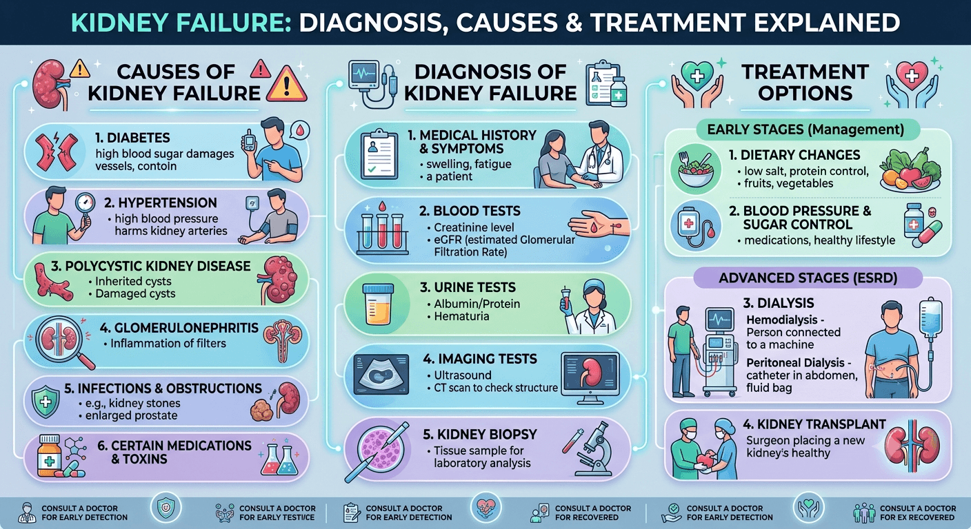
Causes and Risk Factors of Kidney Failure

Kidney failure rarely occurs without an underlying cause. In most cases, it develops as a result of chronic diseases that gradually damage the kidneys over many years or from acute conditions that suddenly impair kidney function. Identifying the cause is essential because early intervention can slow disease progression, prevent complications, and improve long-term outcomes.

1. Diabetes Mellitus

Diabetes is the leading cause of chronic kidney disease (CKD) and kidney failure worldwide.

Persistently elevated blood glucose damages the tiny blood vessels (glomeruli) responsible for filtering blood. Over time, this condition known as diabetic nephropathy reduces the kidneys’ filtering capacity, allowing protein to leak into the urine and eventually leading to irreversible kidney damage.

Warning signs include:

  • Protein in the urine (albuminuria)
  • Swelling of the feet
  • Rising blood pressure
  • Gradual decline in kidney function

Maintaining good blood sugar control significantly lowers the risk of diabetic kidney disease.

2. High Blood Pressure (Hypertension)

High blood pressure (Hypertension) is both a cause and a consequence of kidney disease.

Persistently elevated blood pressure damages the delicate blood vessels inside the kidneys, reducing their ability to filter blood effectively. As kidney function declines, blood pressure often becomes even more difficult to control, creating a harmful cycle of progressive damage.

3. Glomerulonephritis

Glomerulonephritis refers to inflammation of the glomeruli, the tiny filtering units of the kidneys.

It may result from:

  • Autoimmune diseases
  • Infections
  • Certain medications
  • Genetic disorders

Patients may experience:

  • Blood in the urine
  • Foamy urine
  • Swelling
  • High blood pressure

Early diagnosis and treatment can help preserve kidney function.

4. Polycystic Kidney Disease (PKD)

Polycystic kidney disease is an inherited disorder characterized by the development of numerous fluid-filled cysts within the kidneys. Over time, these cysts enlarge, replacing healthy kidney tissue and impairing kidney function.

Symptoms may include:

5. Urinary Tract Obstruction

Any blockage that prevents urine from flowing normally can damage the kidneys if left untreated.

Common causes include:

Prompt relief of the obstruction often improves kidney function.

6. Autoimmune Diseases

Several autoimmune conditions can attack the kidneys directly, including:

  • Systemic lupus erythematosus (SLE)
  • IgA nephropathy
  • Vasculitis
  • Goodpasture syndrome

These disorders cause inflammation that gradually damages kidney tissue.

7. Severe Infections (Sepsis)

Life-threatening infections can dramatically reduce blood flow to the kidneys and trigger acute kidney injury.

Sepsis remains one of the most common causes of acute kidney failure in hospitalized patients.

8. Certain Medications

Long-term or inappropriate use of certain medications may harm the kidneys, including:

  • Nonsteroidal anti-inflammatory drugs (NSAIDs) such as ibuprofen and naproxen
  • Certain antibiotics
  • Some chemotherapy drugs
  • Contrast dyes used during imaging studies
  • Certain herbal supplements

Medications should always be taken as directed by a healthcare professional, especially in individuals with existing kidney disease.

9. Heart and Liver Diseases

Conditions that reduce blood flow to the kidneys can contribute to kidney failure, including:

  • Heart failure
  • Severe liver disease (cirrhosis)
  • Cardiogenic shock

10. Dehydration

Severe dehydration caused by prolonged vomiting, diarrhea, excessive sweating, or inadequate fluid intake can reduce blood flow to the kidneys, leading to acute kidney injury if not corrected promptly.

Who Is at Higher Risk?

Several factors increase the likelihood of developing kidney failure:

  • Age over 60 years
  • Diabetes mellitus
  • High blood pressure
  • Cardiovascular disease
  • Family history of kidney disease
  • Obesity
  • Smoking
  • Chronic use of pain-relieving medications
  • Autoimmune disorders
  • Recurrent urinary tract infections
  • Kidney stones
  • African, Hispanic, Native American, and certain Asian populations have a higher prevalence of chronic kidney disease due to a combination of genetic, environmental, and socioeconomic factors.

People with these risk factors should undergo regular kidney function screening.

Diagnosis of Kidney Failure

Early diagnosis allows healthcare providers to slow disease progression, manage complications, and improve quality of life.

Medical History

The physician will ask about:

  • Symptoms
  • Family history
  • Diabetes
  • Hypertension
  • Medication use
  • Previous kidney disease
  • Recent infections
  • Fluid intake and urine output

Physical Examination

The healthcare provider evaluates:

  • Blood pressure
  • Swelling (edema)
  • Weight changes
  • Heart and lung sounds
  • Signs of dehydration
  • Skin changes
  • Neurological status

Blood Tests

  • Kidney function test
  • CBC
  • LDH
  • Lipid profile
  • Diabetic panel (RBS, HbA1c, INSULIN)

Serum Creatinine

Creatinine is a waste product produced by muscles.

Elevated creatinine usually indicates reduced kidney function.

Estimated Glomerular Filtration Rate (eGFR)

The eGFR estimates how efficiently the kidneys filter blood.

General interpretation:

eGFR (mL/min/1.73 m²)Kidney Function
90 or aboveNormal
60–89Mild decrease
30–59Moderate CKD
15–29Severe CKD
Below 15Kidney failure

Blood Urea Nitrogen (BUN)

High BUN levels often indicate reduced kidney function but can also rise due to dehydration or gastrointestinal bleeding.

Electrolyte Panel

Tests measure:

  • Potassium
  • Sodium
  • Calcium
  • Phosphate
  • Bicarbonate

Abnormal values help guide treatment.

Urine Tests

Urinalysis evaluates:

  • Protein
  • Blood
  • Infection
  • Glucose
  • Microscopic abnormalities

Urine Albumin-to-Creatinine Ratio (UACR)

One of the earliest indicators of kidney damage is excess albumin in the urine, particularly in people with diabetes or hypertension.

Imaging Studies

Imaging helps evaluate kidney size, structure, and blood flow.

Common tests include:

  • Kidney ultrasound
  • CT scan
  • MRI
  • Doppler ultrasound

Kidney Biopsy

In selected patients, a small sample of kidney tissue is examined under a microscope to determine the exact cause of kidney disease.

Treatment Options for Kidney Failure

Treatment depends on:

  • The underlying cause
  • Whether kidney failure is acute or chronic
  • The severity of kidney damage
  • Overall health
  • Associated medical conditions

The primary goals are to preserve remaining kidney function, manage symptoms, prevent complications, and improve quality of life.

1. Treating the Underlying Cause

Whenever possible, the underlying disease should be addressed.

Examples include:

  • Improving blood sugar control in diabetes
  • Managing high blood pressure
  • Treating infections
  • Relieving urinary obstruction
  • Discontinuing nephrotoxic medications
  • Treating autoimmune diseases with immunosuppressive therapy

2. Lifestyle Modifications

Lifestyle changes play a vital role in slowing the progression of chronic kidney disease.

Patients are encouraged to:

  • Follow a kidney-friendly diet
  • Limit sodium intake
  • Avoid smoking
  • Maintain a healthy weight
  • Exercise regularly, as tolerated
  • Stay adequately hydrated (following medical advice)
  • Avoid unnecessary over-the-counter pain medications

3. Dietary Therapy (Medical Nutrition Therapy)

Nutrition is one of the most important components of kidney disease management. A renal dietitian can help create a personalized eating plan based on the stage of kidney disease and laboratory results.

Key dietary recommendations may include:

  • Limiting sodium to help control blood pressure and reduce fluid retention.
  • Adjusting protein intake to reduce the kidneys’ workload while maintaining adequate nutrition.
  • Monitoring potassium intake if blood potassium levels are elevated.
  • Limiting phosphorus-rich foods to protect bone health.
  • Managing fluid intake in advanced kidney disease to prevent fluid overload.

Dietary needs vary significantly between individuals, particularly for those receiving dialysis, so professional guidance is essential.

Therapy for Kidney Failure

Treatment often extends beyond medications and includes supportive therapies aimed at preserving kidney function and improving overall health.

Blood Pressure Management

Strict blood pressure control is one of the most effective ways to slow kidney disease progression. Lifestyle measures combined with prescribed medications help protect the kidneys and reduce cardiovascular risk.

Blood Sugar Management

For people with diabetes, maintaining blood glucose within the target range helps prevent further damage to the kidneys.

Anemia Management

Reduced production of erythropoietin by damaged kidneys can lead to anemia. Treatment may include iron supplementation and erythropoiesis-stimulating agents (ESAs) under medical supervision.

Bone and Mineral Disorder Management

Chronic kidney disease can disrupt calcium, phosphorus, and vitamin D balance. Therapy may involve dietary changes, vitamin D supplements, and phosphate binders to protect bone health.

Dialysis

When kidney function declines to the point where waste products and excess fluid can no longer be adequately removed, dialysis becomes necessary.

There are two main types:

  • Hemodialysis: Blood is filtered through a dialysis machine, usually three times per week at a dialysis center or, in some cases, at home.
  • Peritoneal Dialysis: The lining of the abdomen (peritoneum) acts as a natural filter. Dialysis fluid is infused into and drained from the abdominal cavity, allowing waste removal at home.

Dialysis is a life-sustaining treatment but does not cure kidney failure.

Kidney Transplantation

For eligible patients with end-stage kidney disease, a kidney transplant offers the best long-term outcomes. A healthy kidney from a living or deceased donor is surgically implanted, restoring many normal kidney functions.

After transplantation, lifelong immunosuppressive medications are required to prevent organ rejection.

Medications Used in Kidney Failure

Medication regimens are tailored to each patient’s needs and may include:

  • ACE inhibitors or ARBs: Help control blood pressure and reduce protein loss in the urine.
  • Diuretics: Help remove excess fluid and reduce swelling.
  • Sodium-glucose cotransporter-2 (SGLT2) inhibitors: In selected patients with diabetes or chronic kidney disease, these medications can slow kidney disease progression and reduce cardiovascular risk.
  • Phosphate binders: Lower phosphorus levels in the blood.
  • Vitamin D analogs: Support bone health and calcium balance.
  • Erythropoiesis-stimulating agents (ESAs): Treat anemia associated with chronic kidney disease.
  • Iron supplements: Correct iron deficiency and support red blood cell production.
  • Potassium-lowering medications: Used when high potassium levels pose a risk.

Summary

Kidney failure is a serious condition that develops when the kidneys can no longer effectively remove waste products, excess fluid, and toxins from the blood. It may occur suddenly as acute kidney injury (AKI) or progress gradually as chronic kidney disease (CKD). Early recognition of symptoms, timely diagnosis, and appropriate treatment can slow disease progression, prevent complications, and improve quality of life.

Read More https://kdigo.org
Kidney Failure: 12 Symptoms You Should Never Ignore  

Kidney Failure 12 Symptoms You Should Never Ignore

Learn the Kidney Failure 12 Symptoms You Should Never Ignore, understand kidney anatomy, the different types of kidney failure, and how early diagnosis can help protect your kidney health.

Introduction

Your kidneys are among the hardest-working organs in your body, quietly performing life-sustaining tasks every minute of every day. They filter approximately 180 liters of blood daily, removing waste products, balancing fluids and electrolytes, regulating blood pressure, producing hormones that stimulate red blood cell production, and helping maintain healthy bones. Despite their remarkable resilience, kidney disease often develops silently. By the time noticeable symptoms appear, significant kidney damage may have already occurred.

Kidney failure, also known as renal failure or end-stage kidney disease (ESKD) in its advanced form, occurs when the kidneys lose their ability to effectively filter waste products and excess fluid from the bloodstream. Without prompt diagnosis and appropriate treatment, kidney failure can lead to life-threatening complications affecting nearly every organ system.

The encouraging news is that early recognition of warning signs, timely medical evaluation, and appropriate treatment can often slow or even prevent further kidney damage. Understanding the symptoms and risk factors empowers individuals to seek medical care before irreversible complications develop.

This comprehensive guide explains the Kidney Failure Symptoms You Should Never Ignore, understands kidney anatomy, the different types of kidney failure, and how early diagnosis can help protect your kidney health.

What Is Kidney Failure?

Kidney failure is a serious medical condition in which one or both kidneys can no longer perform their essential functions adequately. Healthy kidneys continuously filter blood, eliminating waste products, excess water, toxins, and medications through urine while maintaining the body’s delicate balance of minerals and fluids.

When kidney function declines significantly:

  • Waste products accumulate in the bloodstream.
  • Fluid builds up in the body.
  • Electrolyte levels become abnormal.
  • Blood pressure often rises.
  • Hormonal functions become impaired.
  • Red blood cell production decreases.
  • Bone metabolism becomes disrupted.

Kidney failure is generally diagnosed when kidney function falls below approximately 15% of normal, although symptoms may begin much earlier depending on the underlying cause and the speed of disease progression.

Without treatment, kidney failure may require dialysis or kidney transplantation to sustain life.

Types of Kidney Failure

Kidney failure is broadly classified into two major categories.

1. Acute Kidney Injury (AKI)

Previously known as acute renal failure, acute kidney injury develops suddenly, often within hours or days.

Common characteristics include:

  • Rapid decline in kidney function
  • Often reversible if treated promptly
  • Frequently occurs during severe illness or hospitalization
  • May result from dehydration, infection, medications, surgery, or reduced blood flow to the kidneys

Many patients recover completely when the underlying cause is identified and treated early.

2. Chronic Kidney Disease (CKD) Leading to Kidney Failure

Chronic kidney disease develops gradually over months or years.

Unlike AKI, chronic kidney disease usually causes:

  • Progressive loss of kidney function
  • Permanent kidney damage
  • Slow onset of symptoms
  • Increased cardiovascular risk

When chronic kidney disease advances to end-stage kidney disease (ESKD), dialysis or kidney transplantation becomes necessary for survival.

Other Clinical Classifications

Healthcare professionals often categorize kidney failure according to its underlying mechanism:

Prerenal Kidney Failure

Occurs when insufficient blood reaches the kidneys.

Examples include:

  • Severe dehydration
  • Major blood loss
  • Heart failure
  • Shock

Intrinsic (Renal) Kidney Failure

Results from direct damage to kidney tissue.

Causes include:

  • Glomerulonephritis
  • Acute tubular necrosis
  • Autoimmune diseases
  • Certain medications
  • Severe infections

Postrenal Kidney Failure

Develops when the urine flow becomes blocked.

Examples include:

Removing the obstruction early can often restore kidney function.

Kidney Anatomy: Understanding How Your Kidneys Work

Understanding kidney anatomy helps explain why kidney disease affects so many body systems.

Location

Most people have two kidneys, located:

  • On either side of the spine
  • Just below the rib cage
  • Behind the abdominal cavity

Each kidney is approximately the size of a clenched fist.

Major Structures of the Kidney

Renal Cortex

The outer layer, where blood filtration begins.

Renal Medulla

The inner portion, containing pyramids responsible for concentrating urine.

Nephrons

Each kidney contains approximately one million nephrons, the microscopic functional units responsible for filtration.

Each nephron consists of:

  • Glomerulus
  • Bowman’s capsule
  • Proximal tubule
  • Loop of Henle
  • Distal tubule
  • Collecting duct

Renal Artery

Supplies oxygen-rich blood to the kidneys.

Renal Vein

Returns filtered blood back to circulation.

Ureters

Carry urine from the kidneys to the bladder.

Functions of Healthy Kidneys

Healthy kidneys perform numerous vital functions:

Blood Filtration

They remove:

  • Urea
  • Creatinine
  • Excess salts
  • Toxins
  • Drug metabolites

Fluid Balance

Kidneys carefully regulate:

  • Water content
  • Sodium
  • Potassium
  • Calcium
  • Phosphate

Blood Pressure Regulation

Kidneys produce renin, an enzyme involved in controlling blood pressure.

Red Blood Cell Production

They release erythropoietin, which stimulates bone marrow to produce red blood cells.

Bone Health

Kidneys activate vitamin D, enabling proper calcium absorption and healthy bone metabolism.

Pathophysiology of Kidney Failure

Kidney failure develops when the kidneys lose functioning nephrons faster than the body can compensate.

Initially, surviving nephrons enlarge and work harder to maintain filtration. This process, known as hyperfiltration, temporarily preserves kidney function. Over time, however, the increased workload damages these remaining nephrons, creating a cycle of progressive kidney injury.

As kidney function declines:

  • Waste products such as urea and creatinine accumulate in the bloodstream.
  • Fluid retention leads to swelling and elevated blood pressure.
  • Potassium levels may rise to dangerous levels, increasing the risk of life-threatening heart rhythm disturbances.
  • Acid builds up in the body, causing metabolic acidosis.
  • Reduced erythropoietin production results in anemia, leading to fatigue and weakness.
  • Impaired activation of vitamin D contributes to weakened bones and disturbances in calcium and phosphate balance.

In advanced stages, nearly every organ system can be affected, including the heart, lungs, nervous system, digestive tract, bones, and immune system.

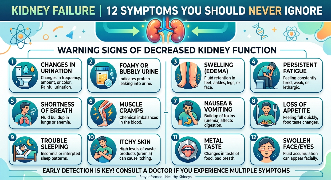
Kidney Failure: 12 Symptoms You Should Never Ignore

Kidney disease is often called a “silent disease” because many people experience no symptoms until significant damage has occurred. Recognizing the following warning signs can lead to earlier diagnosis and better outcomes.

1. Persistent Fatigue and Weakness

One of the earliest and most common symptoms is unexplained tiredness.

As kidney function declines:

  • Waste products accumulate in the blood.
  • Anemia develops due to reduced erythropoietin production.
  • Oxygen delivery to tissues decreases.

Patients often describe:

  • Constant exhaustion
  • Reduced stamina
  • Difficulty concentrating
  • Feeling weak despite adequate sleep

2. Swelling of the Feet, Ankles, Legs, or Face

Healthy kidneys remove excess fluid.

When they fail, fluid accumulates in body tissues, causing swelling (edema).

Common areas include:

  • Ankles
  • Feet
  • Lower legs
  • Hands
  • Eyelids
  • Face

Swelling that persists or worsens should never be ignored.

3. Changes in Urination

Changes in urination are often among the earliest indicators of kidney disease.

Watch for:

  • Urinating more frequently
  • Urinating less than usual
  • Foamy urine
  • Dark-colored urine
  • Blood in urine
  • Difficulty urinating
  • Pain during urination
  • Frequent nighttime urination

4. Shortness of Breath

Kidney failure can cause breathing difficulties because:

  • Fluid accumulates in the lungs.
  • Severe anemia reduces oxygen delivery.
  • Metabolic acidosis increases breathing effort.

Sudden or worsening shortness of breath requires immediate medical evaluation.

5. Persistent High Blood Pressure

High blood pressure is both:

  • A major cause of kidney disease
  • A common consequence of kidney failure

When the kidneys cannot regulate blood pressure effectively, hypertension becomes increasingly difficult to control.

6. Nausea and Vomiting

Accumulation of toxins in the bloodstream (uremia) frequently affects the digestive system.

Symptoms may include:

  • Persistent nausea
  • Vomiting
  • Metallic taste
  • Loss of appetite
  • Feeling full quickly

7. Loss of Appetite and Unintentional Weight Loss

Many patients gradually lose interest in eating because waste products alter taste perception and reduce appetite.

Over time, this may result in:

  • Weight loss
  • Muscle wasting
  • Nutritional deficiencies

8. Persistent Itching (Pruritus)

Advanced kidney disease commonly causes widespread itching due to:

  • Elevated phosphorus levels
  • Dry skin
  • Accumulation of waste products
  • Inflammation

The itching can become severe enough to interfere with sleep and daily activities.

9. Muscle Cramps

Damaged kidneys struggle to maintain a normal electrolyte balance.

Abnormal levels of:

  • Calcium
  • Potassium
  • Magnesium
  • Sodium

can trigger painful muscle cramps, especially at night.

10. Difficulty Concentrating or Confusion

As toxins accumulate in the bloodstream, brain function may be affected.

People may notice:

  • Poor memory
  • Difficulty focusing
  • Confusion
  • Reduced alertness

In severe kidney failure, an altered mental status is a medical emergency.

11. Chest Pain or Pressure

Kidney failure increases the risk of:

  • Fluid around the heart (pericardial effusion)
  • Heart disease
  • Electrolyte-related heart rhythm abnormalities

Chest pain should always be evaluated promptly, particularly in individuals with known kidney disease.

12. Persistent Bad Breath and Metallic Taste

A buildup of urea in the blood can lead to a distinctive metallic taste in the mouth and ammonia-like breath odor (uremic fetor). These symptoms often accompany advanced kidney dysfunction and may contribute to poor appetite and nausea.

When Should You Seek Immediate Medical Care?

Seek emergency medical attention if kidney-related symptoms are accompanied by:

  • Severe shortness of breath
  • Chest pain
  • Confusion or decreased consciousness
  • Complete inability to urinate
  • Rapidly worsening swelling
  • Persistent vomiting with dehydration
  • Very high blood pressure with headache or vision changes

Read More https://www.kidney.org

https://www.niddk.nih.gov

Continue Reading Part 2 Kidney Failure Treatment, Diagnosis & Causes Explained

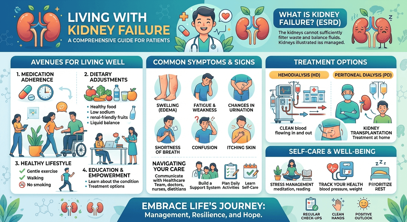
Part 3 Living with Kidney Failure: Expert Tips for Better Kidney Health

Living with Kidney Failure: Expert Tips for Better Kidney Health

living with kidney failure

Living with Kidney Failure: Expert Tips for Better Kidney Health

Living with Kidney Failure can be challenging, but the right care makes a difference. Discover expert tips to protect kidney health, manage symptoms, and improve your quality of life.

Introduction

A diagnosis of kidney failure can feel overwhelming, but it does not mean life has to stop. Advances in medical care, dialysis technology, kidney transplantation, and supportive therapies have significantly improved both survival and quality of life. Many people with kidney failure continue to work, travel, raise families, and enjoy meaningful activities with proper medical care and lifestyle adjustments.

Living with Kidney Failure

Living well with kidney failure requires a partnership between the patient, family members, nephrologists, primary care physicians, nurses, dietitians, pharmacists, and mental health professionals. The goal is not only to manage the disease but also to preserve independence, prevent complications, and maintain physical and emotional well-being.

1. Follow Your Treatment Plan

Kidney failure is a chronic condition that requires ongoing medical care. Adhering to the treatment plan prescribed by your healthcare provider is one of the most effective ways to reduce complications.

This includes:

  • Taking medications exactly as prescribed.
  • Attending all medical appointments.
  • Undergoing recommended blood and urine tests.
  • Following dialysis schedules without missing sessions.
  • Reporting new or worsening symptoms promptly.

Consistency is key. Even if you feel well, skipping medications or dialysis sessions can lead to serious health consequences.

2. Adopt a Kidney-Friendly Diet

Nutrition plays a central role in managing kidney failure. Since damaged kidneys cannot efficiently remove excess minerals and fluids, dietary modifications help reduce the workload on the kidneys and prevent complications.

General Dietary Recommendations

A renal dietitian may recommend:

  • Limiting sodium to help control blood pressure and reduce swelling.
  • Managing protein intake according to the stage of kidney disease.
  • Restricting potassium-rich foods if blood potassium levels are elevated.
  • Reducing phosphorus intake to protect bone health.
  • Controlling fluid intake in advanced kidney disease or for patients on dialysis.

Foods That Are Often Limited

  • Processed and packaged foods
  • Fast food
  • Salty snacks
  • Processed meats
  • Soft drinks containing phosphate additives
  • High-potassium fruits and vegetables (when advised)

Healthier Choices

  • Fresh fruits (based on potassium recommendations)
  • Low-sodium vegetables
  • Whole grains (when appropriate)
  • Lean poultry and fish
  • Healthy fats such as olive oil
  • Adequate calories to maintain a healthy weight

Dietary recommendations vary depending on laboratory results and whether a person is receiving dialysis. Individualized nutrition counseling is strongly recommended.

3. Stay Physically Active

Regular physical activity offers numerous benefits, including:

  • Improved cardiovascular health
  • Better blood pressure control
  • Increased muscle strength
  • Enhanced mood
  • Improved energy levels
  • Better sleep quality

Most individuals benefit from at least 150 minutes of moderate-intensity exercise per week, as tolerated. Activities may include walking, cycling, swimming, yoga, or light resistance training.

Always consult your healthcare provider before beginning a new exercise program.

4. Monitor Blood Pressure and Blood Sugar

Controlling blood pressure (hypertension) and blood glucose (diabetes) is essential for slowing the progression of kidney disease.

Blood Pressure Goals

Many patients with chronic kidney disease aim for a blood pressure target recommended by their healthcare provider, often below 130/80 mmHg, depending on individual circumstances.

Diabetes Management

People with diabetes should:

  • Monitor blood sugar regularly.
  • Take diabetes medications as prescribed.
  • Follow dietary recommendations.
  • Attend regular eye, foot, and kidney screenings.

Good diabetes control helps protect the kidneys from further damage.

5. Avoid Smoking and Limit Alcohol

Smoking damages blood vessels, accelerates kidney disease progression, and increases the risk of heart disease and stroke.

Excessive alcohol consumption may:

  • Increase blood pressure
  • Cause dehydration
  • Interfere with medications
  • Worsen existing kidney disease

Quitting smoking and limiting alcohol intake can substantially improve long-term health outcomes.

6. Stay Up to Date With Vaccinations

People with kidney disease have an increased risk of infections.

Healthcare providers commonly recommend:

  • Annual influenza vaccination
  • COVID-19 vaccination (as appropriate)
  • Pneumococcal vaccination
  • Hepatitis B vaccination, particularly for patients receiving dialysis

Vaccination schedules should always be discussed with a healthcare professional.

Emotional and Mental Health

Kidney failure affects more than physical health. Many individuals experience emotional challenges after diagnosis, including anxiety, depression, fear, frustration, and uncertainty about the future.

Common concerns include:

  • Changes in lifestyle
  • Financial stress
  • Dependence on dialysis
  • Reduced ability to work
  • Fear of complications

Seeking emotional support is an important part of treatment.

Helpful resources include:

  • Family and friends
  • Support groups
  • Mental health counselors
  • Clinical psychologists
  • Social workers
  • Patient advocacy organizations

Open communication with healthcare providers can also help address emotional and practical concerns.

Potential Complications of Kidney Failure

Without appropriate treatment, kidney failure can lead to serious complications affecting multiple organ systems.

These may include:

Cardiovascular Disease

People with chronic kidney disease are at significantly increased risk of:

  • Heart attack
  • Stroke
  • Heart failure
  • Peripheral artery disease

Cardiovascular disease remains the leading cause of death in patients with chronic kidney disease.

Anemia

Reduced production of erythropoietin by damaged kidneys results in fewer red blood cells, causing:

  • Fatigue
  • Weakness
  • Shortness of breath
  • Reduced exercise tolerance

Bone Disease

Impaired vitamin D activation and disturbances in calcium and phosphorus balance can lead to:

  • Weak bones
  • Bone pain
  • Increased fracture risk
  • Secondary hyperparathyroidism

Electrolyte Imbalances

Dangerously high potassium levels (Hyperkalemia) can cause life-threatening heart rhythm abnormalities.

Fluid Overload

Excess fluid may accumulate in:

  • Legs
  • Lungs
  • Abdomen

Severe fluid overload can result in breathing difficulties and heart failure.

Metabolic Acidosis

The inability of the kidneys to eliminate acid can cause metabolic acidosis, affecting muscle function, bone health, and overall metabolism.

Can Kidney Failure Be Prevented?

Although not all cases are preventable, many can be delayed or avoided through healthy lifestyle choices and regular medical care.

Prevention Tips

  • Maintain healthy blood pressure.
  • Control diabetes effectively.
  • Stay physically active.
  • Eat a balanced diet.
  • Maintain a healthy weight.
  • Avoid smoking.
  • Limit alcohol consumption.
  • Drink adequate fluids unless otherwise advised.
  • Avoid unnecessary use of NSAID pain relievers.
  • Manage cholesterol levels.
  • Treat urinary tract infections promptly.
  • Attend regular health checkups if you have risk factors.

Early detection through routine screening is particularly important for individuals with diabetes, hypertension, or a family history of kidney disease.

Frequently Asked Questions (FAQs)

Can kidney failure be reversed?

Acute kidney injury may be reversible if the underlying cause is treated promptly. Chronic kidney disease, however, generally causes permanent damage. While treatment can slow or stabilize progression, advanced kidney failure is usually irreversible.

What are the earliest signs of kidney failure?

Early symptoms may include:
Fatigue
Swelling of the feet or ankles
Changes in urination
High blood pressure
Foamy urine
Loss of appetite
Because symptoms can be subtle, routine blood and urine tests are important for people at risk.

Is dialysis a cure for kidney failure?

No. Dialysis replaces some of the kidneys’ filtering functions by removing waste products and excess fluid from the blood. It helps sustain life but does not restore normal kidney function or cure kidney failure.

Can a person live a normal life with one kidney?

Yes. Many people live healthy, active lives with one functioning kidney, including kidney donors. Regular medical follow-up and a healthy lifestyle help protect the remaining kidney.

How long can someone live with kidney failure?

Life expectancy depends on several factors, including age, overall health, the cause of kidney failure, adherence to treatment, and access to dialysis or transplantation. Many people live for years or even decades with appropriate care.

What foods should people with kidney failure avoid?

Dietary restrictions vary depending on the stage of kidney disease and blood test results. Many people are advised to limit:
High-sodium processed foods
Excess phosphorus
High-potassium foods (when potassium levels are elevated)
Excessive protein (in some stages)
Sugary beverages
A renal dietitian can provide personalized guidance.

When should I see a doctor?

Seek medical attention if you experience:
Persistent swelling
Blood in the urine
Foamy urine
Reduced urine output
Persistent fatigue
Uncontrolled high blood pressure
Shortness of breath
Persistent nausea or vomiting
Early evaluation can prevent further kidney damage.

Who should be screened for kidney disease?

Screening is recommended for individuals with:
Diabetes
High blood pressure
Cardiovascular disease
A family history of kidney disease
Obesity
Age over 60 years
Recurrent kidney stones or urinary tract infections
Routine blood and urine tests can detect kidney disease before symptoms develop.

Conclusion

Kidney failure is a serious but manageable medical condition that affects millions of people worldwide. Because the kidneys play a vital role in filtering waste, regulating blood pressure, maintaining electrolyte balance, supporting red blood cell production, and promoting bone health, their failure can have widespread effects on the body. Fortunately, many cases of kidney disease can be slowed or even prevented from progressing through early diagnosis, appropriate medical treatment, and healthy lifestyle choices.

Recognizing the 12 warning signs discussed in this article, including persistent fatigue, swelling, changes in urination, shortness of breath, nausea, itching, muscle cramps, and difficulty concentrating, can lead to earlier medical evaluation and better outcomes. Individuals with diabetes, high blood pressure, cardiovascular disease, or a family history of kidney disease should undergo regular kidney function screening, even if they feel well.

Modern treatments such as medications, dietary therapy, dialysis, and kidney transplantation have transformed the outlook for people living with kidney failure. Equally important are patient education, adherence to treatment plans, emotional support, and close collaboration with healthcare professionals. With timely intervention and comprehensive care, many individuals with kidney failure can lead productive, fulfilling lives.

Key Takeaway:

Never ignore persistent symptoms that may suggest kidney disease. If you notice changes in your health, particularly swelling, altered urination, unexplained fatigue, or uncontrolled high blood pressure, seek medical evaluation promptly. Early action remains one of the most powerful tools for protecting kidney function and preserving overall health.

Start with Part 1 and Part 2 for a complete understanding of kidney failure

https://www.kidney.org

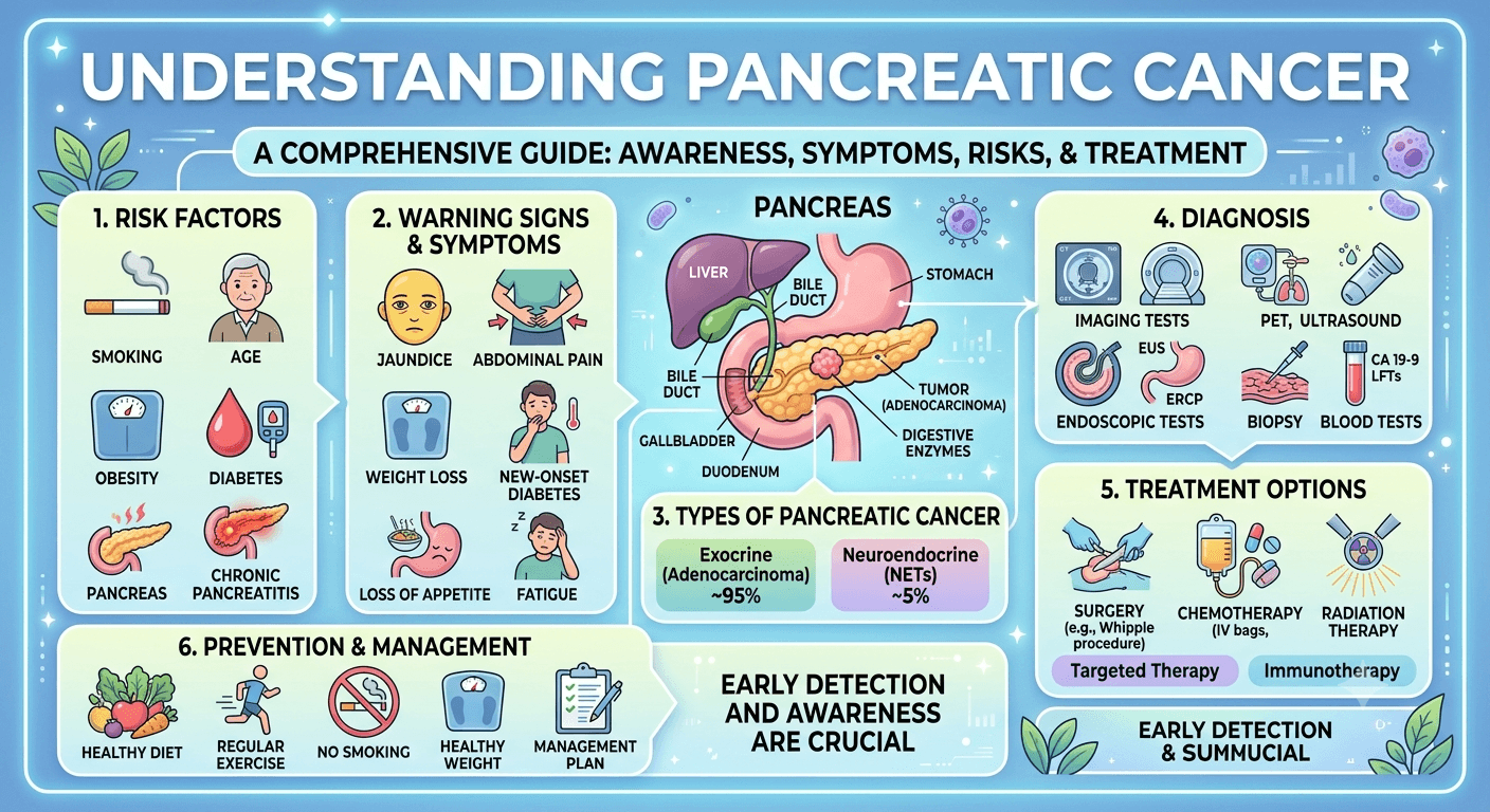
Catching It Early: The Silent Warning Signs of Pancreatic Cancer

warning signs of pancreatic cancer

Catching It Early: The Silent Warning Signs of Pancreatic Cancer

Learn the silent warning signs of pancreatic cancer, early symptoms, risk factors, diagnosis, and treatment options to improve the chances of early detection.

Introduction

Pancreatic cancer is often referred to as the “silent disease” since it starts developing without noticeable symptoms. The lack of early symptoms often leads to late diagnosis of this condition when the patient experiences pain or other complications associated with an already advanced stage of cancer development. Recognizing early warning signs and knowing about possible risk factors will allow diagnosing this cancer at the earliest stage, which would help a lot with choosing the best treatment strategy and increasing chances of successful recovery.

Even though pancreatic cancer is rather uncommon among all cancer types, this disease ranks among the most deadly cancerous conditions. Progress in medical sciences and new diagnostic methods, personalized treatments, and targeted therapies allow gradually improving survival rates for this type of cancer.

The current paper discusses in detail pancreatic cancer, including its causes, anatomy, symptoms, diagnosis, treatment, and the life of patients who deal with this disease.

What Is Pancreatic Cancer?

Pancreatic cancer represents a condition when some cells start growing uncontrollably and forming tumors in the pancreas. Cancerous cells not only invade surrounding tissues but may metastasize and affect other parts of the body, for example, liver, lungs, or abdomen.

The pancreas is an important organ placed behind the stomach. This organ serves two major functions:

  • Production of enzymes that help to digest food
  • Production of hormones, such as insulin and glucagon, that help to control the level of blood sugar

The appearance of cancer in the pancreas may influence both digestion and regulation of blood sugar levels.

One of the main features of pancreatic cancer is the absence of early symptoms.

Types of Pancreatic Cancer

Pancreatic cancer is broadly classified into two major categories.

1. Exocrine Pancreatic Cancer

Approximately 90–95% of pancreatic cancers originate from the exocrine cells that produce digestive enzymes.

The most common type is:

Pancreatic Ductal Adenocarcinoma (PDAC)

  • Begins in the ducts carrying digestive enzymes
  • Most aggressive form
  • Responsible for the majority of pancreatic cancer deaths

Other rare exocrine cancers include:

  • Acinar cell carcinoma
  • Adenosquamous carcinoma
  • Squamous cell carcinoma

2. Neuroendocrine Pancreatic Tumors (PNETs)

These cancers develop from hormone-producing endocrine cells.

Some produce excessive hormones, causing noticeable symptoms, while others remain non-functional.

Examples include:

  • Insulinomas
  • Gastrinomas
  • Glucagonomas
  • VIPomas

Neuroendocrine tumors generally grow more slowly and often have a better prognosis than pancreatic adenocarcinoma.

Anatomy of the Pancreas

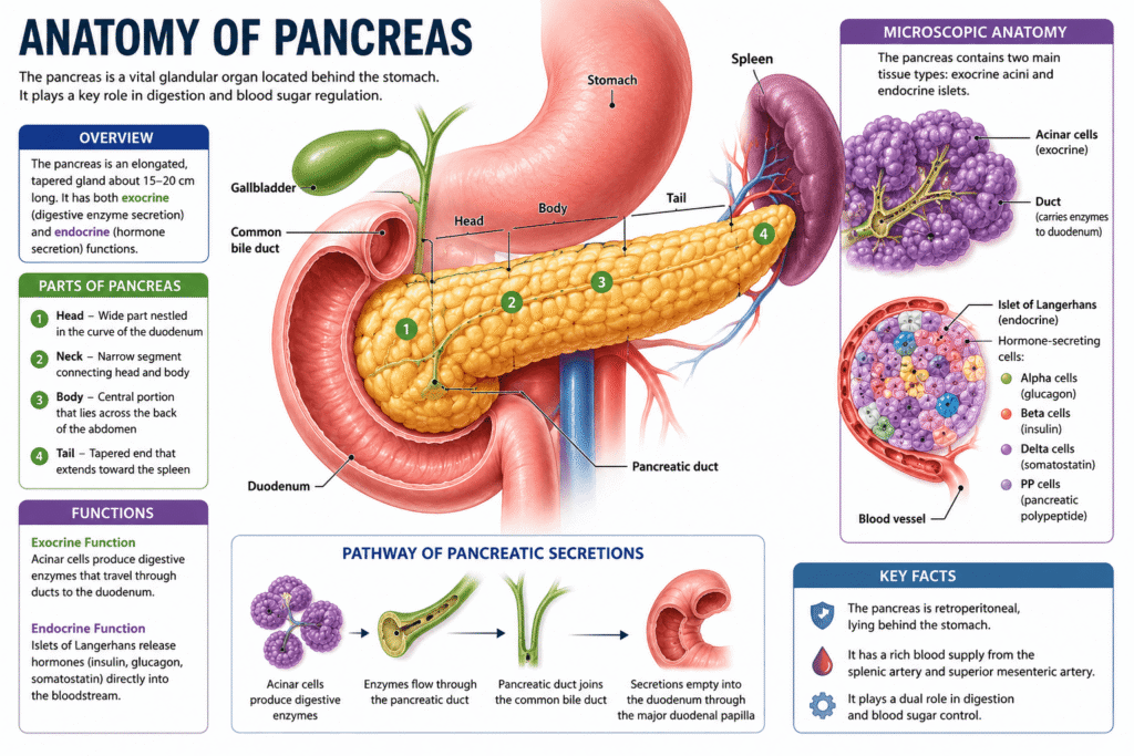
Anatomy of Pancreas

The pancreas is a long, flat gland measuring about 15–20 cm (6–8 inches) in length.

It consists of three major sections:

Head

  • Located near the small intestine (duodenum)
  • Most pancreatic cancers develop here

Body

  • Middle portion extending across the abdomen

Tail

  • Extends toward the spleen
  • Tumors here are often detected later because they cause fewer early symptoms

Functions of the Pancreas

Exocrine Function

Produces digestive enzymes including:

  • Amylase
  • Lipase
  • Proteases

These enzymes help digest carbohydrates, fats, and proteins.

Endocrine Function

Produces hormones including:

  • Insulin
  • Glucagon
  • Somatostatin

These regulate blood glucose and metabolism.

Pathophysiology

Pancreatic cancer develops through a gradual accumulation of genetic mutations that transform healthy pancreatic cells into malignant ones.

The process often begins with precancerous lesions known as:

  • PanIN (Pancreatic Intraepithelial Neoplasia)
  • IPMN (Intraductal Papillary Mucinous Neoplasm)
  • MCN (Mucinous Cystic Neoplasm)

Over time, mutations in genes such as:

  • KRAS
  • TP53
  • CDKN2A
  • SMAD4

allow cells to:

  • Divide uncontrollably
  • Resist normal cell death
  • Invade nearby tissues
  • Spread through blood vessels and lymphatics

Pancreatic tumors also create a dense fibrous environment that limits blood supply and makes chemotherapy less effective.

Silent Warning Signs of Pancreatic Cancer

One reason pancreatic cancer is so dangerous is that symptoms may initially seem mild or unrelated.

Persistent Upper Abdominal Pain

Pain often develops in the upper abdomen and may radiate to the back.

It may worsen:

  • After eating
  • While lying flat

Unexplained Weight Loss

Unexpected weight loss is one of the earliest warning signs.

Patients often notice:

  • Loss of appetite
  • Feeling full quickly
  • Muscle wasting

Jaundice

Tumors in the pancreatic head can block the bile duct.

Symptoms include:

  • Yellow skin
  • Yellow eyes
  • Dark urine
  • Pale stools
  • Itchy skin

Digestive Problems

Many patients develop:

  • Nausea
  • Vomiting
  • Indigestion
  • Bloating
  • Fatty stools
  • Diarrhea

New-Onset Diabetes

Suddenly developing diabetes after age 50 without obvious risk factors can occasionally be an early indicator of pancreatic cancer.

Fatigue

Persistent tiredness often accompanies cancer progression.

Blood Clots

Some patients experience unexplained blood clots before their cancer diagnosis.

Back Pain

Pain spreading into the middle or lower back may occur as tumors enlarge.

Causes and Risk Factors

The exact cause of pancreatic cancer is not always known. It develops when genetic mutations cause pancreatic cells to grow uncontrollably, forming a tumor. Several lifestyle, environmental, and inherited factors can increase the risk of developing the disease.

Common risk factors include:

  • Smoking: The leading preventable risk factor, significantly increasing the likelihood of pancreatic cancer.
  • Older age: Most cases occur in people over 60 years of age.
  • Family history: Having close relatives with pancreatic cancer raises the risk.
  • Inherited genetic syndromes: Mutations in genes such as BRCA1, BRCA2, PALB2, CDKN2A, STK11, and PRSS1 can increase susceptibility.
  • Chronic pancreatitis: Long-term inflammation of the pancreas increases cancer risk, particularly hereditary pancreatitis.
  • Diabetes mellitus: Long-standing type 2 diabetes and newly diagnosed diabetes in older adults may be associated with pancreatic cancer.
  • Obesity: Excess body weight contributes to chronic inflammation and metabolic changes that promote cancer development.
  • Unhealthy diet: Diets high in processed meats, red meat, and saturated fats and low in fruits and vegetables may increase risk.
  • Heavy alcohol consumption: Excessive alcohol intake can lead to chronic pancreatitis, an established risk factor.
  • Physical inactivity: A sedentary lifestyle contributes indirectly through obesity and insulin resistance.
  • Occupational exposure: Long-term exposure to certain chemicals used in the petroleum, metalworking, and pesticide industries may increase risk.
  • Race and ethnicity: Incidence is higher in some populations, including people of African ancestry.
  • Male sex: Men have a slightly higher risk than women, partly due to higher rates of smoking and occupational exposures.

Diagnosis

Because symptoms are often vague, multiple diagnostic tools are used.

Medical History and Physical Examination

Doctors evaluate:

  • Symptoms
  • Weight changes
  • Family history
  • Risk factors

Blood Tests

May include:

  • Liver function tests
  • RBS, HbA1c
  • CRP, ESR
  • Kidney function test
  • S. Amylase and S. Lipase
  • Blood sugar levels
  • CA 19-9 tumor marker

CA 19-9 alone cannot diagnose pancreatic cancer but helps monitor treatment.

Imaging Studies

Common imaging tests include:

  • CT scan
  • MRI
  • MRCP
  • PET scan
  • Ultrasound

Endoscopic Ultrasound (EUS)

Allows detailed imaging while obtaining tissue samples through biopsy.

Biopsy

A biopsy confirms the diagnosis by examining tissue under a microscope.

Treatment Options

Treatment depends on:

  • Stage
  • Tumor location
  • Overall health
  • Cancer type

Surgery

Surgery offers the best chance for cure when cancer is localized.

Common procedures include:

Whipple Procedure

Removes:

  • Pancreatic head
  • Duodenum
  • Gallbladder
  • Part of the bile duct

Distal Pancreatectomy

Removes:

  • Pancreatic body
  • Tail
  • Sometimes the spleen

Total Pancreatectomy

Entire pancreas removed.

Patients require lifelong insulin and digestive enzyme replacement.

Therapy

Chemotherapy

Common chemotherapy regimens include:

  • FOLFIRINOX
  • Gemcitabine
  • Gemcitabine plus nab-paclitaxel

Chemotherapy may be given:

  • Before surgery
  • After surgery
  • For advanced disease

Radiation Therapy

High-energy radiation destroys cancer cells.

It may:

  • Shrink tumors
  • Reduce pain
  • Improve surgical outcomes

Targeted Therapy

Patients with specific genetic mutations may benefit from targeted medications.

Examples include:

  • PARP inhibitors for BRCA mutations
  • NTRK inhibitors for rare gene fusions

Immunotherapy

Although only effective in a small percentage of patients, immunotherapy can be beneficial for tumors with specific genetic characteristics such as mismatch repair deficiency.

Medications

Several medications help manage symptoms and improve quality of life.

These include:

Pain medications

  • Acetaminophen
  • NSAIDs
  • Opioids

Pancreatic enzyme replacement

Improves digestion and nutrient absorption.

Insulin

For diabetes caused by pancreatic dysfunction.

Anti-nausea medications

Help reduce chemotherapy-related nausea.

Appetite stimulants

May improve nutritional intake.

Anticoagulants

Used if blood clots develop.

Living with Pancreatic Cancer

The condition influences one’s physical, psychological, social and economic aspects of life. However, people are able to live an enriched life, retain their connections and relationships, and feel well with proper treatment and assistance.

Eating Right

It is hard to eat properly due to problems with digestion and lack of appetite.

Useful measures comprise:

  • Eating frequently and in small portions
  • Foods with high amount of protein
  • Taking pancreatic enzyme preparations
  • Hydration
  • Meeting with a nutritionist

Dealing with Fatigue

Fatigue is a typical problem.

There are some things that patients may find useful:

  • Smooth physical activity
  • Getting enough rest
  • Conserving energy
  • Balanced nutrition

Being Mentally Healthy

Anxiety, sadness, or fear may be felt. One may use counseling services, take part in discussion groups, engage in meditation and speak to relatives and friends.

Palliative Treatment

It helps to alleviate symptoms, control pain, and improve life quality. It can be used not only when cancer is terminal but also alongside active treatment.

Regular Check-Ups

They help to evaluate one’s progress in treatment, handle side effects and detect early signs of disease recurrence.

Frequently Asked Questions (FAQs)

Is pancreatic cancer hereditary?

Most cases are not inherited, but approximately 10% are associated with inherited genetic mutations or strong family history.

Can pancreatic cancer be cured?

When detected early and completely removed surgically, some patients can achieve long-term survival. However, many cases are diagnosed at an advanced stage.

Who is most at risk?

People over 60 years of age, smokers, individuals with obesity, chronic pancreatitis, diabetes, or a family history of pancreatic cancer have a higher risk.

Does pancreatic cancer always cause pain?

No. Early pancreatic cancer may not cause pain or any noticeable symptoms.

Can lifestyle changes reduce risk?

Maintaining a healthy weight, avoiding tobacco, limiting alcohol consumption, eating a balanced diet, and staying physically active may reduce the risk.

What are the survival rates?

Survival depends on the stage at diagnosis. Patients diagnosed when the disease is localized generally have significantly better outcomes than those diagnosed after the cancer has spread.

Conclusion

Pancreatic cancer continues to be one of the toughest cancers because it develops subtly with symptoms that could be misunderstood as something else. However, increased awareness of its hidden signs including sudden weight loss, constant abdominal or back pain, yellow skin or eyes, digestive problems, and diabetes could help people visit their doctor earlier and perhaps even save their lives.

With the development of better surgical procedures, chemotherapies, radiation therapies, targeted therapies, supportive care, and personalized medicine, many people are surviving longer. Nutritional support, pain relief, mental counseling, and palliative care are equally important during treatment.

If you have symptoms or some risk factors for developing the disease, it is crucial to visit your doctor without delay. Early detection, timely diagnosis, and comprehensive treatment are the best way of fighting this type of cancer.

Read More

Disclaimer: The information provided in this blog post is for educational and informational purposes only and should not be considered medical advice. It is not intended to replace professional medical consultation, diagnosis, or treatment.

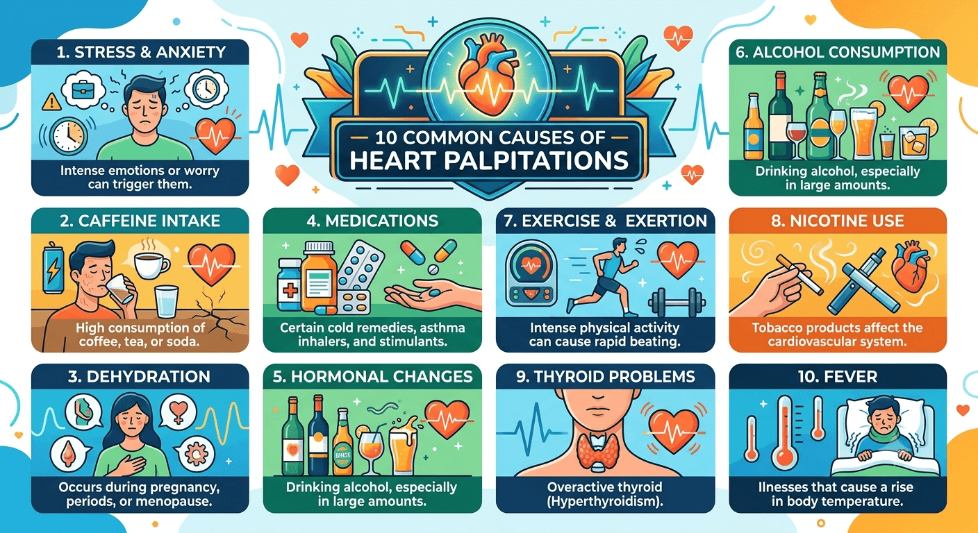
10 Common Causes of Heart Palpitations: You Should Never Ignore

causes of heart palpitations

Discover the 10 common causes of heart palpitations you should never ignore. Learn about symptoms, risk factors, diagnosis, treatment options, and when to seek medical attention for a healthier heart.

Introduction

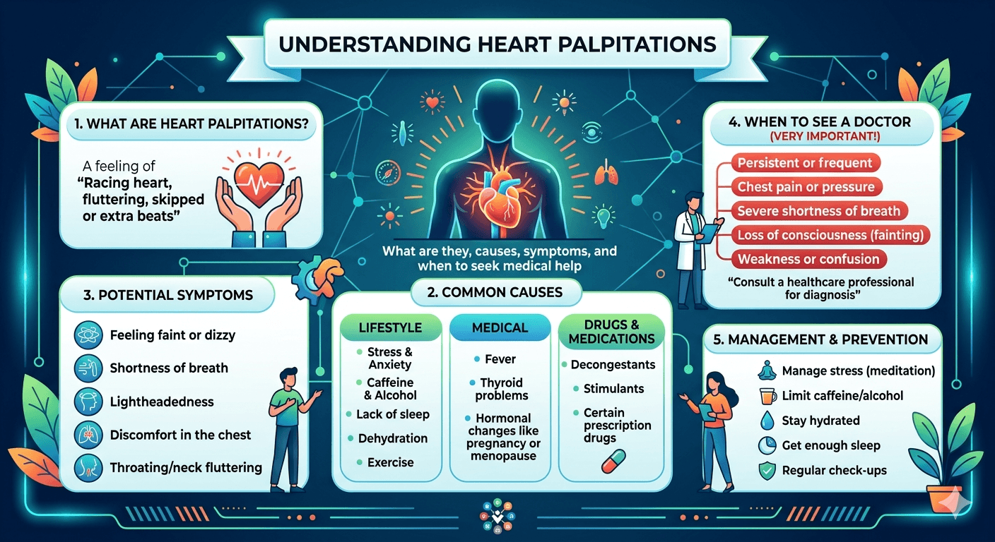
Heart palpitations are a frequent complaint that people suffer from throughout their lives. They occur when there is a sensation of racing, pounding, fluttering, skipping beats, or irregular heartbeats. While palpitations are often innocent, they can also be a sign of a medical problem requiring treatment.

Find out about 10 common reasons for heart palpitations you shouldn’t overlook. Get information about symptoms, risks, diagnosis, treatment and when to seek help for better heart health.

For patients, knowing about the meaning of heart palpitations can help in alleviating worries and getting prompt medical consultation where needed. For medical students, heart palpitations are a clinically relevant sign, which falls into the field of cardiology, endocrinology, psychiatry, and internal medicine.

In this article, we will discuss the physiology, causes, warning signs, diagnostic techniques, and management of heart palpitations with particular attention to 10 causes that shouldn’t be overlooked.

What Are Heart Palpitations?

Heart palpitations are the sensation of one’s heartbeat. Usually, the heart beats in a rhythmic manner, which is unnoticed by patients. However, when the heart beats faster, stronger, irregular, or prematurely, people become aware of such sensations.

Palpitations can be felt in the:

  • Chest
  • Throat
  • Neck
  • Upper abdomen

They can last:

  • Some seconds
  • Minutes
  • Hours
  • Sometimes even longer

The sensation itself does not always indicate a dangerous condition, but understanding the underlying cause is essential.

How the Heart Normally Beats

To understand palpitations, it helps to review normal cardiac conduction.

The heart’s electrical system begins at the sinoatrial (SA) node, often called the heart’s natural pacemaker.

The electrical impulse travels through:

  1. SA node
  2. Atria
  3. Atrioventricular (AV) node
  4. Bundle of His
  5. Purkinje fibers
  6. Ventricles

This coordinated process produces a regular heartbeat, typically between 60 and 100 beats per minute in adults at rest.

Any disturbance in this electrical pathway can result in palpitations.

What Do Heart Palpitations Feel Like?

Patients commonly describe palpitations as:

  • Fluttering in the chest
  • Racing heartbeat
  • Heart “flip-flopping”
  • Skipped beats
  • Thumping sensation
  • Rapid pounding
  • Irregular rhythm

Associated symptoms may include:

  • Lightheadedness
  • Dizziness
  • Shortness of breath
  • Chest discomfort
  • Fatigue
  • Anxiety

The accompanying symptoms often provide important clues about the severity of the underlying condition.

10 Common Causes of Heart Palpitations

1. Cardiac Arrhythmias

One of the most important causes of palpitations is an abnormal heart rhythm, known as an arrhythmia.

Common arrhythmias include:

Atrial Fibrillation (AFib)

AFib causes rapid, chaotic electrical activity in the atria.

Symptoms may include:

  • Irregular heartbeat
  • Fatigue
  • Shortness of breath
  • Reduced exercise tolerance

AFib significantly increases the risk of stroke and should never be ignored.

Supraventricular Tachycardia (SVT)

SVT involves episodes of rapid heart rate originating above the ventricles.

Patients often report:

  • Sudden onset
  • Sudden termination
  • Heart rates exceeding 150 beats per minute

Ventricular Arrhythmias

These arise from the ventricles and can be life-threatening.

Examples include:

  • Ventricular tachycardia
  • Ventricular fibrillation

Immediate medical evaluation is required.

2. Anxiety and Panic Disorders

The relationship between the mind and heart is powerful.

During anxiety or panic attacks, the body releases stress hormones such as:

  • Adrenaline
  • Noradrenaline

These chemicals increase:

  • Heart rate
  • Blood pressure
  • Cardiac contractility

Patients may experience:

  • Racing heartbeat
  • Sweating
  • Trembling
  • Shortness of breath
  • Fear of dying

Although anxiety-related palpitations are often benign, they can closely mimic serious cardiac conditions.

3. Excessive Caffeine Consumption

Caffeine stimulates the central nervous system and can increase heart rate.

Common sources include:

  • Coffee
  • Energy drinks
  • Tea
  • Soda
  • Pre-workout supplements

Excessive intake may trigger:

  • Premature atrial contractions
  • Premature ventricular contractions
  • Tachycardia

Individuals vary significantly in caffeine sensitivity.

4. Hyperthyroidism

An overactive thyroid gland produces excessive thyroid hormones.

These hormones increase metabolic activity throughout the body, including the cardiovascular system.

Symptoms often include:

  • Palpitations
  • Weight loss
  • Heat intolerance
  • Tremors
  • Nervousness
  • Increased appetite

Hyperthyroidism commonly causes:

  • Sinus tachycardia
  • Atrial fibrillation

Medical evaluation and treatment are essential.

5. Anemia

Anemia occurs when there are insufficient healthy red blood cells to deliver adequate oxygen to tissues.

The heart compensates by pumping harder and faster.

Common symptoms include:

  • Palpitations
  • Fatigue
  • Weakness
  • Dizziness
  • Shortness of breath

Causes of anemia may include:

  • Iron deficiency
  • Vitamin B12 deficiency
  • Chronic disease
  • Blood loss

Treating the underlying anemia often resolves the palpitations.

6. Heart Disease

Structural heart disease is a significant cause of concerning palpitations.

Examples include:

Coronary Artery Disease

Reduced blood flow to heart muscle can trigger abnormal rhythms.

Heart Failure

Damaged heart tissue may disrupt normal electrical pathways.

Cardiomyopathy

Diseases affecting heart muscle structure increase arrhythmia risk.

Patients with known heart disease who develop new palpitations should seek prompt evaluation.

7. Electrolyte Imbalances

Normal heart function depends on carefully regulated electrolyte levels.

Key electrolytes include:

  • Potassium
  • Magnesium
  • Calcium
  • Sodium

Abnormal levels can alter electrical conduction.

Potential causes include:

  • Dehydration
  • Severe vomiting
  • Diarrhea
  • Kidney disease
  • Certain medications

Both low and high potassium levels can produce dangerous arrhythmias.

8. Medication Side Effects

Many medications can trigger palpitations.

Examples include:

Respiratory Medications

  • Albuterol inhalers
  • Bronchodilators

Decongestants

  • Pseudoephedrine
  • Phenylephrine

Thyroid Medications

Excessive thyroid hormone replacement may increase heart rate.

Stimulants

  • ADHD medications
  • Weight-loss drugs

A careful medication review is an important part of evaluating palpitations.

9. Alcohol and Recreational Substances

Alcohol can affect cardiac electrical activity.

A phenomenon known as “holiday heart syndrome” occurs when excessive alcohol consumption triggers arrhythmias in otherwise healthy individuals.

Other substances associated with palpitations include:

  • Cocaine
  • Amphetamines
  • Methamphetamine
  • Cannabis (in some individuals)
  • Nicotine

These substances may increase the risk of serious cardiovascular complications.

10. Pregnancy

Pregnancy places significant demands on the cardiovascular system.

Changes include:

  • Increased blood volume
  • Increased heart rate
  • Higher cardiac output

Many pregnant women experience occasional palpitations that are benign.

However, evaluation may be necessary if symptoms are accompanied by:

  • Fainting
  • Severe shortness of breath
  • Chest pain
  • Persistent tachycardia

Underlying conditions such as anemia and thyroid disease should also be considered.

Warning Signs That Require Immediate Medical Attention

Certain symptoms suggest a potentially serious cause and warrant urgent evaluation.

Seek emergency care if palpitations occur with:

  • Chest pain
  • Fainting (syncope)
  • Severe shortness of breath
  • Persistent rapid heartbeat
  • Loss of consciousness
  • Severe dizziness
  • Sudden weakness
  • Signs of stroke

These symptoms may indicate a dangerous arrhythmia or cardiovascular emergency.

How Doctors Diagnose Heart Palpitations

Diagnosis begins with a thorough history and physical examination.

Important questions include:

  • When did symptoms start?
  • How often do they occur?
  • How long do they last?
  • What triggers them?
  • Are there associated symptoms?

Diagnostic Tests

Electrocardiogram (ECG)**

Records the heart’s electrical activity.

Holter Monitor

Continuous ECG recording over 24–48 hours.

Event Monitor

Used for intermittent symptoms occurring less frequently.

Blood Tests

May evaluate:

  • Thyroid function tests
  • CBC
  • Cardio Marker (Troponin I & T), CK-MB
  • LDH
  • Liver function test
  • Electrolytes
  • Infection (CRP, ESR)

Echocardiogram

Assesses heart structure and function.

Exercise Stress Testing

Evaluates symptoms related to physical activity.

Treatment of Heart Palpitations

Treatment depends entirely on the underlying cause.

Lifestyle Measures

Many patients benefit from:

  • Reducing caffeine intake
  • Limiting alcohol consumption
  • Managing stress
  • Staying hydrated
  • Getting adequate sleep
  • Avoiding smoking

Medication

Depending on the diagnosis, treatment may include:

  • Beta blockers
  • Antiarrhythmic drugs
  • Thyroid medications
  • Anxiety treatments

Procedures

Some arrhythmias require:

  • Catheter ablation
  • Pacemaker implantation
  • Implantable cardioverter-defibrillator (ICD)

Prevention Strategies

While not all palpitations can be prevented, healthy lifestyle habits can significantly reduce risk.

Recommended measures include:

  • Maintaining a healthy weight
  • Exercising regularly
  • Managing blood pressure
  • Controlling diabetes
  • Limiting stimulants
  • Practicing stress-reduction techniques
  • Following prescribed treatments

Regular medical checkups can identify risk factors before complications develop.

Frequently Asked Questions (FAQs)

Are heart palpitations always dangerous?

No. Many palpitations are harmless and related to stress, caffeine, or temporary physiological changes. However, some may indicate significant heart disease.

Can dehydration cause palpitations?

Yes. Dehydration can alter electrolyte balance and increase heart rate, leading to palpitations.

When should I see a doctor for palpitations?

You should seek medical evaluation if palpitations are frequent, persistent, worsening, or associated with dizziness, chest pain, or fainting.

Can anxiety alone cause palpitations?

Yes. Anxiety and panic attacks are among the most common non-cardiac causes of palpitations.

Does caffeine affect everyone the same way?

No. Sensitivity to caffeine varies widely among individuals. Some people experience palpitations even with small amounts.

Can young healthy people develop dangerous arrhythmias?

Although uncommon, serious arrhythmias can occur in young individuals, particularly those with inherited heart conditions or structural abnormalities.

Conclusion

Palpitations are a symptom and not a disorder, the importance of which varies from entirely harmless to potentially fatal. Although occasional palpitations due to stress, caffeine, or insomnia might not raise any concerns, recurring or repeated episodes warrant closer scrutiny. Arrhythmias, hyperthyroidism, anemia, structural heart disorders, or electrolyte imbalances are just some of the disorders where palpitations form part of the symptom complex.

As far as patients are concerned, the bottom line here is clear: palpitations should never be ignored if they happen frequently or if they are serious and associated with worrying symptoms. From the medical student’s perspective, palpitations serve as a perfect case study of a single symptom which is caused by a wide variety of physiological and pathophysiological mechanisms.

Early recognition and timely evaluation can make the difference between reassurance and life-saving intervention.

Read More

Disclaimer: The information provided in this blog post is for educational and informational purposes only and should not be considered medical advice. It is not intended to replace professional medical consultation, diagnosis, or treatment.

Obsessive Compulsive Disorder (OCD): Symptoms, Causes, Diagnosis, and Treatment

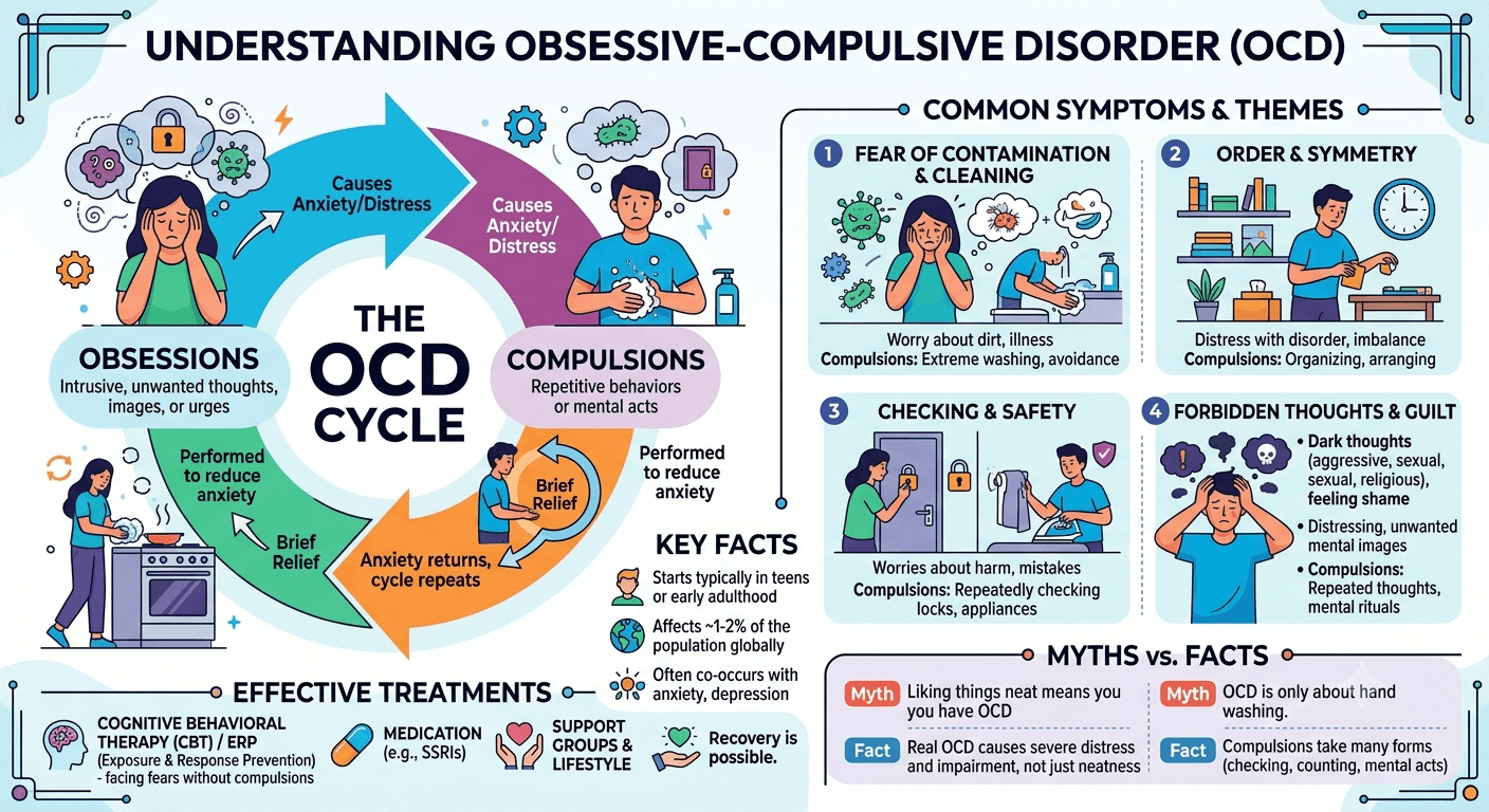
Obsessive compulsive disorder (OCD)

In this article, you will find information on obsessive compulsive disorder (OCD) including its definition, symptoms, causes, treatment and coping strategies.

Introduction

Obsessive compulsive disorder (OCD) is a very widespread mental disorder that affects thousands of people throughout the world. However, although people tend to use the term OCD for describing someone’s obsession with cleanliness or organization, it is a serious condition which can have a huge impact on one’s daily activities, personal and professional relationships, etc.

From my experience as a healthcare provider, I would like to say that obsessive-compulsive disorder can be very stressful for patients and their families. Luckily, it is a condition which can be treated properly if an adequate diagnosis and necessary treatment are received.

Learn about Obsessive-Compulsive Disorder (OCD), including its symptoms, causes, risk factors, diagnosis, and treatment options. Discover how therapy, medications, and lifestyle strategies can help manage OCD and improve quality of life.

What Is Obsessive-Compulsive Disorder (OCD)?

Obsessive compulsive disorder (OCD) is a chronic mental condition characterized by:

  • Obsessions – persistent and intrusive thoughts, urges, images which cause anxiety and distress;
  • Compulsions – repetitive activities and behaviors which people engage in order to alleviate the anxiety or avoid negative consequences.

Patients with OCD usually realize that their obsessions and compulsions are irrational but they cannot control them.

Here are examples of obsessions and compulsions:

  • A patient is constantly thinking about the possibility of having contaminated his/her hands (obsession).
  • The person washes his/her hands dozens of times per day in order to relieve anxiety (compulsion).

The cycle of obsession and compulsion can consume hours each day and significantly impair functioning.

Types of OCD

OCD is unique for each patient; however, there are some typical symptoms of the disease.

1. Contamination OCD

Fear of germs, dirt, chemicals, bodily fluids, illness.

Typical compulsions:

  • Frequent hand washing
  • Frequent showers
  • Multiple cleanings of household items
  • Refusal to go to public places

Example

An individual washes his or her hands for 30 minutes after using a door handle because of the fear of infection.

2. Checking OCD

An obsession with the idea that something terrible can happen because of one’s negligence.

  • Typical compulsions:
  • Re-checking locks
  • Re-checking appliances
  • Checking the email repeatedly
  • Confirmation of the performed security measures

Example

An individual goes back home several times a day to make sure that the stove is switched off.

3. Symmetry and Ordering OCD

The need to keep objects in an exact order or to reach symmetry.

Typical behaviors:

  • Keeping things symmetrical
  • Doing actions until they become “just right”
  • Counting rituals

Example

An individual may spend hours on aligning books correctly.

4. Intrusive Thoughts OCD

This type is characterized by distressing unwanted thoughts connected with:

  • Violence
  • Sexuality
  • Religious issues
  • Self-harm or harm to other people

It should be noted that these thoughts do not correspond to the real intentions and desires of the individual.

Example

An individual experiences intrusive thoughts about harming his or her kid and feels guilty about that without any intention to do it.

5. Hoarding-related OCD

Despite the fact that the hoarding disorder is considered as a different disease, there are still individuals who suffer from OCD that have trouble with discarding items because of their obsessive fears.

Typical fears:

  • Fear of not having the item when you need it
  • Fear of making mistakes
  • Emotional attachment to object

Pathophysiology of OCD

Understanding the biological basis of OCD helps reduce stigma and highlights that OCD is a genuine medical condition rather than a personal weakness.

Brain Circuit Abnormalities

Research suggests that OCD involves dysfunction in specific brain circuits, particularly the:

  • Orbitofrontal cortex
  • Anterior cingulate cortex
  • Basal ganglia
  • Thalamus

These regions are involved in:

  • Decision-making
  • Error detection
  • Emotional regulation
  • Habit formation

When these circuits become overactive, the brain may repeatedly signal that something is wrong, even when no actual danger exists.

Neurotransmitter Imbalances

Neurotransmitters are chemical messengers that help brain cells communicate.

Several neurotransmitters have been implicated in OCD:

Serotonin

Serotonin dysregulation is strongly linked to OCD and forms the basis for many effective medications.

Dopamine

Abnormal dopamine activity may contribute to repetitive behaviors and compulsions.

Glutamate

Emerging research suggests glutamate dysfunction may play a role in OCD symptoms.

Genetic Factors

Family studies indicate a significant hereditary component.

People are at increased risk if:

  • A parent has OCD
  • A sibling has OCD
  • Multiple family members have anxiety disorders

However, genetics alone do not determine whether someone develops OCD.

Symptoms of OCD

Symptoms vary widely but generally involve obsessions, compulsions, or both.

Common Obsessions

  • Fear of contamination
  • Fear of harming others
  • Excessive doubt
  • Need for certainty
  • Intrusive violent thoughts
  • Religious or moral concerns
  • Fear of making mistakes

Common Compulsions

  • Hand washing
  • Cleaning
  • Checking
  • Counting
  • Repeating words or actions
  • Seeking reassurance
  • Arranging objects
  • Mental rituals

Emotional Symptoms

Many individuals experience:

  • Anxiety
  • Shame
  • Guilt
  • Depression
  • Frustration
  • Social withdrawal

Physical Consequences

Severe OCD may lead to:

  • Skin damage from excessive washing
  • Sleep disturbances
  • Fatigue
  • Poor concentration
  • Reduced productivity

Causes and Risk Factors

The exact cause of OCD remains unknown, but several factors contribute to its development.

Genetics

Research consistently shows a hereditary component.

Risk increases when close relatives have OCD or anxiety disorders.

Brain Structure and Function

Differences in brain activity and communication pathways appear to contribute to OCD development.

Environmental Factors

Potential triggers include:

  • Childhood trauma
  • Abuse
  • Stressful life events
  • Major transitions
  • Chronic stress

Infections

In rare cases, children may develop OCD symptoms suddenly following streptococcal infections.

This condition is known as:

  • Pediatric Autoimmune Neuropsychiatric Disorders Associated with Streptococcal Infections (PANDAS)

Research in this area remains ongoing.

Personality Traits

Certain characteristics may increase vulnerability:

  • Perfectionism
  • High responsibility
  • Need for control
  • Intolerance of uncertainty

These traits alone do not cause OCD but may contribute to symptom development.

Diagnosis of OCD

Early diagnosis is essential because untreated OCD often worsens over time.

Clinical Evaluation

Diagnosis is based primarily on:

  • Detailed medical history
  • Psychiatric assessment
  • Symptom evaluation
  • Functional impairment assessment

Mental health professionals assess:

  • Presence of obsessions
  • Presence of compulsions
  • Duration of symptoms
  • Impact on daily life

Diagnostic Criteria

According to modern psychiatric guidelines, OCD is diagnosed when:

  • Obsessions or compulsions are present
  • Symptoms are time-consuming
  • Symptoms cause distress
  • Symptoms interfere with functioning
  • Symptoms are not better explained by another condition

Screening Tools

Healthcare professionals may use:

  • Yale-Brown Obsessive Compulsive Scale (Y-BOCS)
  • Obsessive-Compulsive Inventory (OCI)

These tools help determine symptom severity.

Treatment Options for OCD

OCD treatment typically involves a combination of psychotherapy, medication, education, and lifestyle modifications.

Early intervention often leads to better outcomes.

Therapy for OCD

Cognitive Behavioral Therapy (CBT)

CBT is considered one of the most effective treatments for OCD.

The therapy helps individuals:

  • Identify irrational beliefs
  • Challenge distorted thinking
  • Develop healthier coping strategies
  • Reduce compulsive behaviors

Benefits of CBT

  • Long-lasting symptom improvement
  • Better emotional regulation
  • Increased confidence in managing symptoms

Exposure and Response Prevention (ERP)

ERP is a specialized form of CBT and is considered the gold standard treatment for OCD.

How ERP Works

Patients are gradually exposed to situations that trigger anxiety while refraining from performing compulsions.

Example

A person with contamination OCD may touch a public surface and resist washing their hands immediately.

Over time:

  • Anxiety decreases naturally
  • Fear loses its power
  • Compulsive behaviors diminish

Acceptance and Commitment Therapy (ACT)

ACT teaches individuals to:

  • Accept intrusive thoughts
  • Reduce struggles against anxiety
  • Focus on personal values
  • Increase psychological flexibility

ACT may complement CBT and ERP.

Medications for OCD

Medication is often recommended for moderate to severe OCD.

Selective Serotonin Reuptake Inhibitors (SSRIs)

SSRIs are the first-line medications.

Common examples include:

  • Fluoxetine
  • Sertraline
  • Fluvoxamine
  • Paroxetine
  • Escitalopram

These medications increase serotonin activity in the brain and often reduce obsessive thoughts and compulsive behaviors.

Clomipramine

Clomipramine is another effective medication specifically approved for OCD treatment in many regions.

It may be considered when SSRIs are ineffective.

Treatment Considerations

Patients should understand that:

  • Improvement may take 8–12 weeks or longer
  • Medication works best when combined with therapy
  • Regular follow-up is important
  • Medication should not be stopped abruptly

Living With OCD

Living with OCD can be challenging, but many individuals lead fulfilling, productive lives with proper treatment and support.

Build a Support System

Support from:

  • Family
  • Friends
  • Therapists
  • Support groups

can significantly improve recovery.

Learn About OCD

Education reduces fear and helps individuals understand that intrusive thoughts are symptoms not reflections of character.

Practice Stress Management

Helpful techniques include:

  • Mindfulness
  • Meditation
  • Deep breathing
  • Yoga
  • Regular exercise

Stress often worsens OCD symptoms, making self-care particularly important.

Frequently Asked Questions (FAQs)

Is OCD just about cleanliness?

No. While contamination fears are common, OCD can involve checking, intrusive thoughts, symmetry concerns, and many other symptoms.

Can OCD go away on its own?

Some symptoms may fluctuate, but OCD often requires professional treatment for significant improvement.

Are intrusive thoughts dangerous?

No. Intrusive thoughts are unwanted mental events and do not indicate that a person will act on them.

Can children develop OCD?

Yes. OCD frequently begins during childhood, adolescence, or early adulthood.

Is OCD hereditary?

Genetics play an important role, but environmental factors and brain function also contribute.

What is the most effective treatment for OCD?

Exposure and Response Prevention (ERP), a specialized form of CBT, is considered the most effective psychotherapy for OCD and is often combined with medication when necessary.

Can people with OCD live normal lives?

Absolutely. With appropriate treatment and support, many individuals successfully manage symptoms and enjoy fulfilling personal and professional lives.

Conclusion

Obsessive Compulsive Disorder (OCD) is a difficult yet treatable mental disorder which manifests itself in the form of obsessive thoughts and repetitive behaviors that might disrupt an individual’s day-to-day routine. Current medical research suggests that OCD is associated with physiological, psychological, and environmental triggers, involving the changes in brain circuitry, neurotransmission, genetics, and life experiences.

Identifying the symptoms early on and consulting professionals are essential steps towards healing. Evidence-based practices such as cognitive behavioral therapy, exposure and response prevention, and medications have allowed many to get their lives back in order.

Above all else, patients suffering from OCD need to realize that they are not their symptoms. Recovery is definitely possible with the right approach.

Disclaimer: The information provided in this blog post is for educational and informational purposes only and should not be considered medical advice. It is not intended to replace professional medical consultation, diagnosis, or treatment. Always seek the guidance of a qualified healthcare professional regarding any medical condition or health-related concerns.

Read More

10 Early Warning Signs of Pelvic Inflammatory Disease (PID) You Should Never Ignore

Pelvic Inflammatory Disease (PID)

Pelvic Inflammatory Disease (PID) often develops silently, making it easy to overlook its earliest symptoms. Recognizing the early warning signs of Pelvic Inflammatory Disease, such as mild pelvic discomfort, unusual vaginal discharge, or pain during intercourse, is essential for timely diagnosis. Because untreated PID can lead to serious complications, including infertility, understanding these early warning signs of Pelvic Inflammatory Disease can help you seek prompt medical care and prevent long-term reproductive health problems.

Introduction

As a medical professional, one of the most concerning aspects of PID is that its early symptoms can be subtle. Many women dismiss the initial signs as menstrual discomfort, digestive issues, or a mild urinary infection. Unfortunately, delayed treatment can lead to serious complications, including chronic pelvic pain, infertility, ectopic pregnancy, and recurrent infections.

Recognizing the early warning signs is therefore crucial. This article explores the 10 early warning signs of Pelvic Inflammatory Disease (PID) you should never ignore, providing practical insights for patients and foundational knowledge for medical students.

What Is Pelvic Inflammatory Disease (PID)?

PID refers to inflammation and infection involving the upper female genital tract, including:

  • The uterus (endometrium)
  • Fallopian tubes (salpingitis)
  • Ovaries
  • Surrounding pelvic tissues

The infection usually begins in the lower reproductive tract and gradually spreads upward. The inflammatory response can cause tissue damage, scarring, and adhesions that may permanently affect reproductive health.

Common Risk Factors

Risk factors include:

  • Multiple sexual partners
  • Unprotected sexual intercourse
  • Previous history of PID
  • Previous STI infection
  • Recent insertion of an intrauterine device (IUD)
  • Douching
  • Age younger than 25 years with active sexual activity

10 Early Warning Signs of Pelvic Inflammatory Disease (PID) You Should Never Ignore

1. Persistent Lower Abdominal or Pelvic Pain

The most common early symptom of PID is pain in the lower abdomen or pelvis.

Why It Happens

As bacteria spread into the upper reproductive organs, inflammation develops. This inflammatory process irritates surrounding tissues and nerves, resulting in discomfort or pain.

Typical Characteristics

  • Dull, aching pain
  • Mild to moderate intensity initially
  • May affect one or both sides of the pelvis
  • Often worsens over time

Many patients mistake this symptom for menstrual cramps, delaying medical evaluation.

Medical Student Note

Pelvic pain is considered a hallmark symptom of PID and is part of the minimal clinical criteria often used for diagnosis.

2. Unusual Vaginal Discharge

Changes in vaginal discharge frequently appear early in the disease process.

What to Look For

Discharge may become:

  • Yellow or green
  • Thick or purulent
  • Foul-smelling
  • Excessively abundant

Clinical Significance

The abnormal discharge reflects infection in the cervix and reproductive tract. Patients often report noticing a change in odor or consistency before other symptoms become obvious.

Differential Diagnosis

Medical students should remember that abnormal discharge may also occur in:

  • Bacterial vaginosis
  • Yeast infections
  • Cervicitis
  • Certain STIs

The presence of pelvic pain alongside discharge increases suspicion for PID.

3. Pain During Sexual Intercourse

Painful intercourse, medically known as dyspareunia, is another early warning sign.

Mechanism

Inflamed reproductive organs become sensitive to pressure and movement during sexual activity.

Patient Experience

Women may describe:

  • Deep pelvic pain during intercourse
  • Pain that persists afterward
  • Progressive worsening over weeks

This symptom should never be dismissed as a normal variation in sexual experience.

4. Irregular Menstrual Bleeding

Abnormal uterine bleeding can occur early in PID and PCOS.

Possible Manifestations

  • Spotting between periods
  • Heavier-than-normal periods
  • Bleeding after intercourse
  • Unexpected bleeding patterns

Underlying Cause

Inflammation affects the lining of the uterus and cervix, disrupting normal bleeding patterns.

Clinical Importance

Any new menstrual irregularity accompanied by pelvic discomfort warrants medical assessment.

5. Painful Urination

Many women with early PID experience discomfort while urinating.

Symptoms Include

  • Burning sensation
  • Increased urgency
  • Frequent urination
  • Mild pelvic pressure

Why It Can Be Misleading

Patients often assume they have a urinary tract infection (UTI). While UTIs are common, persistent urinary symptoms combined with pelvic pain should raise suspicion for PID.

Urinary symptoms in PID may occur due to inflammation of adjacent pelvic structures (cystitis) rather than direct urinary tract infection.

6. Low-Grade Fever

A mild fever is a sign that the body’s immune system is responding to infection.

Typical Presentation

  • Temperature between 37.8°C and 38.5°C (100°F–101.3°F)
  • Accompanied by malaise
  • May fluctuate throughout the day

Why It Matters

Even a low-grade fever should not be ignored when occurring with pelvic symptoms.

Clinical Relevance

Fever is considered an additional supportive criterion for PID diagnosis.

7. Fatigue and General Malaise

Many infectious diseases cause systemic symptoms, and PID is no exception.

Common Complaints

Patients frequently report:

  • Feeling unusually tired
  • Lack of energy
  • Reduced concentration
  • Generalized weakness

Pathophysiology

Inflammatory cytokines released during infection contribute to fatigue and a sense of illness.

Because fatigue is nonspecific, it is often overlooked despite being an important clue.

8. Nausea and Digestive Discomfort

As inflammation progresses, gastrointestinal symptoms may emerge.

Possible Symptoms

  • Nausea
  • Loss of appetite
  • Mild abdominal bloating
  • Occasional vomiting

Diagnostic Challenge

PID may mimic:

  • Gastroenteritis
  • Appendicitis
  • Irritable bowel syndrome
  • Ovarian disorders

For this reason, clinicians should always consider reproductive tract pathology in women presenting with abdominal complaints.

9. Lower Back Pain

Pelvic infections frequently produce referred pain.

Characteristics

  • Dull ache in the lower back
  • Persistent discomfort
  • Associated pelvic symptoms

Why It Occurs

Inflammation within the pelvis can irritate nerves that also supply structures in the lower back.

Clinical Pearl

Lower back pain alone is rarely diagnostic, but in combination with vaginal discharge and pelvic pain, it becomes more significant.

10. Cervical Motion Tenderness

This is a key clinical sign identified during a pelvic examination.

What Is It?

When a healthcare provider gently moves the cervix during examination, the patient experiences significant pain.

Why It Happens

Inflamed reproductive organs become extremely sensitive, and movement transmits pressure throughout the infected tissues.

Cervical motion tenderness is one of the classic findings associated with PID and is often remembered as part of the diagnostic triad:

  • Cervical motion tenderness
  • Uterine tenderness
  • Adnexal tenderness

Although patients may not recognize this sign themselves, it remains an important early indicator identified by clinicians.

Potential Complications of Untreated PID

Ignoring early symptoms can have serious consequences.

Infertility

Repeated inflammation may scar the fallopian tubes, preventing fertilization.

Ectopic Pregnancy

Tubal damage increases the risk of implantation outside the uterus, most commonly within a fallopian tube.

Chronic Pelvic Pain

Persistent inflammation and scarring can lead to long-term pain that significantly affects quality of life.

Tubo-Ovarian Abscess

Severe infections may form abscesses requiring hospitalization or surgery.

Recurrent PID

A previous episode increases susceptibility to future infections.

How PID Is Diagnosed

Diagnosis often combines:

Clinical Assessment

Healthcare providers evaluate:

  • Symptoms
  • Sexual history
  • Risk factors
  • Pelvic examination findings

Laboratory Testing

Tests may include:

  • STI screening (HIV, Hepatitis B & C, VDRL)
  • Cervical swabs
  • Pregnancy testing
  • Blood tests for inflammation(ESR,CRP)
  • Kidney function test (KFT)
  • Urine Test

Imaging

Pelvic ultrasound may help identify:

  • Tubo-ovarian abscesses
  • Fluid collections
  • Alternative diagnoses
  • TVS (transvaginal ultrasound)

Treatment of PID

Early treatment is essential.

Antibiotics

Most patients receive broad-spectrum antibiotics that target likely bacterial causes.

Vaginal pessary tablets

Treatment often begins before laboratory confirmation because delaying therapy increases the risk of complications.

Partner Treatment

Sexual partners may also require evaluation and treatment to prevent reinfection.

Follow-Up Care

Patients should:

  • Complete the full antibiotic course
  • Attend follow-up appointments
  • Avoid sexual activity until treatment is completed

Frequently Asked Questions (FAQs)

Can PID occur without an STI?

Yes. Although STIs are common causes, PID can also result from other bacteria that naturally inhabit the vagina.

Can PID go away on its own?

No. Untreated PID can progress and cause permanent reproductive damage. Medical treatment is necessary.

Is PID always painful?

Not always. Some women experience mild or even silent infections, which is why routine screening is important.

Can PID cause infertility?

Yes. Scarring of the fallopian tubes can interfere with conception and increase the risk of infertility.

How quickly does PID develop?

The progression varies. Symptoms may appear gradually over days to weeks following infection.

Can PID return after treatment?

Yes. Reinfection can occur, especially if sexual partners are not treated or if new infections develop.

Is hospitalization always required?

No. Many cases are treated successfully with outpatient antibiotics, although severe infections may require hospitalization.

Can PID affect pregnancy?

Previous PID increases the risk of infertility and ectopic pregnancy. Active infection during pregnancy requires immediate medical evaluation.

Conclusion

Pelvic inflammatory disease is a very serious problem that usually starts with mild symptoms. These are the 10 first symptoms of Pelvic Inflammatory Disease (PID) which you should pay attention to: pain in pelvis, vaginal discharge, pain during sexual intercourse, irregular menstruation, pain when urinating, fever, tiredness, nausea, pain in the lower back and pain on examination of cervix. Timely identification of these symptoms allows preventing serious problems such as infertility, chronic pelvic pain and ectopic pregnancy.

It is important for patients to understand these symptoms as a way to prevent reproductive problems. For medical students who need to learn about this problem, these symptoms will be an excellent background for identification and management of PID.

Disclaimer: The information provided in this blog post is for educational and informational purposes only and should not be considered medical advice. It is not intended to replace professional medical consultation, diagnosis, or treatment. Always seek the guidance of a qualified healthcare professional regarding any medical condition or health-related concerns.

What Causes Vertigo? Common Triggers and Warning Signs

what causes vertigo

What Causes Vertigo? Common Triggers and Warning Signs

This article reveals the essence of vertigo, its cause, most common triggers, symptoms that never should be ignored.

Introduction

Think about exiting a merry-go-round that spins too fast. Despite standing on the floor, everything feels dizzy and chaotic. However, now imagine yourself feeling that way while walking, working at your desk or even resting in bed. That is precisely what many vertigo sufferers go through.

Learn more about what causes vertigo, its potential triggers, medical disorders leading to vertigo, and red flags that need immediate medical intervention. Understand the role of vertigo in balance control, learn how vertigo is diagnosed and treated.

Vertigo is not a medical condition but rather a symptom of some other condition. It may be alarming, confusing, and interfere with the everyday routine of a sufferer. Being a physician, I saw plenty of patients arriving with concerns about stroke or some severe neurological disease. Vertigo does not always indicate an acute situation; however, it is often a result of an easily treatable condition involving either the inner ear or balance mechanism.

This article reveals the essence of vertigo, its cause, most common triggers, symptoms that never should be ignored, and ways healthcare providers evaluate and treat this condition.

Disclaimer: The information provided in this blog post is for educational and informational purposes only and should not be considered medical advice. It is not intended to replace professional medical consultation, diagnosis, or treatment.

What Is Vertigo?

Vertigo is the false sensation that you or your surroundings are moving when no actual movement is occurring. Most people describe it as a spinning sensation, but it may also feel like:

  • Tilting
  • Rocking
  • Swaying
  • Floating
  • Being pulled in one direction

Vertigo differs from general dizziness. Dizziness is a broad term that may include lightheadedness, weakness, or feeling faint. Vertigo specifically involves an illusion of movement.

The Balance System: A Brief Overview

To understand vertigo, it helps to know how the body maintains balance.

Three major systems work together:

  1. The inner ear (vestibular system) detects head movement and position.
  2. The eyes provide visual information about surroundings.
  3. The brain and nervous system integrate signals and coordinate responses.

When these systems send conflicting information to the brain, vertigo can occur.

Think of the balance system as a three-legged stool. If one leg becomes unstable, the entire stool wobbles. Similarly, disruption in any component of the balance system can produce vertigo.

How Common Is Vertigo?

Vertigo turns out to be quite common.

Several studies show that a considerable number of people develop vertigo at least once in their lives. With increasing age, vertigo becomes even more common especially in older individuals.

Women suffer from vertigo more often than men because some vestibular diseases and migraines occur in women more frequently than in men.

Major Causes of Vertigo

Healthcare professionals generally divide vertigo into two broad categories:

Peripheral Vertigo

Peripheral vertigo originates from problems in the inner ear or vestibular nerve.

It accounts for the majority of vertigo cases.

Central Vertigo

Central vertigo results from disorders affecting the brain, particularly the brainstem or cerebellum.

Although less common, central causes may be more serious and require urgent evaluation.

Common Causes of Peripheral Vertigo

1. Benign Paroxysmal Positional Vertigo (BPPV)

BPPV is the most common cause of vertigo.

What Happens?

Tiny calcium carbonate crystals called otoconia normally reside in a part of the inner ear called the utricle. Sometimes these crystals become dislodged and migrate into the semicircular canals.

When the head changes position, the misplaced crystals stimulate balance sensors incorrectly, creating the sensation of spinning.

Typical Triggers

  • Rolling over in bed
  • Looking upward
  • Bending down
  • Sudden head movements

Symptoms

  • Brief episodes of spinning
  • Symptoms lasting seconds to minutes
  • Nausea
  • Loss of balance

Clinical Importance

BPPV is benign but can significantly affect quality of life and increase fall risk, especially in older adults.

2. Vestibular Neuritis

Vestibular neuritis occurs when the vestibular nerve becomes inflamed, often after a viral infection.

Symptoms

  • Sudden severe vertigo
  • Difficulty walking
  • Nausea and vomiting
  • Balance problems

Unlike some other conditions, hearing is usually preserved.

Why It Happens

Viruses may damage or inflame the nerve responsible for transmitting balance information from the inner ear to the brain.

3. Labyrinthitis

Labyrinthitis is similar to vestibular neuritis but affects both balance and hearing structures.

Symptoms

  • Severe vertigo
  • Hearing loss
  • Tinnitus (ringing in the ears)
  • Nausea

Because hearing changes occur, distinguishing labyrinthitis from vestibular neuritis is important clinically.

4. Ménière’s Disease

Ménière’s disease is a chronic inner ear disorder believed to involve abnormal fluid accumulation.

Classic Symptom Triad

  • Episodic vertigo
  • Fluctuating hearing loss
  • Tinnitus

Many patients also report a feeling of pressure or fullness in the ear.

Duration

Episodes may last from 20 minutes to several hours.

Potential Triggers

  • High salt intake
  • Stress
  • Fatigue
  • Caffeine
  • Alcohol

5. Vestibular Migraine

Many people are surprised to learn that migraines can cause vertigo.

Characteristics

  • Episodes of vertigo lasting minutes to hours
  • Sensitivity to light
  • Sensitivity to sound
  • Headache may or may not be present

Vestibular migraine is increasingly recognized as a common cause of recurrent vertigo.

Common Triggers

  • Lack of sleep
  • Hormonal changes
  • Stress
  • Certain foods
  • Bright lights

Central Causes of Vertigo

1. Stroke

One of the most important conditions to recognize is stroke involving the brainstem or cerebellum.

Warning Signs

Vertigo accompanied by:

  • Double vision
  • Slurred speech
  • Weakness
  • Facial drooping
  • Difficulty swallowing
  • Severe imbalance

These symptoms require immediate emergency evaluation.

2. Multiple Sclerosis (MS)

Multiple sclerosis can damage pathways involved in balance and coordination.

Associated Symptoms

  • Numbness
  • Vision changes
  • Weakness
  • Coordination difficulties

Vertigo may occasionally be one of the earliest manifestations.

3. Brain Tumors

Although uncommon, tumors affecting balance centers can produce vertigo.

Additional Symptoms

  • Progressive headaches
  • Hearing loss
  • Neurological deficits
  • Coordination problems

4. Cerebellar Disorders

The cerebellum plays a critical role in maintaining balance.

Diseases affecting this region may cause:

  • Persistent vertigo
  • Unsteady gait
  • Poor coordination
  • Difficulty with precise movements

Common Triggers That Can Worsen Vertigo

Even when an underlying condition exists, certain factors may provoke or worsen symptoms.

Head Position Changes

Especially common in BPPV.

Examples include:

  • Turning in bed
  • Looking upward
  • Bending forward

Stress and Anxiety

Stress and Anxiety does not directly cause most vestibular disorders but can intensify symptoms.

Many patients notice that episodes become more frequent during emotionally difficult periods.

Sleep Deprivation

Lack of sleep can worsen:

  • Vestibular migraine
  • Balance disturbances
  • Perceived dizziness severity

Dietary Factors

Certain foods and beverages may trigger attacks in susceptible individuals.

Potential culprits include:

  • Excess salt
  • Alcohol
  • Caffeine
  • Processed foods
  • Foods containing monosodium glutamate (MSG)

Dehydration

Inadequate hydration may contribute to dizziness and worsen balance problems.

Maintaining adequate fluid intake is especially important during illness or hot weather.

Certain Medications

Several medications can affect balance.

Examples include:

  • Sedatives
  • Antiseizure medications
  • Some antibiotics
  • Certain blood pressure medications

Medical students should remember the term ototoxicity, which refers to medication-induced damage to the inner ear.

Recognizing the Warning Signs

Most vertigo is not life-threatening, but some symptoms demand urgent medical attention.

Seek emergency care if vertigo occurs alongside:

Neurological Symptoms

  • Facial weakness
  • Arm or leg weakness
  • Difficulty speaking
  • Confusion
  • Vision loss

Severe Headache

A sudden, intense headache may indicate a neurological emergency.

Chest Pain

Vertigo combined with chest pain requires immediate assessment.

Persistent Vomiting

Severe dehydration can develop rapidly.

Inability to Walk

Extreme imbalance may signal a serious central nervous system problem.

How Doctors Diagnose Vertigo

Diagnosis begins with a detailed history.

As physicians, we often gain crucial clues from how patients describe their symptoms.

Important questions include:

  • When did symptoms begin?
  • How long do episodes last?
  • What triggers them?
  • Are hearing changes present?
  • Are there neurological symptoms?

Physical Examination

A comprehensive evaluation may include:

Eye Movement Testing

Abnormal eye movements called nystagmus can help identify the cause.

Balance Assessment

Doctors may evaluate:

  • Gait
  • Coordination
  • Postural stability

Positional Testing

The Dix-Hallpike maneuver is commonly used to diagnose BPPV.

Additional Tests

Depending on findings, physicians may order:

  • Hearing tests
  • MRI scans
  • CT scans
  • Blood tests (CBC, Lipid profile, CRP,
  • Thyroid profile
  • Liver function test
  • KFT with Electrolytes
  • Vit D
  • RBS, HbA1c
  • Vestibular function testing

Treatment Approaches

Treatment depends on the underlying cause.

BPPV Treatment

Special repositioning maneuvers, such as the Epley maneuver, often provide rapid relief.

Vestibular Rehabilitation

Physical therapy exercises help the brain adapt to altered balance signals.

These exercises can be remarkably effective for many vestibular disorders.

Medications

Depending on the cause, treatment may include:

  • Antiemetics for nausea
  • Vestibular suppressants
  • Migraine medications
  • Steroids in selected cases

Lifestyle Modifications

Patients may benefit from:

  • Good sleep hygiene
  • Stress management
  • Adequate hydration
  • Dietary adjustments
  • Regular exercise

Frequently Asked Questions (FAQs)

Is vertigo the same as dizziness?

No. Vertigo specifically refers to the sensation of movement or spinning, while dizziness is a broader term that includes lightheadedness and feeling faint.

Can anxiety cause vertigo?

Anxiety can worsen dizziness and may contribute to sensations similar to vertigo, but true vertigo usually has an underlying vestibular or neurological cause.

How long does a vertigo episode last?

It varies. BPPV episodes often last seconds, while Ménière’s disease and vestibular migraine attacks may last hours.

Is vertigo dangerous?

Most cases are not dangerous, but vertigo accompanied by neurological symptoms may indicate a stroke or other serious condition.

Can dehydration trigger vertigo?

Yes. Dehydration can worsen dizziness and may aggravate existing balance disorders.

Does vertigo go away on its own?

Some forms resolve spontaneously, while others require treatment or ongoing management.

Can young people develop vertigo?

Absolutely. Although more common in older adults, vertigo can occur at any age.

When should I see a doctor for vertigo?

Seek medical evaluation if symptoms are severe, recurrent, associated with hearing changes, or accompanied by neurological symptoms.

Conclusion

Vertigo is an indication that stems from an impairment within the complex system of the body that regulates balance. Some of the main causes of vertigo are benign paroxysmal positional vertigo (BPPV), vestibular neuritis, labyrinthitis, Ménière’s disease, and vestibular migraine. In some cases, however, vertigo might be indicative of more serious medical problems like stroke and multiple sclerosis.

Knowing the common causes like changes in head position, stress, lack of sleep, dehydration, and diet will enable patients to control their vertigo more effectively. On the other hand, recognizing those situations where vertigo indicates something more serious and requires immediate professional assistance is also important, for example, weakness, trouble speaking, a very bad headache, or not being able to walk.

For patients, knowledge will turn fear into confidence. For medical students, vertigo provides an interesting example of interaction of different sensory systems for keeping balance.

Read More https://www.ncbi.nlm.nih.gov/books/NBK482356/

Blood in Vomit: 10 Causes, Risks, and Warning Signs

Blood in vomit

Blood in Vomit: 10 Causes, Risks, and Warning Signs

Is blood in vomit an emergency? Learn 10 potential causes, understand the risks, and find out exactly which warning signs require immediate medical attention.

Introduction

Bleeding in vomit may cause considerable anxiety in many patients. It may appear in different forms such as bright red, dark clots or like coffee grounds and it always indicates the development of a bleed somewhere in the upper gastrointestinal tract. Though sometimes bleeding is rather benign, in other cases, it may indicate serious conditions and requires immediate medical intervention.

As a healthcare specialist, I usually make my patients understand that it is essential to know about possible causes and warning signs to obtain adequate treatment. From the viewpoint of a medical student, hematemesis is an important symptom which may indicate certain disorders in the esophagus, stomach, or duodenum.

This paper will consider 10 main causes of blood in vomit, their risks, warning signs, methods of diagnostics and treatment.

Disclaimer: The information provided in this blog post is for educational and informational purposes only and should not be considered medical advice.

Understanding Blood in Vomit

Blood in vomit occurs when bleeding develops within the:

  • Esophagus
  • Stomach
  • Duodenum (first part of the small intestine)
  • Occasionally the mouth, throat, or respiratory tract

The appearance of blood can provide important diagnostic clues:

AppearancePossible Meaning
Bright red bloodActive or recent bleeding
Dark red bloodSlower bleeding
Coffee-ground appearanceBlood exposed to stomach acid
Blood mixed with foodStomach-related bleeding

Not all red-colored vomit contains blood. Certain foods, drinks, or medications may mimic the appearance of blood, but any suspicion of true bleeding warrants medical evaluation.

10 Causes of Blood in Vomit

1. Peptic Ulcers

Peptic ulcers are among the most common causes of hematemesis.

These sores develop in the lining of the:

  • Stomach
  • Duodenum

Common causes include:

  • Infection with Helicobacter pylori
  • Long-term use of NSAIDs (such as ibuprofen or naproxen)
  • Excessive smoking
  • Chronic alcohol consumption

Why Ulcers Bleed

An ulcer can erode into nearby blood vessels. When this happens, bleeding may range from mild to severe.

Symptoms

  • Burning abdominal pain
  • Nausea
  • Bloating
  • Black stools
  • Vomiting blood

2. Gastritis

Gastritis refers to inflammation of the stomach lining.

When severe, inflamed tissues may bleed, resulting in blood-stained vomit.

Common Causes

  • Alcohol abuse
  • NSAID medications
  • Stress-related illness
  • Autoimmune disorders
  • Infections

Symptoms

  • Upper abdominal discomfort
  • Nausea
  • Loss of appetite
  • Vomiting
  • Blood in vomit

Mild gastritis may resolve with treatment, but severe bleeding requires urgent care.

3. Esophageal Varices

Esophageal varices are enlarged veins within the esophagus.

They commonly occur in patients with:

Why They Are Dangerous

These veins have thin walls and can rupture suddenly.

Clinical Features

  • Massive vomiting of blood
  • Dizziness
  • Rapid heart rate
  • Low blood pressure
  • Shock

This condition represents a true medical emergency and carries significant mortality if untreated.

4. Mallory-Weiss Tear

A Mallory-Weiss tear is a laceration at the junction of the esophagus and stomach.

It often occurs after:

  • Severe vomiting
  • Persistent retching
  • Excessive coughing
  • Heavy alcohol use

Symptoms

  • Bright red blood in vomit
  • Chest discomfort
  • Abdominal pain
  • History of repeated vomiting

Most tears heal naturally, though severe bleeding occasionally requires endoscopic treatment.

5. Esophagitis

Esophagitis is inflammation of the esophagus.

Potential Causes

  • Acid reflux disease (GERD)
  • Certain medications
  • Infections
  • Radiation therapy

Chronic irritation may damage blood vessels and lead to bleeding.

Symptoms

  • Painful swallowing
  • Heartburn
  • Chest discomfort
  • Blood in vomit

6. Stomach Cancer

Although less common than ulcers or gastritis, stomach cancer can cause gastrointestinal bleeding.

Risk Factors

  • Smoking
  • Family history
  • Chronic gastritis
  • H. pylori infection

Warning Signs

  • Unexplained weight loss
  • Persistent stomach pain
  • Early fullness after meals
  • Fatigue
  • Blood in vomit

Early diagnosis significantly improves outcomes.

7. Esophageal Cancer

Cancer of the esophagus may cause progressive bleeding as tumors invade nearby tissues.

Symptoms

  • Difficulty swallowing
  • Weight loss
  • Chest pain
  • Chronic reflux
  • Blood in vomit

Bleeding is often gradual but can occasionally become severe.

8. Severe Gastroenteritis

Intense vomiting caused by viral or bacterial infections may damage the lining of the upper gastrointestinal tract.

Repeated forceful vomiting can:

  • Irritate tissues
  • Cause minor tears
  • Trigger bleeding

Associated Symptoms

  • Diarrhea
  • Fever
  • Dehydration
  • Abdominal cramps

Small streaks of blood may appear after repeated episodes of vomiting.

9. Swallowed Blood

Sometimes the blood originates outside the digestive tract.

Examples Include

  • Nosebleeds
  • Dental bleeding
  • Oral injuries
  • Throat injuries

Blood may be swallowed unknowingly and later appear in vomit.

Healthcare providers distinguish this from true gastrointestinal bleeding through history-taking and examination.

10. Blood-Clotting Disorders

Conditions affecting normal blood clotting can increase bleeding risk throughout the body, including the digestive tract.

Examples

  • Hemophilia
  • Liver failure
  • Low platelet counts
  • Anticoagulant medication use

Even minor irritation may produce noticeable bleeding in susceptible individuals.

Risk Factors for Vomiting Blood

Several factors increase the likelihood of hematemesis.

Lifestyle Factors

  • Heavy alcohol use
  • Smoking
  • Poor diet
  • Chronic NSAID use

Medical Conditions

  • Liver disease
  • Portal Hypertension
  • Type 2 Diabetes
  • Acid reflux
  • Peptic ulcer disease
  • Blood-clotting disorders

Medication Risks

Certain drugs increase bleeding risk:

  • Aspirin
  • Ibuprofen
  • Naproxen
  • Warfarin
  • Apixaban
  • Rivaroxaban

Medical students should always obtain a detailed medication history when evaluating gastrointestinal bleeding.

Warning Signs That Require Immediate Medical Attention

Some symptoms indicate potentially severe blood loss and should never be ignored.

Seek Emergency Care If Blood in Vomit Is Accompanied By:

  • Large amounts of blood
  • Black, tarry stools
  • Severe abdominal pain
  • Chest pain
  • Difficulty breathing
  • Fainting
  • Confusion
  • Pale skin
  • Rapid heartbeat
  • Low blood pressure

These symptoms may indicate significant internal bleeding requiring urgent intervention.

How Doctors Diagnose the Cause

Accurate diagnosis requires a systematic approach.

Medical History

Doctors typically ask about:

  • Timing of bleeding
  • Quantity of blood
  • Associated symptoms
  • Medication use
  • Alcohol consumption

Physical Examination

Assessment includes:

  • Vital signs
  • Abdominal examination
  • Signs of liver disease
  • Evidence of anemia

Laboratory Tests

Common investigations include:

  • Complete blood count (CBC)
  • Coagulation studies
  • Liver function tests
  • Lipid profile
  • LDH
  • KFT
  • Viral markers for Hepatitis
  • Blood chemistry panels

Upper Endoscopy

Upper gastrointestinal endoscopy remains the most valuable diagnostic tool.

It allows physicians to:

  • Visualize the bleeding source
  • Diagnose ulcers
  • Identify varices
  • Obtain biopsies
  • Deliver treatment

Treatment Options

Treatment depends on the underlying cause and severity.

Emergency Stabilization

For significant bleeding:

  • Intravenous fluids
  • Blood transfusions
  • Oxygen support
  • Monitoring in hospital
  • Endoscopic Band Ligation (EVL)

Medications

Doctors may prescribe:

  • Proton pump inhibitors
  • Beta Blockers (carvedilol, nadolol)
  • Antibiotics for H. pylori
  • Acid-reducing medications
  • Drugs to control portal hypertension (beta blockers)
  • Vasoactive Drugs (terlipressin or Octreotide)

Endoscopic Therapy

Endoscopy can stop bleeding through:

  • Clipping
  • Injection therapy
  • Thermal coagulation
  • Band ligation of varices

Surgery

Surgery may be necessary when:

  • Endoscopic treatment fails
  • Cancer is present
  • Severe ulcers continue to bleed

Frequently Asked Questions (FAQs)

Is blood in vomit always serious?

Not always. Small amounts of blood may result from irritation or minor tears. However, because it can signal severe bleeding, medical evaluation is recommended.

What does coffee-ground vomit mean?

Coffee-ground vomit usually indicates blood that has been partially digested by stomach acid, suggesting upper gastrointestinal bleeding.

Can stress cause blood in vomit?

Stress alone rarely causes bleeding, but severe illness-related stress can contribute to gastritis and ulcer formation.

When should I go to the emergency room?

Seek emergency care immediately if vomiting blood is accompanied by dizziness, fainting, rapid heartbeat, breathing difficulty, or large-volume bleeding.

Can acid reflux cause blood in vomit?

Yes. Severe reflux can cause esophageal inflammation and bleeding, although major bleeding is uncommon.

How is vomiting blood different from coughing up blood?

Vomiting blood originates from the digestive tract, while coughing up blood usually comes from the lungs or airways.

Can children vomit blood?

Yes. Causes may include swallowed blood, infections, gastritis, or rare gastrointestinal disorders. Pediatric evaluation is important.

Is blood in vomit a sign of cancer?

Sometimes, but cancer is a less common cause than ulcers, gastritis, or tears. Persistent symptoms require investigation.

Conclusion

Bloody vomit is an alarming sign that must not be taken lightly. Whereas gastritis, small tears, or swallowed blood might lead to minor bleeding, peptic ulcers, esophageal varices, liver diseases, and gastrointestinal cancers are among some serious illnesses.

For patients, the most critical point is that any hematemesis is to be checked immediately, especially in case of accompanying weakness, dizzy spells, black stools, or ongoing bleeding. For medical students, hematemesis will always be one of the critical clinical presentations.

Timely diagnosis and therapy are vital, which means knowing causes, risks, and warning signs is essential for everyone.

Read More

Blood in Urine (Hematuria): Is It an Emergency?

Blood in urine

Blood in Urine (Hematuria): Is It an Emergency?

Blood in Urine (Hematuria): Is It an Emergency? Learn the common causes, warning signs, diagnosis, and treatment options for hematuria, and discover when blood in the urine requires immediate medical attention.

Introduction

Discovering blood in your urine can be alarming. Many patients immediately fear the worst, while medical students often wonder about the broad range of conditions that can cause this symptom. As a physician, one of the most common questions I hear is: Blood in Urine (Hematuria): Is It an Emergency?

The answer depends on the underlying cause and associated symptoms. While some cases are harmless and temporary, others may signal serious conditions requiring urgent medical attention. Understanding hematuria is essential for both patients and healthcare trainees because it can range from a minor issue to an important warning sign of disease.

Finding blood in your urine can be alarming Learn the common causes, warning signs, diagnosis, and treatment options for hematuria, and discover when blood in the urine requires immediate medical attention.

Understanding Hematuria

Hematuria refers to the presence of red blood cells in the urine. Normally, urine should not contain visible blood. When blood enters the urinary tract, it can alter the appearance of urine or be detected only through laboratory testing.

The urinary system includes:

  • Kidneys
  • Ureters
  • Bladder
  • Urethra

Bleeding can originate from any part of this system. The challenge for healthcare providers is determining where the blood is coming from and why it is occurring.

For patients, seeing blood in urine often causes anxiety. Fortunately, many causes are treatable when diagnosed early.

How Common Is Blood in Urine?

Hematuria is more common than many people realize.

Studies suggest that microscopic hematuria may be found in up to 13% of adults during routine testing. Gross hematuria, where blood is visible to the naked eye, is less common but usually prompts immediate medical evaluation.

The likelihood of serious disease increases with:

  • Age over 40 years
  • Smoking history
  • Occupational chemical exposure
  • Family history of urinary tract cancers

Types of Hematuria

Understanding the different forms of hematuria helps guide diagnosis and treatment.

Gross Hematuria

Gross hematuria occurs when blood is visible in the urine.

Urine may appear:

  • Pink
  • Red
  • Brown
  • Tea-colored
  • Cola-colored

Even a small amount of blood can dramatically change urine color.

Patients frequently notice gross hematuria immediately and seek medical care.

Microscopic Hematuria

Microscopic hematuria occurs when blood cells are present but cannot be seen without a microscope.

This type is often discovered during:

  • Routine health examinations
  • Employment screenings
  • Annual physicals
  • Evaluation of unrelated symptoms

Although microscopic hematuria may seem less concerning, it still requires investigation because serious diseases can present this way.

How Urine Normally Appears

Healthy urine is generally:

  • Pale yellow
  • Straw-colored
  • Clear

Color variations can occur due to:

  • Hydration status
  • Foods
  • Vitamins
  • Medications

Certain substances may mimic blood in urine:

  • Beets
  • Blackberries
  • Rhubarb
  • Some antibiotics
  • Phenazopyridine

Laboratory testing helps distinguish true hematuria from harmless discoloration.

Why Blood Appears in Urine

Blood enters urine when there is disruption somewhere along the urinary tract.

Possible mechanisms include:

  • Infection-induced inflammation
  • Stone-related injury
  • Tumor bleeding
  • Glomerular damage
  • Trauma
  • Vascular abnormalities

The kidneys normally filter blood while retaining red blood cells. When this filtration barrier becomes damaged, blood may leak into the urine.

Common Causes of Hematuria

Urinary Tract Infections (UTIs)

UTIs are among the most frequent causes of blood in urine.

Symptoms may include:

  • Burning during urination
  • Frequent urination
  • Urgency
  • Lower abdominal discomfort
  • Cloudy urine

Inflammation of the bladder lining can lead to bleeding.

Women experience UTIs more often than men because of anatomical differences.

Kidney Stones

Kidney stones are a classic cause of hematuria.

As stones move through the urinary tract, they may scratch delicate tissues, causing bleeding.

Typical symptoms include:

  • Severe flank pain
  • Nausea
  • Vomiting
  • Blood in urine
  • Pain radiating to the groin

The pain is often described as one of the most intense pains a person can experience.

Enlarged Prostate

In older men, benign prostatic hyperplasia (BPH) frequently contributes to hematuria.

An enlarged prostate may:

  • Obstruct urine flow
  • Irritate nearby blood vessels
  • Increase bladder pressure

Symptoms often include:

  • Weak urine stream
  • Frequent nighttime urination
  • Difficulty starting urination

Kidney Disease

Many kidney disorders can cause microscopic or gross hematuria.

Hematuria may occur in patients with chronic kidney disease.

Examples include:

  • Glomerulonephritis
  • IgA nephropathy
  • Lupus nephritis
  • Hereditary kidney diseases

These conditions often involve inflammation or damage to kidney filtration units.

Associated findings may include:

  • Protein in urine
  • High blood pressure
  • Swelling
  • Reduced kidney function

Cancer of the Urinary Tract

One of the most important causes of painless blood in urine is cancer.

Potential malignancies include:

  • Bladder cancer
  • Kidney cancer
  • Ureteral cancer
  • Urethral cancer

Painless visible blood in urine should never be ignored, especially in older adults and smokers.

Early detection significantly improves outcomes.

Less Common Causes

Additional causes include:

  • Vigorous exercise
  • Trauma
  • Blood clotting disorders
  • Certain medications
  • Radiation therapy
  • Sickle cell disease
  • Polycystic kidney disease
  • Endometriosis affecting the urinary tract

Exercise-induced hematuria may occur after long-distance running and typically resolves within 48 to 72 hours.

Risk Factors

Several factors increase the likelihood of developing hematuria:

Risk FactorImpact
SmokingIncreases cancer risk
Older ageHigher incidence of urinary diseases
Family historyRaises genetic risk
Kidney diseaseDirect cause of bleeding
Recurrent UTIsCauses chronic inflammation
Certain medicationsMay increase bleeding tendency
Chemical exposureAssociated with bladder cancer

Recognizing these factors helps healthcare providers prioritize investigations.

Is Hematuria an Emergency?

This is the central question: Blood in Urine (Hematuria): Is It an Emergency?

Not every case requires emergency treatment, but some situations demand immediate evaluation.

Seek Emergency Medical Care If You Have:

  • Large amounts of blood in urine
  • Blood clots causing urinary blockage
  • Inability to urinate
  • Severe flank or abdominal pain
  • High fever with urinary symptoms
  • Significant trauma
  • Dizziness or fainting
  • Rapid blood loss symptoms

These may indicate serious conditions such as:

  • Obstructing kidney stones
  • Severe infection
  • Major urinary tract injury
  • Significant bleeding disorders

When It Is Less Urgent

You should still schedule prompt medical evaluation if:

  • Blood appears without pain
  • Microscopic hematuria is discovered
  • Symptoms persist
  • Hematuria recurs

Even painless bleeding may signal bladder or kidney cancer.

Red Flag Symptoms

Medical students should remember these warning signs that increase concern:

Painful Hematuria

Often associated with:

  • Stones
  • Infections
  • Trauma

Painless Hematuria

Raises concern for:

  • Malignancy
  • Certain kidney diseases

Systemic Symptoms

Watch for:

  • Weight loss
  • Night sweats
  • Fatigue
  • Fever

These may indicate serious underlying illness.

Diagnostic Evaluation

A systematic evaluation is essential.

Medical History

Healthcare providers ask about:

  • Duration
  • Pain
  • Smoking history
  • Medications
  • Family history

Physical Examination

Assessment may include:

  • Blood pressure
  • Abdominal examination
  • Prostate examination
  • Signs of kidney disease

Urinalysis

Urinalysis helps identify:

  • Red blood cells
  • Protein
  • Infection
  • Crystals

Blood Tests

Blood work may assess:

  • Kidney function
  • Liver function test
  • CRP
  • ESR
  • PT INR
  • Autoimmune markers

Imaging Studies

Common imaging methods include:

  • Ultrasound
  • CT scan
  • MRI

Cystoscopy

A cystoscope allows direct visualization of the bladder and urethra.

This procedure is particularly valuable when evaluating unexplained hematuria in adults.

Treatment Approaches

Treatment depends entirely on the cause.

For Urinary Tract Infections

  • Antibiotics
  • Increased hydration
  • Follow-up urine testing

For Kidney Stones

  • Pain management
  • Hydration
  • Medical expulsive therapy
  • Surgical removal if necessary

For Enlarged Prostate

  • Medications
  • Minimally invasive procedures
  • Surgery

For Kidney Disease

Management may involve:

  • Blood pressure control
  • Immunosuppressive therapy
  • Lifestyle modifications

For Cancer

Treatment options include:

  • Surgery
  • Chemotherapy
  • Radiation therapy
  • Immunotherapy

Prevention Strategies

Not all causes are preventable, but several strategies can reduce risk.

Stay Hydrated

Adequate fluid intake helps:

  • Prevent stones
  • Reduce infections
  • Support kidney health

Avoid Smoking

Smoking is a major risk factor for bladder cancer and kidney cancer.

Treat Infections Promptly

Early treatment reduces complications.

Manage Chronic Diseases

Good control of:

helps protect kidney function.

Frequently Asked Questions (FAQs)

Can dehydration cause blood in urine?

Dehydration alone rarely causes hematuria but can contribute to stone formation, which may lead to bleeding.

Is blood in urine always a sign of cancer?

No. Most cases are caused by infections, stones, or benign conditions. However, cancer must be ruled out, especially in older adults.

Can exercise cause hematuria?

Yes. Intense exercise, particularly long-distance running, can result in temporary hematuria.

How much blood is concerning?

Any visible blood should be evaluated by a healthcare provider, even if only a small amount is present.

Can hematuria go away on its own?

Occasionally. However, spontaneous resolution does not eliminate the need for medical assessment.

Should microscopic hematuria be investigated?

Yes. Persistent microscopic hematuria often requires further evaluation to exclude serious disease.

Can medications cause blood in urine?

Yes. Blood thinners and certain medications may increase bleeding risk or reveal underlying urinary tract problems.

Conclusion

From a physician’s perspective, the safest approach is prompt evaluation whenever blood appears in the urine. Patients should seek immediate medical attention if hematuria is accompanied by severe pain, fever, urinary obstruction, or signs of significant blood loss. For medical students, hematuria serves as an important clinical sign that requires a structured and thorough diagnostic approach.

Remember: early evaluation leads to earlier diagnosis, better treatment options, and improved outcomes. If you notice blood in your urine, consult a healthcare professional without delay.

Read More National Kidney Foundation

Disclaimer: The information provided in this blog post is for educational and informational purposes only and should not be considered medical advice. It is not intended to replace professional medical consultation, diagnosis, or treatment.

What Is Diverticulitis? Signs, Risk Factors, and Treatment options

what is Diverticulitis

What Is Diverticulitis? Signs, Risk Factors, and Treatment options

Learn about what is diverticulitis, including its symptoms, causes, diagnosis, treatment options, complications, and prevention strategies in this comprehensive guide.

Introduction

Diverticulitis is a digestive disease that can result in significant pain in the abdomen and, at times, lead to severe complications. In patients, knowledge of the disease helps minimize their anxiety levels and directs them towards the right treatment. In medical students, on the other hand, diverticulitis is a good example of how structural changes in the colon can lead to inflammation and infection.

Learn about diverticulitis, including its causes, symptoms, risk factors, diagnosis, treatment options, and prevention strategies. Discover when to seek medical care and how lifestyle changes can help reduce the risk of complications.

As a health practitioner, I have observed that, although most patients know what diverticulitis is, they do not understand its nature, causative agents, and treatment measures. This article presents an in-depth overview of diverticulitis, covering aspects such as causes, symptoms, risk factors, diagnosis, treatment modalities, and preventive measures for the disease.

Understanding Diverticulitis

Diverticulitis develops when small pouches forming on the wall of the colon become inflamed or infected.

For you to understand diverticulitis better, it is important to learn about diverticulosis.

What Is Diverticulosis?

Diverticulosis is a disease characterized by the formation of small, bulging sacs referred to as diverticula along the lining of the large intestine (colon). These sacs are most prevalent in the sigmoid colon, the lower section of the large intestine.

Diverticulosis is a very common disease and becomes more prevalent as people age. Most individuals with diverticula do not experience any symptoms.

What Is Diverticulitis?

If there is inflammation or infection of the diverticula, the condition is referred to as diverticulitis.

There may be different stages of the inflammation, ranging from simple irritation to a serious infection; complications of the condition include abscess formation, perforation, or blockage of the intestines.

Anatomy and Pathophysiology

Anatomy and pathophysiology of diverticulitis

To understand diverticulitis, it is essential to first understand the anatomy of the colon.

The colon (large intestine), is the final portion of the gastrointestinal tract. It consists of:

  • Cecum
  • Ascending colon
  • Transverse colon
  • Descending colon
  • Sigmoid colon
  • Rectum

Diverticulitis most commonly affects the sigmoid colon, the S-shaped segment located just before the rectum. This region experiences relatively high intraluminal pressure during bowel movements, making it particularly susceptible to the formation of diverticula.

Development of Diverticula

Diverticula form due to herniation of the colon wall through weak spots at high pressure.

Increased intra-colonic pressure plays an important role in the development of the disease.

Usually, they are formed in places where there is a penetration of blood vessels into the intestinal wall.

Mechanism of Developing Diverticulitis

Although the mechanism of development is unknown, several factors could be contributing:

  • Obstruction of the diverticulum with stools;
  • Inflammation;
  • Gut microflora changes;
  • Tiny hole formation in the wall of the diverticulum;
  • Decreased blood supply to the damaged area.

Signs and Symptoms of Diverticulitis

The presentation of diverticulitis can vary from mild to severe.

Common Symptoms

The most frequent symptoms include:

  • Abdominal pain
  • Tenderness over the affected area
  • Fever
  • Nausea
  • Vomiting
  • Loss of appetite
  • Changes in bowel habits

Characteristic Pain Location

In Western populations, diverticulitis most commonly affects the sigmoid colon, causing:

However, right-sided diverticulitis may occur and is relatively more common in some Asian populations.

Gastrointestinal Symptoms

Patients may experience:

  • Constipation
  • Diarrhea
  • Bloating
  • Excessive gas
  • Difficulty passing stool

Systemic Symptoms

More significant inflammation may produce:

  • Fever
  • Chills
  • Fatigue
  • General malaise

When Symptoms Suggest a Medical Emergency

Certain symptoms may indicate serious complications and require immediate medical attention.

These include:

  • Severe abdominal pain
  • High fever
  • Persistent vomiting
  • Signs of sepsis
  • Abdominal rigidity
  • Inability to pass stool or gas
  • Rectal bleeding
  • Dizziness or fainting

Prompt evaluation is essential because delayed treatment can increase the risk of life-threatening complications.

Risk Factors for Diverticulitis

Not everyone with diverticulosis develops diverticulitis. Several factors increase the likelihood of inflammation.

Age

Risk increases with advancing age.

Although diverticulosis is increasingly recognized in younger adults, it remains most common in individuals over 50 years old.

Dietary Factors

A diet low in dietary fiber may contribute to:

  • Increased colonic pressure
  • Altered bowel habits
  • Changes in gut microbial composition

Low consumption of fruits, vegetables, legumes, and whole grains has been associated with a greater risk of diverticular disease.

Obesity

Excess body weight is a significant risk factor.

Obesity is associated with:

  • Increased inflammation throughout the body
  • Higher rates of complicated diverticulitis
  • Increased likelihood of recurrence

Physical Inactivity

Regular exercise supports healthy bowel function and may reduce the risk of diverticular disease complications.

Sedentary lifestyles are associated with a higher incidence of diverticulitis.

Smoking

Smoking appears to increase:

  • Risk of diverticulitis
  • Risk of perforation
  • Risk of recurrent disease

Medication Use

Certain medications may increase susceptibility, including:

  • Nonsteroidal anti-inflammatory drugs (NSAIDs)
  • Corticosteroids
  • Opioids
  • Some immunosuppressive agents

Genetic Factors

Family history plays a role.

Studies suggest that genetic predisposition contributes significantly to the development of diverticular disease and its complications.

Complications of Diverticulitis

While many cases are uncomplicated, some patients develop serious complications.

Abscess

An abscess is a localized collection of pus near the infected diverticulum.

Symptoms may include:

  • Persistent fever
  • Ongoing abdominal pain
  • Elevated inflammatory markers

Perforation

A perforation occurs when the intestinal wall ruptures.

This can lead to:

  • Peritonitis
  • Severe infection
  • Septic shock

Perforation is a medical emergency.

Fistula Formation

A fistula is an abnormal connection between the colon and another structure.

Common examples include:

  • Colon-to-bladder fistula
  • Colon-to-vagina fistula
  • Colon-to-skin fistula

Patients may experience recurrent infections or passage of gas through unusual routes.

Bowel Obstruction

Repeated inflammation can cause scarring and narrowing of the colon, resulting in partial or complete obstruction.

Recurrent Diverticulitis

Some patients experience repeated episodes, which may affect quality of life and influence treatment decisions.

How Diverticulitis Is Diagnosed

Accurate diagnosis is important because many other conditions can mimic diverticulitis.

Medical History

Clinicians assess:

  • Location of pain
  • Duration of symptoms
  • Previous episodes
  • Associated gastrointestinal symptoms
  • Medication use

Physical Examination

The examination often reveals:

  • Localized abdominal tenderness
  • Guarding
  • Signs of systemic infection

Laboratory Tests

Common investigations include:

  • Complete blood count (CBC)
  • C-reactive protein (CRP)
  • ESR
  • Electrolytes
  • Kidney function tests

Elevated white blood cell counts and inflammatory markers often support the diagnosis.

Computed Tomography (CT) Scan

CT imaging is considered the gold standard for diagnosing diverticulitis.

A CT scan can identify:

  • Inflamed diverticula
  • Abscesses
  • Perforations
  • Obstruction
  • Fistulas

It also helps determine disease severity and guides treatment planning.

Colonoscopy

Colonoscopy is generally avoided during an acute attack because of the risk of perforation.

However, it is often recommended several weeks after recovery to:

  • Exclude colorectal cancer
  • Evaluate the extent of diverticular disease
  • Investigate persistent symptoms

Classification of Diverticulitis

Uncomplicated Diverticulitis

This form involves localized inflammation without major complications.

Characteristics include:

  • Mild to moderate symptoms
  • No abscess
  • No perforation
  • No obstruction

Most cases fall into this category.

Complicated Diverticulitis

Complicated disease includes:

  • Abscess formation
  • Perforation
  • Fistula
  • Obstruction
  • Generalized peritonitis

These patients often require hospitalization and more aggressive treatment.

Treatment Options for Diverticulitis

Treatment depends on disease severity, patient characteristics, and the presence of complications.

Management of Mild Uncomplicated Diverticulitis

Many patients can be treated at home.

Treatment may include:

  • Rest
  • Adequate hydration
  • Pain management
  • Temporary dietary modifications

Current evidence suggests that antibiotics may not always be necessary for carefully selected patients with mild uncomplicated diverticulitis.

Clinical judgment remains essential.

Dietary Recommendations During Recovery

During acute symptoms, physicians may recommend:

  1. Clear liquids initially
  2. Gradual advancement to low-fiber foods
  3. Return to a balanced high-fiber diet after recovery

Examples of clear liquids include:

  • Water
  • Broth
  • Gelatin
  • Oral rehydration solutions

Antibiotic Therapy

Antibiotics may be prescribed when:

  • Symptoms are moderate to severe
  • Patients are immunocompromised
  • Significant systemic infection is present
  • Complications are suspected

The choice of antibiotic depends on local guidelines and patient-specific factors.

Hospital Treatment

Hospitalization may be necessary for patients with:

  • Severe pain
  • High fever
  • Inability to tolerate oral intake
  • Significant comorbidities
  • Complicated diverticulitis

Hospital-based treatment often includes:

  • Intravenous fluids
  • Intravenous antibiotics
  • Pain control
  • Monitoring for complications

Interventional Procedures

Percutaneous Abscess Drainage

For larger abscesses, radiologists may insert a drainage catheter under CT or ultrasound guidance.

Benefits include:

  • Control of infection
  • Reduced need for emergency surgery

Faster recovery

Surgical Treatment

Surgery is reserved for selected situations.

Indications for Surgery

Potential indications include:

  • Perforation
  • Generalized peritonitis
  • Large abscesses not responding to drainage
  • Recurrent severe attacks
  • Obstruction
  • Fistula formation

Types of Surgical Procedures

Colon Resection

The diseased segment of colon is removed and healthy sections are reconnected.

Hartmann Procedure

In emergency situations, surgeons may:

  • Remove the affected colon segment
  • Create a temporary colostomy
  • Restore bowel continuity later if feasible

The specific operation depends on patient stability and disease severity.

Frequently Asked Question (FAQ)

What is the difference between diverticulosis and diverticulitis?

Diverticulosis refers to the presence of small pouches (diverticula) in the wall of the colon. Most people with diverticulosis have no symptoms. Diverticulitis occurs when one or more of these pouches become inflamed or infected, leading to symptoms such as abdominal pain, fever, and digestive disturbances.

What are the most common symptoms of diverticulitis?

The most common symptom is persistent pain in the lower left side of the abdomen. Other symptoms may include:
Fever and chills
Nausea or vomiting
Constipation or diarrhea
Abdominal tenderness
Bloating and gas
Symptoms can range from mild discomfort to severe illness requiring hospitalization.

What causes diverticulitis and who is at risk?

The exact cause is not fully understood, but factors that increase the risk include:
Age over 40 years
A low-fiber diet
Obesity
Physical inactivity
Smoking
Regular use of certain medications, such as NSAIDs and steroids
A family history of diverticular disease
These factors may contribute to inflammation or infection of diverticula.

How is diverticulitis diagnosed?

Doctors diagnose diverticulitis using a combination of:
Medical history and physical examination
Blood tests to look for signs of infection or inflammation
Imaging studies, especially a CT scan of the abdomen and pelvis, which is the most accurate test for confirming diverticulitis and identifying complications
A colonoscopy is usually performed after recovery to evaluate the colon and rule out other conditions.

What are the treatment options for diverticulitis?

Treatment depends on the severity of the condition:
Mild cases: Rest, increased fluid intake, and dietary modifications
Moderate cases: Oral antibiotics may be prescribed when appropriate
Severe cases: Hospitalization, intravenous antibiotics, and close monitoring
Complicated cases: Surgery may be necessary if there is an abscess, perforation, fistula, or recurrent severe attacks

Conclusion

Fortunately, most patients are diagnosed with uncomplicated diverticulitis and manage to get cured without serious complications. New scientific research helps to understand the condition better and allows individual approaches to treatment with the use of medication, imaging tests, and surgery. Early diagnosis, appropriate evaluation, and a healthy way of life are considered the keys to success.

Disclaimer: The information provided in this blog post is for educational and informational purposes only and should not be considered medical advice. It is not intended to replace professional medical consultation, diagnosis, or treatment.

Read more https://www.niddk.nih.gov/health-information/digestive-diseases/diverticulosis-diverticulitis

Proteinuria: Why Is There Protein in Your Urine? A Comprehensive Guide

proteinuria

Proteinuria: Why Is There Protein in Your Urine? A Comprehensive Guide

Learn about what proteinuria is, why proteins appear in your urine, their causes, symptoms, diagnosis, and treatment methods.

Introduction

Learning that you have protein in your urine can be scary. Patients often become concerned about having some type of kidney disease, whereas medical students want to know the physiological background of this condition.

Learn about what proteinuria is, why proteins appear in your urine, their causes, symptoms, diagnosis, and treatment methods. This article will help both patients learn more about their condition and medical students to obtain knowledge about kidney diseases and kidney physiology.

As a doctor, I always make sure to tell my patients that proteinuria is not the disease but a symptom which may indicate various diseases.

What Is Proteinuria?

Proteinuria means that there is an abnormal level of protein in the urine.

Healthy kidneys filter out protein from the urine under normal conditions. However, if the filtration process of the kidneys becomes impaired or overloaded, proteins start appearing in the urine.

The criteria of significant proteinuria in most laboratories are:

• More than 150 mg of protein per day in 24 hour collection

• Elevated protein/creatinine ratio (PCR)

• Elevated albumin/creatinine ratio (ACR)

The main protein in urine is albumin. Therefore, the terms albuminuria and proteinuria are mostly interchangeable, especially in diabetes and chronic kidney disease.

Understanding Normal Kidney Function

To understand proteinuria, it helps to review basic kidney physiology.

The Nephron: The Functional Unit of the Kidney

Each kidney contains approximately one million nephrons.

A nephron consists of:

  1. Glomerulus—the filtering unit
  2. Bowman’s capsule
  3. Renal tubules

Blood enters the glomerulus under pressure, allowing water and small molecules to pass through while retaining larger molecules such as proteins and blood cells.

The Three-Layer Filtration Barrier

The glomerular filtration barrier includes:

LayerFunction
EndotheliumPrevents passage of cells
Basement membraneBlocks large proteins
PodocytesProvide selective filtration

Together, these structures ensure that proteins remain in the bloodstream.

When any part of this barrier is damaged, proteins can leak into the urine.

Why Are Proteins Important in the Body?

Proteins perform numerous essential functions:

  • Maintain fluid balance
  • Transport hormones and medications
  • Support immune function
  • Assist blood clotting
  • Build and repair tissues

Albumin, the most abundant blood protein, helps keep fluid inside blood vessels.

When excessive amounts are lost through urine, complications may develop, including:

  • Swelling (edema)
  • Malnutrition
  • Increased cardiovascular risk
  • Progression of kidney disease

Types of Proteinuria

Proteinuria can be classified according to its cause.

1. Glomerular Proteinuria

This is the most common type.

It occurs when the glomerular filtration barrier becomes damaged.

Examples include:

  • Diabetic kidney disease
  • Glomerulonephritis
  • Lupus nephritis
  • Focal segmental Glomerulosclerosis (FSGS)
  • Membranous nephropathy

The primary protein lost is albumin.

2. Tubular Proteinuria

Normally, small proteins filtered by the kidney are reabsorbed by the renal tubules.

Tubular injury reduces this reabsorption.

Causes include:

  • Acute tubular injury
  • Interstitial nephritis
  • Drug-induced kidney damage
  • Heavy metal toxicity

3. Overflow Proteinuria

Protein production exceeds the kidney’s ability to reabsorb filtered proteins.

Examples include:

  • Multiple myeloma
  • Hemolysis
  • Rhabdomyolysis

In these conditions, abnormal proteins enter the urine despite relatively normal filtration barriers.

4. Post-Renal Proteinuria

Protein enters the urine after it leaves the kidney.

Causes include:

  • Urinary tract infections
  • Kidney stones
  • Bladder inflammation
  • Urinary tract tumors

Transient vs. Persistent Proteinuria

One of the most important distinctions in clinical practice is whether proteinuria is temporary or persistent.

Transient Proteinuria

Temporary proteinuria may occur due to:

  • Fever
  • Intense exercise
  • Dehydration
  • Emotional stress
  • Acute illness

This form often resolves once the triggering factor disappears.

Persistent Proteinuria

Persistent proteinuria is more concerning and may indicate:

Repeated testing is necessary to confirm persistence.

Orthostatic Proteinuria

A fascinating and generally benign condition is orthostatic (postural) proteinuria.

This occurs when:

  • Protein appears in urine while standing
  • Protein disappears during sleep

It is most common in:

  • Adolescents
  • Young adults

The long-term prognosis is usually excellent.

Common Causes of Proteinuria

Diabetes Mellitus

Diabetes is the leading cause of chronic kidney disease worldwide.

High blood sugar damages glomerular capillaries, allowing albumin leakage.

Microalbuminuria often represents the earliest sign of diabetic kidney disease.

Hypertension

Long-standing high blood pressure damages renal blood vessels.

Over time, this leads to:

  • Reduced kidney function
  • Increased protein leakage
  • Progressive Nephron loss

Glomerulonephritis

Glomerulonephritis refers to inflammation of the glomeruli.

It may occur due to:

  • Infections
  • Autoimmune diseases
  • Immune complex deposition

Patients may experience:

  • Proteinuria
  • Blood in urine
  • Swelling
  • High blood pressure

Autoimmune Diseases

Conditions such as:

  • Systemic Lupus Erythematosus
  • Vasculitis
  • IgA nephropathy

can damage kidney tissue and produce significant proteinuria.

Pregnancy

Proteinuria during pregnancy requires careful evaluation.

It may indicate:

  • Preeclampsia
  • Underlying kidney disease
  • Transient physiological changes

New-onset proteinuria accompanied by elevated blood pressure warrants urgent medical attention.

Infections

Examples include:

  • Urinary tract infections
  • Hepatitis
  • HIV infection
  • Endocarditis

These conditions can produce temporary or persistent proteinuria.

Symptoms of Proteinuria

Proteinuria itself often causes no symptoms.

Many individuals discover it during routine health screening.

When protein loss becomes substantial, symptoms may include:

Foamy Urine

Excess protein can cause urine to appear unusually frothy or bubbly.

Although not always diagnostic, persistent foamy urine deserves evaluation.

Swelling (Edema)

Protein loss lowers blood albumin levels.

This allows fluid to leak into tissues, causing swelling of:

  • Feet
  • Ankles
  • Legs
  • Face
  • Eyelids

Weight Gain

Fluid retention can cause rapid increases in body weight.

Fatigue

Patients may experience tiredness due to:

  • Kidney dysfunction
  • Inflammation
  • Protein loss

How Proteinuria Is Diagnosed

Urine Dipstick Test

The simplest screening tool.

Advantages:

  • Fast
  • Inexpensive
  • Widely available

Limitations:

  • Less sensitive for small amounts of albumin
  • Can be affected by urine concentration

Urine Albumin-to-Creatinine Ratio (ACR)

This is the preferred screening test for many patients.

Especially useful for:

  • Diabetes
  • Hypertension
  • Chronic kidney disease monitoring

Protein-to-Creatinine Ratio (PCR)

Provides an estimate of daily protein excretion without requiring a 24-hour collection.

24-Hour Urine Collection

Historically considered the gold standard.

Measures total daily protein excretion directly.

Blood Tests

Important investigations include:

  • Serum creatinine
  • Estimated glomerular filtration rate (eGFR)
  • Albumin level
  • Electrolytes

These help assess overall kidney function.

The Clinical Significance of Proteinuria

For clinicians and medical students, proteinuria is more than a laboratory abnormality.

It serves as:

  • A marker of kidney injury
  • A predictor of kidney disease progression
  • A cardiovascular risk indicator

Research consistently shows that increasing levels of proteinuria are associated with:

  • Higher risk of chronic kidney disease
  • Increased cardiovascular events
  • Greater mortality

Even mild albuminuria may indicate endothelial dysfunction throughout the body.

Nephrotic Syndrome and Heavy Proteinuria

When protein loss becomes severe, nephrotic syndrome may develop.

Classic diagnostic features include:

  • Proteinuria greater than 3.5 g/day
  • Low serum albumin
  • Generalized edema
  • Hyperlipidemia

Common causes include:

  • Minimal change disease
  • Membranous nephropathy
  • FSGS
  • Diabetic nephropathy

Patients often present with dramatic swelling and significant fluid retention.

Treatment of Proteinuria

Treatment depends on the underlying cause.

Blood Pressure Control

Controlling blood pressure is one of the most effective interventions.

Preferred medications include:

  • ACE inhibitors
  • Angiotensin receptor blockers (ARBs)

These drugs reduce protein leakage and protect kidney function.

Diabetes Management

Optimal glucose control helps prevent progression of diabetic kidney disease.

Key strategies include:

  • Blood sugar monitoring
  • Medication adherence
  • Lifestyle modification

Lifestyle Changes

Patients are often advised to:

  • Reduce sodium intake
  • Maintain healthy weight
  • Exercise regularly
  • Stop smoking
  • Limit excessive alcohol use

These measures improve both kidney and cardiovascular health.

Treating Underlying Diseases

Specific therapies may include:

  • Immunosuppressive medications
  • Antibiotics
  • Antiviral treatment
  • Cancer therapy

depending on the underlying diagnosis.

Can Proteinuria Be Reversed?

The answer depends on the cause.

Temporary proteinuria often resolves completely.

Examples include:

  • Fever-related proteinuria
  • Exercise-induced proteinuria
  • Dehydration-associated proteinuria

In chronic kidney diseases, complete reversal may not be possible. However, early diagnosis and treatment can significantly reduce proteinuria and slow disease progression.

When Should Patients Seek Medical Attention?

Medical evaluation is recommended if you experience:

  • Persistent foamy urine
  • Swelling of legs or face
  • High blood pressure
  • Blood in urine
  • Reduced urine output
  • Known diabetes with abnormal urine testing

Early intervention frequently leads to better outcomes.

Frequently Asked Questions (FAQs)

Is protein in urine always a sign of kidney disease?

No. Temporary proteinuria can occur after exercise, fever, stress, or dehydration. Persistent proteinuria requires further evaluation.

Can dehydration cause proteinuria?

Yes. Dehydration can temporarily concentrate urine and produce mild proteinuria.

What foods cause protein in urine?

Dietary protein itself usually does not cause significant proteinuria. Persistent proteinuria generally results from kidney-related conditions rather than food intake.

Is foamy urine always caused by protein?

No. Rapid urination, concentrated urine, and toilet cleaning agents can also create foam. Persistent foamy urine should be assessed medically.

Can proteinuria be prevented?

Not all cases can be prevented, but controlling diabetes, hypertension, maintaining a healthy lifestyle, and avoiding kidney-toxic medications can reduce risk.

What level of proteinuria is considered serious?

Higher levels mostly indicate greater concern. Protein loss exceeding 3.5 grams per day suggests nephrotic-range proteinuria and requires specialist evaluation.

Does proteinuria mean kidney failure is inevitable?

No. Many patients with proteinuria never develop kidney failure, especially when the underlying cause is identified and treated early.

Conclusion

Proteinuria is among the most valuable markers of kidney damage or increased stress on the kidneys. While presence of protein in the urine is alarming in itself, it should not be forgotten that proteinuria is just an indicator rather than a disease. It can occur in temporary conditions and in severe kidney pathologies requiring treatment.

From the perspective of patients, knowledge about proteinuria will help them to ease their fears and go through medical tests. From the perspective of medical students, proteinuria is a basic marker connecting the physiology and pathology of the kidneys and clinical practice. Whatever the cause of the condition is, early detection is essential. Investigation and monitoring of the underlying diseases can make a great difference.

Read more

Dysphagia: Causes, Symptoms, Diagnosis, Treatment, and When to Worry

dysphagia

Dysphagia: Causes, Symptoms, Diagnosis, Treatment, and When to Worry

This article describes the phenomenon of dysphagia in detail, explaining its causes, possible symptoms, diagnostic methods, potential complications, available treatments, and the criteria requiring immediate medical assistance.

Introduction

Dysphagia is an extremely frightening condition for many people. Indeed, most people can relate to occasionally feeling like food went down the wrong pipe or getting stuck in their throats. On the other hand, the repeated occurrence, progression, or worsening of swallowing difficulties may suggest dysphagia.

As a healthcare professional, I would want to assure my patients that dysphagia is a condition rather than a disease. This condition may appear because of various conditions ranging from minor issues, such as gastroesophageal reflux disease, to major neurological disorders or cancers. Being aware of dysphagia is especially crucial since swallowing is a complicated process that one may underestimate until experiencing some issues.

This article describes the phenomenon of dysphagia in detail, explaining its causes, possible symptoms, diagnostic methods, potential complications, available treatments, as well as the criteria requiring immediate medical assistance. Thus, this article will not only provide patients with essential information concerning dysphagia but also give medical students a comprehensive foundation of knowledge.

What Is Dysphagia?

https://www.youtube.com/watch?v=d6Hmq3prOIM

The word dysphagia means difficulty swallowing. This condition implies any problems related to the movement of food, beverages, and even saliva from the mouth to the stomach.

Swallowing involves the coordination of more than 30 muscles and several nerves, which makes this action a rather complex process that might be disrupted, leading to dysphagia.

People with dysphagia may experience numerous symptoms, including:

  • Food being trapped in the throat area
  • Sticking of food in the area below the breastbone
  • Difficulties starting swallowing
  • Choking on food
  • Coughing during meals
  • Feeling of pain during swallowing
  • Ground-up regurgitation of food

Although anyone can have dysphagia, this problem usually affects older people owing to age-specific factors and higher prevalence of neurological diseases among older adults.

Types of Dysphagia

Healthcare providers generally classify dysphagia into two major categories.

Oropharyngeal Dysphagia

This type involves difficulty initiating a swallow.

Patients may experience:

  • Coughing during meals
  • Choking episodes
  • Nasal regurgitation
  • Difficulty moving food from the mouth into the throat

Common causes include neurological disorders and muscle diseases.

Esophageal Dysphagia

This occurs when food has difficulty passing through the esophagus into the stomach.

Patients often describe:

  • Food sticking in the chest
  • Pressure behind the breastbone
  • Regurgitation after swallowing

Structural abnormalities and esophageal motility disorders are common causes.

Common Causes of Dysphagia

The causes of dysphagia can be divided into neurological, muscular, structural, inflammatory, and functional disorders.

Neurological Causes

The brain and nervous system play a crucial role in swallowing.

Common neurological causes include:

  • Stroke
  • Parkinson’s disease
  • Multiple sclerosis
  • Dementia
  • Amyotrophic lateral sclerosis (ALS)
  • Brain tumors
  • Traumatic brain injury

Stroke remains one of the most common causes of dysphagia worldwide.

Muscular Disorders

Muscles responsible for swallowing may become weak or dysfunctional.

Examples include:

  • Myasthenia gravis
  • Muscular dystrophy
  • Polymyositis
  • Dermatomyositis

These conditions often cause fatigue-related swallowing difficulties.

Structural Causes

Physical narrowing or blockage of the swallowing pathway can impair food passage.

Examples include:

  • Esophageal strictures
  • Esophageal rings
  • Webs of tissue
  • Tumors
  • Enlarged thyroid gland
  • Cervical osteophytes (bone spurs)

Gastroesophageal Reflux Disease (GERD)

Chronic acid reflux can damage the esophagus.

Repeated inflammation may lead to:

  • Scarring
  • Narrowing (stricture formation)
  • Difficulty swallowing solid foods

Achalasia

Achalasia is a rare disorder in which the lower esophageal sphincter fails to relax properly.

Symptoms include:

  • Difficulty swallowing solids and liquids
  • Regurgitation
  • Chest discomfort
  • Weight loss

Esophageal Cancer

Cancer of the esophagus is an important cause of progressive dysphagia.

Typically:

  • Difficulty swallowing solids appears first
  • Progresses to difficulty swallowing liquids
  • Significant weight loss may occur

Any progressively worsening dysphagia should be investigated promptly.

Eosinophilic Esophagitis

This chronic inflammatory condition is increasingly recognized, especially among younger individuals.

Patients often report:

  • Food sticking in the esophagus
  • Recurrent food impactions
  • History of allergies or asthma

Risk Factors for Dysphagia

Several factors increase the likelihood of developing swallowing difficulties.

Age

Older adults are more vulnerable due to:

  • Reduced muscle strength
  • Neurological disease
  • Medication side effects

Neurological Disease

Conditions affecting the brain and nerves significantly increase risk.

Head and Neck Cancer

Both cancer and its treatment can impair swallowing.

Radiation Therapy

Radiation can cause scarring and stiffness in swallowing structures.

Chronic Reflux

Long-standing GERD can lead to strictures and esophageal damage.

Symptoms of Dysphagia

Symptoms vary depending on the underlying cause.

Typical Symptoms

  • Difficulty swallowing
  • Sensation of food sticking
  • Choking during meals
  • Coughing while eating
  • Excessive throat clearing
  • Regurgitation of food
  • Drooling
  • Hoarseness

Associated Symptoms

Some symptoms provide clues to the underlying diagnosis.

SymptomPossible Cause
HeartburnGERD
Weight lossCancer, severe dysphagia
Nasal regurgitationNeurological disorders
Chest painEsophageal spasm, achalasia
Progressive symptomsStructural blockage or cancer
Recurrent pneumoniaAspiration

Complications of Dysphagia

Untreated dysphagia can lead to serious health problems.

Aspiration

Food or liquid enters the airway instead of the esophagus.

This may result in:

  • Coughing
  • Breathing difficulties
  • Lung infections

Aspiration Pneumonia

Aspiration pneumonia is one of the most dangerous complications.

It occurs when bacteria-containing material enters the lungs.

Symptoms include:

Malnutrition

Patients may avoid eating due to fear of choking.

Consequences include:

  • Weight loss
  • Muscle wasting
  • Weakness
  • Vitamin deficiencies

Dehydration

Difficulty swallowing liquids can result in inadequate fluid intake.

How Dysphagia Is Diagnosed

A careful evaluation is essential.

Medical History

The clinician will ask questions such as:

  • When did symptoms begin?
  • Are solids, liquids, or both affected?
  • Is swallowing painful?
  • Is there weight loss?
  • Are choking episodes occurring?

The answers often narrow the diagnostic possibilities.

Physical Examination

Examination focuses on:

  • Neurological function
  • Oral cavity
  • Neck
  • Voice quality
  • Signs of malnutrition

Diagnostic Tests

Barium Swallow Study

The patient swallows a contrast material while X-rays are taken.

This test can identify:

  • Narrowing
  • Obstructions
  • Motility disorders

Videofluoroscopic Swallow Study

Often performed by speech-language pathologists.

This test evaluates:

  • Oral phase function
  • Aspiration risk
  • Coordination of swallowing muscles

Upper Endoscopy (EGD)

A flexible camera is inserted through the mouth into the esophagus.

It allows physicians to identify:

  • Inflammation
  • Ulcers
  • Tumors
  • Strictures

Biopsies can be obtained if needed.

Esophageal Manometry

This test measures pressure and muscle contractions within the esophagus.

It is particularly useful for diagnosing:

  • Achalasia
  • Esophageal spasms
  • Motility disorders

CT Scan or MRI

Imaging may be necessary when tumors or neurological disorders are suspected.

Treatment of Dysphagia

Treatment depends entirely on the underlying cause.

Swallowing Therapy

Speech-language pathologists play a central role.

Therapy may include:

  • Swallowing exercises
  • Postural techniques
  • Compensatory strategies
  • Muscle-strengthening exercises

Dietary Modifications

Food consistency may be adjusted.

Examples include:

  • Pureed foods
  • Soft foods
  • Thickened liquids

These modifications help reduce aspiration risk.

Medications

Certain medications may improve symptoms.

Examples:

  • Proton pump inhibitors for GERD
  • Steroids for eosinophilic esophagitis
  • Medications targeting muscle disorders

Endoscopic Procedures

Some structural abnormalities can be treated endoscopically.

Examples include:

  • Dilation of strictures
  • Removal of food impactions
  • Treatment of esophageal rings

Surgery

Surgical intervention may be required for:

  • Cancer
  • Severe achalasia
  • Large diverticula
  • Certain structural abnormalities

Feeding Tubes

In severe cases, temporary or permanent feeding tubes may be necessary to maintain nutrition and hydration.

Dysphagia in Older Adults

Swallowing difficulties are particularly common among elderly individuals.

Contributing factors include:

  • Reduced muscle strength
  • Frailty
  • Stroke
  • Parkinson’s disease
  • Dementia

Management often requires a multidisciplinary approach involving physicians, speech therapists, dietitians, and caregivers.

Dysphagia in Children

Children can also experience dysphagia.

Possible causes include:

  • Congenital abnormalities
  • Neurological disorders
  • Developmental delays
  • Gastroesophageal reflux

Warning signs include:

  • Poor weight gain
  • Feeding difficulties
  • Frequent respiratory infections

Early evaluation is essential.

When to Worry About Dysphagia

Many cases of dysphagia require medical evaluation, but certain symptoms demand urgent attention.

Seek immediate medical care if you experience:

Food Completely Stuck in the Esophagus

This may represent an emergency requiring endoscopic removal.

Difficulty Breathing

Breathing problems combined with swallowing difficulties require urgent assessment.

Rapid Weight Loss

Unexplained weight loss may indicate a serious underlying condition.

Progressively Worsening Symptoms

Increasing difficulty swallowing over weeks or months should never be ignored.

Recurrent Aspiration or Pneumonia

Repeated lung infections suggest food or liquid is entering the airway.

Blood in Vomit or Stools

These symptoms may indicate significant gastrointestinal disease.

Persistent Pain While Swallowing

Painful swallowing can signal infection, inflammation, or malignancy.

Prevention and Lifestyle Measures

Although not all cases can be prevented, certain strategies may reduce risk.

Eat Slowly

Taking smaller bites improves swallowing safety.

Chew Thoroughly

Proper chewing reduces choking risk.

Stay Upright During Meals

Remain seated upright while eating and for at least 30 minutes afterward.

Manage Acid Reflux

Early treatment of GERD can prevent complications.

Maintain Good Oral Hygiene

This reduces bacterial load and lowers aspiration pneumonia risk.

Regular Medical Follow-Up

Patients with neurological disorders should undergo periodic swallowing assessments.

Frequently Asked Questions (FAQs)

Is dysphagia a disease or a symptom?

Dysphagia is a symptom indicating an underlying problem affecting swallowing.

Can stress cause dysphagia?

Stress and anxiety can contribute to a sensation of difficulty swallowing, often called globus sensation, but true dysphagia requires medical evaluation.

Is dysphagia always serious?

Not always. Some causes are relatively benign, but persistent or worsening symptoms should be assessed by a healthcare professional.

Can dysphagia be cured?

Many causes are treatable, and some are completely reversible. Outcomes depend on the underlying condition.

Which specialist treats dysphagia?

Depending on the cause, treatment may involve gastroenterologists, neurologists, otolaryngologists (ENT specialists), speech-language pathologists, and surgeons.

Can acid reflux cause swallowing problems?

Yes. Chronic acid reflux can damage the esophagus and lead to narrowing that makes swallowing difficult.

What foods are easier to swallow?

Soft foods such as yogurt, mashed potatoes, soups, oatmeal, and pureed foods are often easier for people with dysphagia.

Can dysphagia lead to pneumonia?

Yes. Aspiration of food or liquids into the lungs can cause aspiration pneumonia, a potentially serious complication.

Conclusion

Prompt diagnosis is crucial. In cases of coughing, difficulty in passing solid foods through the chest area, recurrent pneumonia, unexplained weight loss, and other possible symptoms, one needs to undergo an examination by a professional as soon as possible. Thankfully, nowadays, it is possible to treat many patients successfully, with the help of swallowing therapy, drugs, changes in diet, endoscopy, or surgery.

In terms of patients, knowing about dysphagia is necessary to receive the needed assistance. In terms of medical students, dysphagia should be considered an interesting clinical symptom combining gastroenterology, neurology, otorhinolaryngology, and physical rehabilitation medicine. No matter what caused this issue, the proper approach is always required.

Disclaimer: The information provided in this blog post is for educational and informational purposes only and should not be considered medical advice. It is not intended to replace professional medical consultation, diagnosis, or treatment.

Heart Palpitations: Causes, Symptoms, Diagnosis, and When to Worry

Heart palpitations

Heart Palpitations: Causes, Symptoms, Diagnosis, and When to Worry

Learn about heart palpitations, including their causes, symptoms, diagnosis, treatment options, and warning signs that require medical attention. A comprehensive guide for patients and medical students.

Introduction

Heart palpitations are quite common and can be worrying for many individuals. Patients tend to describe palpitations as the sensation of racing heart, fluttering, pounding, skipping, or irregular heartbeat. Most cases of heart palpitations are harmless; however, sometimes they indicate some underlying disease that should be evaluated immediately by a doctor.

Being a healthcare worker who has helped numerous individuals cope with palpitations, I understand how distressing such sensations are. When dealing with patients suffering from palpitations, the very first question they ask is, Is there anything wrong with my heart?  This issue depends on several factors such as causes, additional symptoms, and individual health risks.

This article provides both patients and future healthcare workers with essential information about heart palpitations.

What Are Heart Palpitations?

Heart palpitations imply one’s awareness of heartbeat. Normally, one does not pay attention to his/her heartbeat. However, some health issues can make palpitations appear.

Some common descriptions are:

  • Fluttering in the chest area
  • Fast heartbeat
  • Missing heartbeats
  • Extra heartbeats
  • Pounding sensation
  • Flip-flopping sensation in the chest area
  • Thumping sensation in the neck or throat area

Palpitations might happen at rest, during exercise, when lying down, and even in dreams. Such sensations might last from a few seconds to minutes and even longer periods of time.

How Does the Heart Usually Function?

To understand what happens during palpitations, it is necessary to consider normal functioning of the cardiac system.

The cardiac cycle starts with the sinoatrial node, a natural pacemaker of the heart. Electrical impulses run through the atria making them contract. After passing the atrioventricular node, the signal reaches ventricles.

A properly coordinated electrical activity of the heart leads to sinus rhythm.

For healthy adults:

  • Normal heart rate at rest is 60-100 bpm
  • In case with athletes, this indicator can be lower than 60 bpm
  • Normally, one’s heart rate increases during physical and mental efforts

Any disruptions in the described above electrical system can lead to palpitations.

Common Causes of Heart Palpitations

1. Stress and Anxiety

One of the most frequent causes of palpitations is psychological stress.

When a person experiences stress, fear, or anxiety, the body releases:

  • Adrenaline
  • Noradrenaline
  • Cortisol

These hormones stimulate the heart, increasing both heart rate and contractility.

Common triggers include:

  • Work-related stress
  • Financial concerns
  • Panic attacks
  • Emotional trauma
  • Sleep deprivation

Many patients notice palpitations during periods of heightened emotional stress.

2. Caffeine and Stimulants

Stimulants can increase sympathetic nervous system activity.

Common examples include:

  • Coffee
  • Energy drinks
  • Tea
  • Cola beverages
  • Certain supplements

Excessive caffeine intake may lead to:

  • Increased heart rate
  • Premature beats
  • Sensations of fluttering

Not everyone is equally sensitive. Some individuals experience palpitations after relatively small amounts of caffeine.

3. Nicotine and Tobacco Products

Nicotine stimulates the release of catecholamines, increasing heart rate and blood pressure.

Sources include:

  • Cigarettes
  • Cigars
  • Vaping products
  • Smokeless tobacco

Long-term tobacco use also increases the risk of cardiovascular disease, making evaluation particularly important.

4. Alcohol Consumption

Alcohol can trigger rhythm disturbances, especially in susceptible individuals.

A well-known phenomenon called holiday heart syndrome refers to arrhythmias occurring after heavy alcohol consumption, even in otherwise healthy people.

Alcohol-related palpitations may occur:

  • During drinking
  • Several hours afterward
  • The next day

5. Exercise

Exercise naturally increases heart rate to meet the body’s oxygen demands.

Most exercise-related palpitations are normal.

However, evaluation may be necessary if symptoms occur with:

  • Chest pain
  • Dizziness
  • Syncope (fainting)
  • Unexplained shortness of breath

6. Hormonal Changes

Hormonal fluctuations frequently affect heart rhythm perception.

Examples include:

Pregnancy

During pregnancy:

  • Blood volume increases
  • Heart rate rises
  • Cardiac output increases

Many pregnant women notice occasional palpitations.

Menopause

Declining estrogen levels may contribute to:

  • Palpitations
  • Hot flashes
  • Sleep disturbances

Thyroid Disorders

An overactive thyroid (hyperthyroidism) is a classic cause of palpitations.

Symptoms often include:

  • Weight loss
  • Tremors
  • Heat intolerance
  • Anxiety
  • Rapid heartbeat

Medical Conditions That Can Cause Palpitations

Cardiac Arrhythmias

An arrhythmia is an abnormal heart rhythm.

Common arrhythmias include:

Premature Atrial Contractions (PACs)

These are extra beats originating in the atria.

Characteristics:

  • Common
  • Usually benign
  • Often felt as a skipped beat

Premature Ventricular Contractions (PVCs)

Extra beats arising from the ventricles.

Patients frequently describe:

  • A pause
  • Followed by a stronger heartbeat

Occasional PVCs are common in healthy individuals.

Atrial Fibrillation

One of the most critical arrhythmias.

Features include:

  • Irregular rhythm
  • Rapid heartbeat
  • Increased stroke risk

Symptoms may include:

  • Fatigue
  • Breathlessness
  • Palpitations

Supraventricular Tachycardia (SVT)

A rapid rhythm originating above the ventricles.

Heart rates often exceed:

  • 150 beats per minute

Patients frequently report:

  • Sudden onset
  • Sudden termination
  • Rapid pounding heartbeat

Ventricular Tachycardia

A potentially life-threatening rhythm arising from the ventricles.

This condition requires urgent medical attention.

Structural Heart Disease

Palpitations can occur in patients with:

  • Heart valve disease
  • Cardiomyopathy
  • Congenital heart defects
  • Prior heart attacks

Structural abnormalities may alter electrical conduction and increase arrhythmia risk.

Anemia

Low hemoglobin reduces oxygen delivery to tissues.

To compensate, the heart works harder.

Symptoms may include:

  • Fatigue
  • Weakness
  • Shortness of breath
  • Palpitations

Electrolyte Imbalances

Electrolytes play critical roles in cardiac electrical activity.

Important electrolytes include:

  • Potassium
  • Magnesium
  • Calcium
  • Sodium

Abnormal levels can trigger arrhythmias and palpitations.

Medications Associated with Palpitations

Certain medications may contribute to symptoms.

Examples include:

  • Asthma inhalers
  • Decongestants
  • ADHD medications
  • Thyroid hormone replacement
  • Some antidepressants
  • Weight-loss medications

A careful medication review is an essential part of clinical evaluation.

Symptoms Associated with Heart Palpitations

Palpitations may occur alone or alongside other symptoms.

Common Accompanying Symptoms

  • Rapid heartbeat
  • Irregular pulse
  • Chest discomfort
  • Mild shortness of breath
  • Anxiety
  • Sweating

More Concerning Symptoms

  • Severe chest pain
  • Syncope
  • Near-fainting
  • Significant breathlessness
  • Confusion
  • Persistent dizziness

These symptoms may indicate a serious cardiovascular issue.

How Doctors Diagnose Heart Palpitations

Medical History

Diagnosis begins with a detailed history.

Important questions include:

  • When did symptoms start?
  • How often do they occur?
  • How long do they last?
  • What triggers them?
  • Are there associated symptoms?
  • Is there a family history of heart disease?

The patient’s description often provides valuable diagnostic clues.

Physical Examination

Healthcare providers assess:

  • Heart rate
  • Blood pressure
  • Cardiac murmurs
  • Signs of thyroid disease
  • Signs of heart failure

A thorough examination can reveal important underlying conditions.

Electrocardiogram (ECG)

The ECG is typically the first diagnostic test.

It evaluates:

  • Heart rhythm
  • Heart rate
  • Conduction abnormalities
  • Evidence of prior heart damage

However, a normal ECG does not exclude intermittent arrhythmias.

Holter Monitor

A Holter monitor continuously records heart rhythms for 24–48 hours.

Useful when symptoms occur daily.

Event Monitor

For infrequent symptoms, event monitors may be worn for weeks.

These devices help capture intermittent rhythm disturbances.

Echocardiogram

An echocardiogram uses ultrasound to evaluate:

  • Heart chambers
  • Heart valves
  • Pumping function
  • Structural abnormalities

This test is particularly valuable when structural heart disease is suspected.

Laboratory Testing

Common laboratory studies include:

TestPurpose
Complete Blood CountDetect anemia
Thyroid Function TestsIdentify hyperthyroidism
ElectrolytesEvaluate mineral imbalances
Kidney FunctionAssess metabolic causes
Cardiac BiomarkersEvaluate acute cardiac injury.

Treatment of Heart Palpitations

Treatment depends on the underlying cause.

Lifestyle Modifications

Many patients improve with simple lifestyle changes.

Recommendations include:

  • Reducing caffeine intake
  • Limiting alcohol
  • Quitting smoking
  • Managing stress
  • Staying hydrated
  • Improving sleep quality

Stress Reduction Techniques

Helpful strategies include:

  • Deep breathing exercises
  • Meditation
  • Yoga
  • Cognitive behavioral therapy
  • Regular physical activity

Managing anxiety can significantly reduce symptom frequency.

Medication Therapy

Depending on the diagnosis, medications may include:

  • Beta blockers
  • Calcium channel blockers
  • Antiarrhythmic drugs
  • Thyroid medications
  • Treatments for underlying conditions

Medication selection depends on the specific rhythm disorder and patient characteristics.

Catheter Ablation

For certain arrhythmias, catheter ablation can provide a cure.

This minimally invasive procedure:

  1. Identifies abnormal electrical pathways.
  2. Uses energy to destroy problematic tissue.
  3. Restores normal rhythm.

Success rates are often excellent for conditions such as SVT.

When Should You Worry About Heart Palpitations?

Most palpitations are benign. However, some situations require prompt medical attention.

Seek immediate medical care if palpitations occur with:

  • Chest pain
  • Fainting
  • Severe shortness of breath
  • Sudden weakness
  • Confusion
  • Persistent rapid heart rate
  • Symptoms of stroke

These may indicate serious cardiovascular disease.

Schedule a Medical Evaluation If:

  • Symptoms are recurrent
  • Episodes are increasing in frequency
  • There is a family history of sudden cardiac death
  • Known heart disease is present
  • Palpitations interfere with daily activities

Early assessment often provides reassurance and identifies treatable conditions.

Heart Palpitations in Special Populations

Children and Adolescents

Common causes include:

  • Anxiety
  • Dehydration
  • Excess caffeine
  • Benign arrhythmias

Persistent symptoms should still be evaluated.

Athletes

Athletes often have unique cardiac adaptations.

While many rhythm changes are benign, symptoms accompanied by fainting or exercise intolerance require specialist evaluation.

Older Adults

In older individuals, palpitations are more likely to be associated with:

  • Atrial fibrillation
  • Structural heart disease
  • Medication effects

Prompt assessment is particularly important.

Frequently Asked Questions (FAQs)

Are heart palpitations always a sign of heart disease?

No. Many palpitations are caused by stress, caffeine, lack of sleep, or other benign factors and are not related to serious heart disease.

Can anxiety cause heart palpitations?

Yes. Anxiety and panic attacks are among the most common causes of palpitations due to increased adrenaline release.

What do skipped heartbeats feel like?

Many people describe them as a brief pause followed by a stronger heartbeat, often caused by premature beats.

Is caffeine dangerous if I have palpitations?

Not necessarily. However, some individuals are highly sensitive to caffeine and may notice symptom improvement by reducing intake.

Can dehydration trigger palpitations?

Yes. Dehydration can affect heart rate and electrolyte balance, increasing the likelihood of palpitations.

When should I go to the emergency room for palpitations?

Seek emergency care if palpitations occur with chest pain, fainting, severe shortness of breath, or neurological symptoms such as weakness or difficulty speaking.

Can heart palpitations be cured?

Many causes are reversible or treatable. Some arrhythmias can be permanently corrected through catheter ablation.

Are occasional palpitations normal?

Occasional palpitations are common and often harmless, especially when not accompanied by concerning symptoms.

Conclusion

Heart palpitations are a very common cause of physician visits, which vary from benign sensations to serious indications of heart disease. A grasp of the causes and risk factors involved allows patients to be better informed on how to seek help if necessary.

As far as the patient goes, he or she must realize that palpitations may seem serious yet be absolutely harmless, but if they worsen, persist, or occur in association with other symptoms, it is worth seeking a doctor’s assistance. For the medical student, heart palpitations constitute an interesting manifestation that requires a thorough examination using a systematic approach.

In conclusion, a careful investigation of the patient’s problem should assist in making the distinction between innocent palpitations and cardiac problems requiring further treatment.

Disclaimer: The information provided in this blog post is for educational and informational purposes only and should not be considered medical advice. It is not intended to replace professional medical consultation, diagnosis, or treatment.

Suggested Reading: American Heart Association

How Anxiety and Depression Affect the Brain and Body? A Comprehensive Guide

Anxiety and Depression

How Anxiety and Depression Affect the Brain and Body: A Comprehensive Guide

This guide explores how anxiety and depression affect the brain and body, why symptoms occur, and how treatment can help restore balance.

Introduction

Anxiety and depression are two of the most widespread forms of mental illnesses, which afflict millions of people regardless of age or background. Although these are referred to as mental or emotional illnesses, they have far-reaching impacts that go beyond just affecting one’s thoughts and emotions. Such disorders affect brain activity, change body systems, have implications on one’s physical well-being, and may even have profound effects on one’s everyday life.

Knowing what is really going on inside the brain and body helps lessen fear and self-blame on the patient’s side, while being informed about the biological and physiological processes involved is crucial for medical students. This manual seeks to explain the brain and bodily processes associated with such disorders.

Understanding Anxiety and Depression

Although anxiety and depression are distinct conditions, they frequently occur together and share several biological mechanisms.

Anxiety and depression

What Is Anxiety?

Anxiety is a state of excessive worry, fear, or anticipation of potential threats. While some anxiety is normal and even protective, anxiety disorders involve persistent and disproportionate fear that interferes with daily functioning.

Common symptoms include:

  • Excessive worrying
  • Restlessness
  • Muscle tension
  • Racing thoughts
  • Rapid heartbeat
  • Difficulty concentrating
  • Sleep disturbances

What Is Depression?

Depression is more than sadness. It is a medical condition characterized by persistent low mood and loss of interest or pleasure in activities.

Common symptoms include:

  • Persistent sadness
  • Fatigue
  • Loss of motivation
  • Feelings of worthlessness
  • Changes in appetite
  • Sleep disturbances
  • Difficulty concentrating
  • Thoughts of death or suicide

Both conditions involve complex interactions among genetics, brain chemistry, environmental stressors, and life experiences.

The Brain: Command Center of Emotion and Behavior

To understand anxiety and depression, it is important to first understand the major brain regions involved in emotional regulation.

Several key structures work together to process emotions, memory, decision-making, and stress responses:

Brain RegionPrimary Function
AmygdalaFear and threat detection
Prefrontal CortexDecision-making and emotional control
HippocampusMemory formation and learning
HypothalamusStress response regulation
BrainstemBasic survival functions
Anterior Cingulate CortexEmotional processing and attention

When these regions become dysregulated, symptoms of anxiety and depression can emerge.

How Anxiety Affects the Brain

The Amygdala Becomes Overactive

The amygdala acts as the brain’s alarm system.

In people with anxiety disorders, the amygdala often becomes hyperactive, causing the brain to perceive danger even when no real threat exists.

This heightened activity leads to:

  • Increased fear responses
  • Hypervigilance
  • Excessive worrying
  • Panic symptoms

The individual may feel constantly on edge because the brain repeatedly activates its danger-detection systems.

Reduced Control from the Prefrontal Cortex

The prefrontal cortex helps regulate emotions and evaluate whether a perceived threat is genuine.

In anxiety disorders:

  • Emotional regulation becomes less effective
  • Fear responses become harder to control
  • Logical reasoning may be overridden by worry

Patients often describe knowing their fears are irrational but being unable to stop them.

Changes in the Hippocampus

The hippocampus plays a critical role in memory and contextual understanding.

Chronic anxiety can impair hippocampal functioning, leading to:

  • Difficulty distinguishing real threats from perceived threats
  • Impaired memory formation
  • Increased sensitivity to stress

Over time, prolonged stress may contribute to structural changes within this region.

How Depression Affects the Brain

Alterations in Neurotransmitters

Neurotransmitters are chemical messengers that allow brain cells to communicate.

Several neurotransmitters are closely linked to depression:

Serotonin

Serotonin helps regulate:

  • Mood
  • Sleep
  • Appetite
  • Emotional stability

Reduced serotonin activity has long been associated with depressive symptoms.

Dopamine

Dopamine is involved in:

  • Motivation
  • Reward
  • Pleasure

Low dopamine activity may contribute to:

  • Loss of interest
  • Lack of motivation
  • Emotional numbness

Norepinephrine

Norepinephrine influences:

  • Energy
  • Alertness
  • Focus

Reduced activity can lead to fatigue and concentration difficulties.

It is important to note that depression cannot be explained solely by a chemical imbalance. Modern research shows that it involves complex changes in neural networks, inflammation, genetics, and environmental influences.

Shrinkage of the Hippocampus

One of the most consistently observed findings in major depression is reduced hippocampal volume.

Possible causes include:

  • Chronic stress
  • Elevated cortisol levels
  • Reduced neuroplasticity

Consequences may include:

  • Memory difficulties
  • Poor concentration
  • Increased vulnerability to future depressive episodes

Encouragingly, effective treatment may partially reverse these changes through neuroplasticity.

Reduced Activity in the Prefrontal Cortex

The prefrontal cortex helps us:

  • Plan
  • Solve problems
  • Regulate emotions

Depression is often associated with reduced activity in this region, leading to:

  • Difficulty making decisions
  • Slowed thinking
  • Reduced emotional regulation
  • Negative thought patterns

This helps explain why even simple daily tasks can feel overwhelming during a depressive episode.

The Stress System: A Common Link

One of the most important biological systems involved in both anxiety and depression is the hypothalamic-pituitary-adrenal (HPA) axis.

This system controls the body’s response to stress.

When a threat is perceived:

  1. The hypothalamus sends signals to the pituitary gland.
  2. The pituitary activates the adrenal glands.
  3. The adrenal glands release stress hormones.

The most important stress hormone is cortisol.

Chronic Cortisol Elevation

Short-term cortisol release is beneficial.

However, prolonged activation can cause:

  • Anxiety
  • Sleep disruption
  • Impaired memory
  • Immune dysfunction
  • Increased inflammation

Many individuals with chronic anxiety or depression experience persistent activation of this stress-response system.

How Anxiety Affects the Body

Anxiety is often experienced physically because the brain constantly prepares the body for danger.

Cardiovascular Effects

Common symptoms include:

  • Rapid heartbeat
  • Palpitations
  • Elevated blood pressure
  • Chest discomfort

These occur because the sympathetic nervous system activates the fight-or-flight response.

Although uncomfortable, these symptoms are usually not dangerous in otherwise healthy individuals.

Respiratory Effects

Anxiety frequently alters breathing patterns.

People may experience:

  • Rapid breathing
  • Hyperventilation
  • Shortness of breath
  • Sensation of choking

These symptoms can be frightening and sometimes mimic serious medical conditions.

Gastrointestinal Effects

The gut and brain communicate continuously through what is often called the gut-brain axis.

Anxiety can cause:

Many patients notice worsening digestive symptoms during stressful periods.

Musculoskeletal Effects

Persistent muscle tension may lead to:

  • Neck pain
  • Shoulder pain
  • Headaches
  • Jaw clenching
  • Chronic fatigue

This occurs because muscles remain partially activated during prolonged states of stress.

How Depression Affects the Body

Depression affects nearly every organ system.

Sleep Disturbances

Sleep problems are extremely common.

Some individuals experience:

  • Insomnia
  • Early morning awakening
  • Frequent nighttime waking

Others may experience:

  • Excessive sleeping
  • Difficulty getting out of bed

Both patterns can worsen depressive symptoms.

Changes in Appetite and Weight

Depression may lead to:

  • Loss of appetite
  • Weight loss
  • Reduced interest in food

Alternatively, some individuals experience:

  • Increased appetite
  • Emotional eating
  • Weight gain

Both responses are common.

Reduced Energy and Fatigue

One of the hallmark symptoms of depression is profound fatigue.

Patients often describe:

  • Feeling physically heavy
  • Lack of stamina
  • Difficulty initiating tasks
  • Exhaustion despite adequate rest

This fatigue is not simply laziness or lack of willpower; it reflects genuine biological changes in brain and body function.

Increased Pain Sensitivity

Depression can amplify pain perception.

Common complaints include:

  • Back pain
  • Joint pain
  • Headaches
  • Generalized body aches

Research suggests that depression alters pain-processing pathways in the nervous system.

How Treatment Helps the Brain and Body

Psychotherapy

Therapies such as cognitive behavioral therapy (CBT) help patients:

  • Identify unhelpful thought patterns
  • Develop coping skills
  • Reduce stress responses
  • Improve emotional regulation

Brain imaging studies show measurable changes in neural circuits following successful therapy.

Medications

Common antidepressants and anti-anxiety medications may help regulate neurotransmitter systems involved in mood and stress.

These medications can:

  • Reduce symptoms
  • Improve functioning
  • Support recovery when combined with therapy

Exercise

Regular physical activity can:

  • Increase dopamine and serotonin activity
  • Improve sleep
  • Reduce inflammation
  • Lower stress hormone levels

Exercise is one of the most effective non-pharmacological interventions for both anxiety and depression.

Sleep Restoration

Healthy sleep supports:

  • Memory consolidation
  • Emotional regulation
  • Stress resilience

Improving sleep often produces meaningful improvements in mental health symptoms.

Frequently Asked Questions (FAQs)

Can anxiety physically damage the brain?

Chronic, severe anxiety may contribute to changes in brain structure and function, particularly in areas involved in stress and memory. However, many of these changes can improve with appropriate treatment and recovery.

Is depression caused only by low serotonin?

No. Depression is far more complex than a simple serotonin deficiency. Genetics, stress, inflammation, neural networks, life experiences, and multiple neurotransmitters all contribute.

Why do anxiety and depression often occur together?

They share overlapping brain circuits, neurotransmitter systems, genetic influences, and stress-response pathways, making co-occurrence common.

Can depression cause physical pain?

Yes. Depression can increase pain sensitivity and contribute to headaches, back pain, joint pain, and generalized body aches.

Can the brain recover from anxiety and depression?

In many cases, yes. Through neuroplasticity, the brain can adapt and recover. Effective treatment often improves both symptoms and underlying brain function.

Why does anxiety affect the stomach?

The brain and gastrointestinal system communicate through the gut-brain axis. Stress and anxiety can directly alter digestive function, causing nausea, cramping, diarrhea, and other symptoms.

Does exercise really help mental health?

Yes. Exercise has been shown to improve mood, reduce anxiety, support neuroplasticity, and enhance overall brain health.

Conclusion

These conditions cannot be viewed only in terms of changes in the patient’s mood; on the contrary, they are whole body disorders affecting the brain, nervous system, immune system, hormone levels, heart and blood circulation, digestion, and sleep. The knowledge of these facts may assist patients in realizing the serious nature of their illness and its medical importance.

As far as medical students are concerned, studying such phenomena helps in comprehending the complex interconnection of neurology, psychology, endocrinology, and physiological processes. For patients, it may be an important discovery that may give them peace of mind.

Above all, it should be pointed out that anxiety and depression are illnesses that have high chances of being successfully treated. Thanks to proper care and treatment, patients will manage to recover both psychologically and physically, although recovery takes time and effort.

Chest Pain Causes, Diagnosis, and a Practical Clinical Approach

Chest Pain

Chest Pain Causes, Diagnosis, and a Practical Clinical Approach

Learn about chest pain causes, diagnosis, and a practical clinical approach. This comprehensive guide explains symptoms, evaluation, investigations, and management strategies for both patients and medical students.

Introduction

Chest pain is probably one of the most common and worrying signs seen in medicine. For patients, it may provoke fear about having a myocardial infarction. As far as medical doctors and interns are concerned, chest pain poses a diagnostic issue since it may have both benign and lethal causes.

As a doctor, one of the first things I tell my intern colleagues is that not all chest pains are of cardiac origin; however, any complaint of chest pain must be approached thoughtfully, which may help save a person’s life while avoiding false worries and expensive tests.

This article describes an accessible yet informative guide to chest pain, outlining possible causes and the process of diagnosing it.

Understanding Chest Pain

Chest pain refers to any discomfort, pressure, tightness, burning, or pain felt between the neck and upper abdomen. The sensation can vary significantly among individuals.

Patients may describe chest pain as:

  • Sharp or stabbing
  • Dull aching
  • Crushing pressure
  • Burning sensation
  • Tightness or squeezing
  • Heaviness on the chest
  • Discomfort radiating to the arm, jaw, back, or neck

The character of pain often provides valuable diagnostic clues but should never be used in isolation to determine the cause.

Why Chest Pain Occurs

The chest contains multiple structures capable of generating pain:

StructurePossible Cause of Pain
HeartAngina, heart attack, pericarditis
Blood vesselsAortic dissection, pulmonary embolism
LungsPneumonia, pneumothorax
EsophagusAcid reflux, esophageal spasm
Muscles and ribsMuscle strain, costochondritis
NervesShingles, nerve irritation
Psychological factorsAnxiety, panic attacks

Understanding the anatomy of the chest helps clinicians systematically evaluate potential sources of pain.

Life-Threatening Causes of Chest Pain

The first responsibility of any healthcare provider is identifying dangerous conditions that require immediate intervention.

1. Acute Coronary Syndrome (ACS)

Acute coronary syndrome includes:

  • Unstable angina
  • Non-ST elevation myocardial infarction (NSTEMI)
  • ST-elevation myocardial infarction (STEMI)

Typical Features

  • Central chest pressure
  • Tightness or heaviness
  • Radiation to left arm, jaw, or shoulder
  • Associated sweating
  • Nausea
  • Shortness of breath

Risk Factors

Patients with ACS may not always present classically. Women, older adults, and diabetic patients frequently have atypical symptoms.

2. Aortic Dissection

Aortic dissection occurs when a tear develops within the wall of the aorta.

Classic Presentation

  • Sudden severe chest pain
  • Tearing or ripping sensation
  • Pain radiating to the back
  • Blood pressure differences between arms

This condition is a true medical emergency with high mortality if untreated.

3. Pulmonary Embolism (PE)

Pulmonary embolism results from a blood clot blocking arteries in the lungs.

Common Symptoms

  • Sudden chest pain
  • Shortness of breath
  • Rapid heart rate
  • Coughing blood
  • Low oxygen levels

Risk Factors

  • Recent surgery
  • Prolonged immobility
  • Cancer
  • Pregnancy
  • Deep vein thrombosis

4. Pneumothorax

A pneumothorax occurs when air enters the pleural space surrounding the lung.

Symptoms

  • Sudden sharp chest pain
  • Difficulty breathing
  • Reduced breath sounds on examination

Young, tall individuals and patients with chronic lung disease are at increased risk.

5. Cardiac Tamponade

Cardiac tamponade results from fluid accumulation around the heart.

Key Findings

  • Chest discomfort
  • Low blood pressure
  • Elevated neck veins
  • Shortness of breath

Prompt recognition is essential because untreated tamponade can rapidly become fatal.

Common Non-Life-Threatening Causes of Chest Pain

Fortunately, many cases of chest pain arise from less dangerous conditions.

Gastroesophageal Reflux Disease (GERD)

Acid reflux is one of the most frequent causes of chest discomfort.

Symptoms

  • Burning behind the breastbone
  • Sour taste in the mouth
  • Symptoms after meals
  • Worse when lying down

GERD often mimics cardiac chest pain, making careful evaluation important.

Costochondritis

Costochondritis is inflammation of the cartilage connecting ribs to the breastbone.

Clinical Features

  • Localized chest pain
  • Pain reproducible by pressing the chest wall
  • Worse with movement

This condition is generally benign and self-limiting.

Muscle Strain

Chest wall muscles may become strained following:

  • Heavy lifting
  • Intense exercise
  • Persistent coughing
  • Trauma

Pain usually worsens with movement and improves with rest.

Panic Attacks and Anxiety

Psychological causes frequently contribute to chest pain.

Associated Symptoms

  • Rapid heartbeat
  • Hyperventilation
  • Dizziness
  • Fear of dying
  • Tingling sensations

Although anxiety-related chest pain is not usually dangerous, a medical assessment should exclude physical causes before making this diagnosis.

Pleuritis

Inflammation of the pleura, the membrane covering the lungs, causes:

  • Sharp chest pain
  • Pain with deep breathing
  • Pain during coughing

Viral infections are common causes.

Clinical Evaluation of Chest Pain

A structured clinical approach is critical.

Step 1: Immediate Assessment

First, the clinician determines whether the patient is stable.

Assess:

  • Airway
  • Breathing
  • Circulation
  • Oxygen saturation
  • Blood pressure
  • Heart rate

Any instability requires urgent intervention.

Step 2: Focused History Taking

A detailed history often provides the most essential diagnostic clues.

Medical students frequently use the mnemonic OPQRST.

O – Onset

  • When did the pain start?
  • Sudden or gradual?

P – Provocation

  • What makes it better or worse?

Q – Quality

  • Sharp?
  • Burning?
  • Crushing?

R – Radiation

  • Does it spread anywhere?

S – Severity

  • Pain scale from 0 to 10

T – Timing

  • Constant or intermittent?

Additional Important Questions

Associated Symptoms

Ask about:

  • Shortness of breath
  • Sweating
  • Nausea
  • Palpitations
  • Fever
  • Cough
  • Syncope

Risk Factors

Assess for:

  • Smoking
  • Hypertension
  • Diabetes
  • Hyperlipidemia
  • Family history

Physical Examination

A careful examination follows.

General Appearance

Observe whether the patient appears:

  • Comfortable
  • Anxious
  • Distressed
  • Sweaty

Vital Signs

Document:

  • Temperature
  • Pulse
  • Respiratory rate
  • Blood pressure
  • Oxygen saturation

Abnormal vital signs often provide diagnostic clues.

Cardiovascular Examination

Evaluate for:

  • Heart murmurs
  • Extra heart sounds
  • Pericardial rub
  • Jugular venous distension

Respiratory Examination

Assess:

  • Breath sounds
  • Crackles
  • Wheezing
  • Reduced air entry

Chest Wall Examination

Palpate the chest wall.

Pain reproducible by palpation often suggests a musculoskeletal source, although this finding does not completely exclude cardiac disease.

Diagnostic Investigations

Investigations should be guided by clinical suspicion.

Electrocardiogram (ECG)

The ECG is one of the most important initial tests.

It can identify:

  • Heart attacks
  • Arrhythmias
  • Pericarditis
  • Ischemia

An ECG should ideally be obtained within minutes of presentation when cardiac causes are suspected.

Cardiac Biomarkers

Troponin

Troponin is the preferred biomarker for myocardial injury.

Elevated levels suggest:

  • Heart attack
  • Myocarditis
  • Severe cardiac stress

Serial measurements improve diagnostic accuracy.

Chest X-Ray

Chest radiography can reveal:

  • Pneumonia
  • Pneumothorax
  • Heart enlargement
  • Pleural effusions

Blood Tests

Common laboratory investigations include:

  • Complete blood count
  • Electrolytes
  • Kidney function
  • Lipid profile
  • Trop T and Trop I
  • RBS, HbA1c
  • D-dimer
  • Inflammatory markers

Echocardiography

Echocardiography evaluates:

  • Cardiac function
  • Valve disease
  • Pericardial effusion
  • Structural abnormalities

CT Imaging

CT scans are especially useful for:

CT Pulmonary Angiography

Used to diagnose pulmonary embolism.

CT Aortography

Used to diagnose aortic dissection.

Stress Testing

Stress tests help evaluate coronary artery disease when immediate danger has been excluded.

Methods include:

  • Exercise treadmill testing
  • Stress echocardiography
  • Nuclear imaging

A Practical Clinical Approach for Medical Students

When evaluating chest pain, think systematically.

First Question: Is the Patient Stable?

Check:

  • Blood pressure
  • Oxygenation
  • Mental status

Instability demands immediate action.

Second Question: Could This Be One of the Deadly Five?

Always consider:

  1. Acute coronary syndrome
  2. Aortic dissection
  3. Pulmonary embolism
  4. Pneumothorax
  5. Cardiac tamponade

These conditions must be excluded first.

Third Question: What Does the History Suggest?

Seek patterns.

Symptom PatternPossible Diagnosis
Exertional pressureAngina
Burning after mealsGERD
Sharp pain with inspirationPleuritis
Reproducible tendernessCostochondritis
Sudden tearing painAortic dissection

Fourth Question: What Investigations Are Needed?

Tailor testing based on clinical probability rather than ordering every available investigation.

This principle improves efficiency and patient safety.

When Should Patients Seek Emergency Care?

Patients should seek immediate medical attention if chest pain is associated with:

  • Shortness of breath
  • Sweating
  • Fainting
  • Radiation to arm or jaw
  • Sudden severe onset
  • Known heart disease
  • Persistent symptoms

When in doubt, it is safer to seek emergency evaluation.

Preventing Serious Causes of Chest Pain

Many cardiac causes are preventable through lifestyle modifications.

Key Preventive Measures

Healthy Diet

Focus on:

  • Fruits
  • Vegetables
  • Whole grains
  • Lean proteins

Regular Exercise

Aim for at least 150 minutes of moderate exercise weekly.

Smoking Cessation

Stopping smoking significantly reduces cardiovascular risk.

Blood Pressure Control

Regular blood pressure monitoring and, if hypertension is present, appropriate treatment are essential.

Diabetes Management

Proper control of diabetes helps preserve blood vessel function and lowers the risk of heart disease.

Frequently Asked Questions (FAQs)

Is all chest pain a heart attack?

No. Many causes of chest pain are unrelated to the heart, including acid reflux, muscle strain, and anxiety. However, serious cardiac causes must always be considered.

Can anxiety really cause chest pain?

Yes. Anxiety and panic attacks can produce significant chest discomfort, palpitations, and shortness of breath that closely mimic heart disease.

What is the first test performed for suspected cardiac chest pain?

An electrocardiogram (ECG) is typically the first diagnostic test because it can rapidly identify life-threatening cardiac conditions.

How can I tell if chest pain is muscular?

Musculoskeletal pain is often localized, worsens with movement, and may be reproducible when pressing on the affected area.

Why do doctors measure troponin levels?

Troponin is released when heart muscle is damaged. Elevated levels help diagnose heart attacks and other cardiac conditions.

Should young, healthy individuals worry about chest pain?

Although serious causes are less common in younger individuals, chest pain should still be evaluated when severe, persistent, or accompanied by concerning symptoms.

Conclusion

Despite all advances in clinical medicine, chest pain continues to be one of the vital signs that may signify conditions starting from innocuous musculoskeletal problems up to potentially fatal pathologies. From the patient’s perspective, knowledge of the variety of factors underlying this condition helps alleviate any concerns but at the same time motivates one to see the doctor if needed. For a medical student, however, the development of such an approach in which he starts with assessing his patient’s stability, identifying life-threatening pathologies, taking a history, examining a patient, and conducting relevant investigations is crucial for competent work.

This basic rule is quite straightforward: first you exclude serious pathology, and then you continue narrowing down the list of differential diagnoses.

Additional Resources

Disclaimer: The information provided in this blog post is for educational and informational purposes only and should not be considered medical advice. It is not intended to replace professional medical consultation, diagnosis, or treatment.

Gastropathy vs. Gastritis: Key Differences Explained

Gastropathy vs. Gastritis

Gastropathy vs. Gastritis: Key Differences Explained

Gastropathy vs. Gastritis: Key Differences Explained clears up the frequent confusion between these two stomach conditions. While they share similar symptoms like nausea and pain, gastritis involves active stomach lining inflammation (often from infections), whereas gastropathy refers to lining damage without significant inflammation (typically caused by NSAIDs or alcohol).

Introduction

The terms Gastropathy and Gastritis are frequently used interchangeably in clinical practice and by patients, but they represent fundamentally different pathological processes. Understanding the distinction is important because the causes, microscopic findings, treatment strategies, and long-term implications may differ significantly.

Gastropathy vs. Gastritis: Key Differences Explained clears up the frequent confusion between these two stomach conditions. While they share similar symptoms like nausea and pain.

Gastritis refers specifically to inflammation of the gastric (stomach) mucosa, characterized by inflammatory cell infiltration within the stomach lining. It is therefore a disease process defined by inflammation.

Gastropathy, in contrast, refers to damage or injury to the gastric mucosa with little or no significant inflammation. It is primarily a morphological (structural) description based on endoscopic and histological findings rather than a specific inflammatory disease.

The purpose of this explanation is to clarify the key differences between gastropathy and gastritis, including their pathophysiology, causes, symptoms, diagnosis, and management.

Anatomy & Pathophysiology

Stomach Anatomy

Normal Anatomy of the Stomach

The stomach is a muscular organ located between the esophagus and the small intestine. Its primary functions include:

  • Storage of food
  • Mechanical digestion
  • Secretion of gastric acid and digestive enzymes
  • Regulation of food passage into the duodenum

The stomach wall consists of several layers:

  1. Mucosa—the innermost layer containing epithelial cells, gastric glands, and protective mucus-producing cells.
  2. Submucosa—contains blood vessels, lymphatics, and nerves.
  3. Muscularis propria—responsible for stomach contractions.
  4. Serosa—the outer covering.

The gastric mucosa is protected by a sophisticated defense system that includes:

  • Mucus secretion
  • Bicarbonate production
  • Adequate blood flow
  • Tight epithelial junctions
  • Rapid cell regeneration

When these protective mechanisms fail, mucosal injury develops.

YouTube

Pathophysiology of Gastritis

Gastritis is fundamentally an inflammatory disorder.

When the gastric mucosa is exposed to damaging stimuli, the immune system responds by recruiting inflammatory cells such as:

  • Neutrophils
  • Lymphocytes
  • Plasma cells
  • Macrophages

The resulting inflammation may be:

Acute Gastritis

Characterized by:

  • Sudden onset
  • Neutrophilic infiltration
  • Mucosal edema
  • Surface erosions
  • Potential bleeding

Chronic Gastritis

Characterized by:

  • Persistent inflammation
  • Lymphocyte and plasma cell infiltration
  • Progressive glandular damage
  • Mucosal atrophy
  • Intestinal metaplasia in some cases

Chronic inflammation can increase the risk of gastric ulcers and, in some patients, gastric cancer.

Pathophysiology of Gastropathy

Gastropathy involves epithelial injury and mucosal damage with little or no inflammatory response.

Instead of inflammation, pathological findings often include:

  • Foveolar hyperplasia (elongation of gastric pits)
  • Mucosal edema
  • Vascular congestion
  • Surface epithelial degeneration
  • Capillary dilation

The injury typically results from:

  • Chemical irritation
  • Bile reflux
  • Medications
  • Alcohol
  • Vascular abnormalities

The classic example is reactive (chemical) gastropathy, where the stomach lining becomes damaged but inflammatory cells are largely absent. Thus, the central distinction is

GastritisGastropathy
Inflammatory processNon-inflammatory mucosal injury
Inflammatory cells presentMinimal or absent inflammation
Disease diagnosisMorphological description
Often immune-mediated or infectiousOften chemical, vascular, or mechanical

Causes

Causes of Gastritis

Helicobacter pylori Infection

The most common cause worldwide is infection with the bacterium:

Helicobacter pylori Infection

This organism colonizes the gastric mucosa and triggers chronic inflammation.

Autoimmune Gastritis

An autoimmune disorder in which antibodies attack:

  • Parietal cells
  • Intrinsic factor

This can lead to:

  • Vitamin B12 deficiency
  • Pernicious anemia
  • Gastric atrophy

Nonsteroidal Anti-inflammatory Drugs (NSAIDs)

Examples include:

  • Aspirin
  • Ibuprofen
  • Naproxen

These medications impair mucosal protection and may induce inflammation.

Alcohol

Heavy alcohol consumption can directly damage the gastric mucosa and provoke gastritis.

Stress-Related Gastritis

Seen in critically ill patients with:

  • Severe burns
  • Trauma
  • Sepsis
  • Major surgery

Radiation and Chemotherapy

These therapies may injure gastric tissues and trigger inflammation.

Less Common Causes

  • Viral infections
  • Fungal infections
  • Parasitic infections
  • Crohn’s disease
  • Sarcoidosis
  • Eosinophilic disorders

Causes of Gastropathy

Reactive (Chemical) Gastropathy

The most common form.

Typically caused by:

  • Bile reflux into the stomach
  • Chronic NSAID exposure
  • Alcohol use

Portal Hypertensive Gastropathy

Occurs in patients with:

Portal Hypertension

Most often due to:

  • Cirrhosis
  • Advanced liver disease

The increased pressure alters gastric blood flow and causes characteristic mucosal changes.

Congestive Gastropathy

Associated with:

  • Heart failure
  • Venous congestion

Radiation Gastropathy

Develops after radiation exposure to the stomach.

Ischemic Gastropathy

Results from reduced blood supply to gastric tissues.

Medication-Induced Gastropathy

Can occur with:

  • NSAIDs
  • Potassium supplements
  • Iron tablets
  • Certain chemotherapy agents

Symptoms

Symptoms of Gastritis

Many patients are asymptomatic, but common symptoms include:

  • Upper abdominal pain
  • Burning epigastric discomfort
  • Nausea
  • Vomiting
  • Bloating
  • Loss of appetite
  • Early satiety

More severe cases may present with:

Chronic gastritis may also lead to:

  • Fatigue
  • Vitamin B12 deficiency
  • Anemia

Symptoms of Gastropathy

Symptoms can overlap considerably with gastritis.

Common manifestations include:

  • Indigestion
  • Upper abdominal discomfort
  • Bloating
  • Nausea
  • Early fullness after meals
  • Mild epigastric pain

Some patients experience no symptoms at all.

Portal hypertensive gastropathy may present with:

  • Chronic blood loss
  • Iron-deficiency anemia
  • Occult gastrointestinal bleeding

Acute bleeding can occur in severe cases.

Diagnosis

Clinical Evaluation

Diagnosis begins with:

  • Medical history
  • Medication review
  • Alcohol intake assessment
  • Evaluation of symptoms
  • Physical examination

However, symptoms alone cannot reliably distinguish gastritis from gastropathy.

Laboratory Testing

Depending on the clinical situation, testing may include:

  • Complete blood count (CBC)
  • Iron studies
  • Vitamin B12 levels
  • Vitamin D3
  • Liver function tests
  • LDH
  • Lipid profile
  • Viral marker
  • H. pylori testing

Methods for detecting H. pylori include:

  • Urea breath test
  • Stool antigen testing
  • Biopsy testing
  • Blood antibody testing

Upper Endoscopy

The most important diagnostic procedure is:

Esophagogastroduodenoscopy (EGD)

During endoscopy, physicians can evaluate:

  • Erythema
  • Erosions
  • Ulcers
  • Bleeding
  • Mucosal abnormalities

Visual appearance alone may not distinguish gastritis from gastropathy.

Histopathological Examination

Biopsy is the gold standard for differentiation.

Gastritis Biopsy Findings

Demonstrates:

  • Neutrophils
  • Lymphocytes
  • Plasma cells
  • Mucosal inflammation
  • Atrophy or metaplasia

Gastropathy Biopsy Findings

Shows:

  • Foveolar hyperplasia
  • Mucosal edema
  • Vascular congestion
  • Surface epithelial damage

Inflammatory cells are absent or minimal.

This histological distinction is what definitively separates gastritis from gastropathy.

Treatment

Treatment of Gastritis

Treatment depends on the underlying cause.

H. pylori Eradication

Patients with H. pylori infection require antibiotic therapy combined with acid suppression.

Acid Suppression

Common medications include:

  • Proton pump inhibitors (PPIs)
  • Histamine-2 receptor blockers

Examples include:

  • Omeprazole
  • Pantoprazole

Autoimmune Gastritis Management

Treatment focuses on:

  • Vitamin B12 replacement
  • Monitoring for complications
  • Surveillance when indicated

Lifestyle Measures

Patients are advised to:

  • Avoid excessive alcohol
  • Stop smoking
  • Limit gastric irritants
  • Avoid unnecessary NSAID use

Treatment of Gastropathy

Management focuses on removing or correcting the underlying source of injury.

Reactive Gastropathy

Treatment includes:

  • Discontinuing offending medications
  • Reducing alcohol intake
  • Managing bile reflux
  • Acid suppression therapy when appropriate

Portal Hypertensive Gastropathy

Management focuses on reducing portal pressure.

Treatment options may include:

  • Nonselective beta-blockers
  • Endoscopic therapy
  • Management of underlying liver disease

Ischemic or Congestive Gastropathy

Treatment addresses:

  • Cardiovascular disease
  • Blood flow abnormalities
  • Underlying systemic conditions

Supportive Measures

General recommendations include:

  • Balanced nutrition
  • Avoidance of irritants
  • Monitoring for bleeding
  • Treatment of anemia if present

Living with Gastropathy & Gastritis

Living with either condition often requires long-term attention to digestive health.

Dietary Considerations

Patients may benefit from:

  • Smaller, more frequent meals
  • Avoiding spicy foods if symptoms worsen
  • Limiting alcohol
  • Reducing highly acidic foods when poorly tolerated
  • Maintaining adequate hydration

Medication Awareness

Patients should discuss chronic use of:

  • Aspirin
  • NSAIDs
  • Steroids

with their healthcare provider.

Monitoring Symptoms

Seek medical attention if any of the following occur:

  • Vomiting blood
  • Black stools
  • Unintentional weight loss
  • Persistent vomiting
  • Severe abdominal pain
  • Progressive anemia

Emotional and Quality-of-Life Impact

Chronic digestive symptoms can affect:

  • Appetite
  • Sleep
  • Daily functioning
  • Social activities

Working closely with a gastroenterologist can help optimize symptom control and prevent complications.

Key Differences Summary

FeatureGastritisGastropathy
DefinitionInflammation of gastric mucosaGastric mucosal injury without significant inflammation
NatureDisease processMorphological/histological description
Inflammatory CellsPresentMinimal or absent
Common CausesH. pylori, autoimmune disease, infectionsNSAIDs, bile reflux, alcohol, portal hypertension
HistologyInflammatory infiltratesEpithelial injury and reactive changes
Cancer RiskMay increase in chronic casesUsually lower unless associated with another disorder
Treatment FocusEliminate inflammation causeRemove the source of mucosal injury.

Conclusion

Although the terms are sometimes used interchangeably, gastritis and gastropathy are distinct entities with different pathological foundations. Accurate diagnosis, frequently requiring endoscopic biopsy, is essential because treatment strategies depend on identifying the underlying cause. Recognizing these differences enables clinicians to provide targeted therapy, reduce complications, and improve long-term gastrointestinal health and quality of life for affected patients.

What Is Guillain-Barré Syndrome (GBS)? Symptoms and recovery

Guillain-Barré Syndrome (GBS)

What Is Guillain-Barré Syndrome (GBS)? Symptoms and recovery

Wondering what Guillain-Barré Syndrome (GBS) is? Learn about this rare neurological disorder where the immune system attacks the nerves. Discover key GBS symptoms like weakness and tingling and find insights on treatment and the recovery timeline. Stay informed with expert, location-accurate medical insights near you today.

Introduction

Guillain-Barré Syndrome (GBS) is a rare but serious neurological disorder in which the body’s immune system mistakenly attacks the peripheral nervous system. This condition can develop rapidly and may lead to muscle weakness, paralysis, and, in severe cases, life-threatening complications affecting breathing and heart function.

Wondering what Guillain-Barré Syndrome (GBS) is? Learn about this rare neurological disorder where the immune system attacks the nerves. Discover key GBS symptoms like weakness and tingling and find insights on treatment and the recovery timeline. Stay informed with expert, location-accurate medical insights near you today.

Although Guillain-Barré Syndrome is uncommon, it is considered a neurological emergency because symptoms can worsen within days or weeks. Fortunately, advances in medical care have significantly improved outcomes, and most patients experience substantial recovery over time.

This article provides a comprehensive overview of GBS, including its causes, symptoms, diagnosis, treatment options, recovery process, and long-term outlook.

Understanding Guillain-Barré Syndrome

What Is GBS?

Guillain-Barré Syndrome is an autoimmune condition that affects the peripheral nerves, the network of nerves outside the brain and spinal cord. In GBS, the immune system attacks the protective covering of nerves (myelin sheath) or, in some cases, the nerve fibers themselves.

When nerves become damaged, communication between the brain and muscles is disrupted, resulting in weakness, numbness, and impaired movement.

How Common Is Guillain-Barré Syndrome?

GBS is considered rare.

Key Statistics

FactDetails
Annual incidenceApproximately 1–2 cases per 100,000 people
Age affectedCan occur at any age
GenderSlightly more common in males
RiskIncreases with age

Most people develop GBS only once in their lifetime.

What Causes Guillain-Barré Syndrome?

The exact cause remains unclear. However, GBS often occurs after an infection that triggers an abnormal immune response.

Common Triggers

1. Respiratory Infections

Many patients report having a cold, flu-like illness, or respiratory infection in the weeks preceding symptoms.

2. Gastrointestinal Infections

One of the strongest associations is with:

Campylobacter jejuni

This bacterium commonly causes food poisoning and diarrhea.

3. Viral Infections

GBS has been linked to:

  • Influenza
  • COVID-19
  • Epstein-Barr Virus Infection
  • Cytomegalovirus Infection
  • Zika Virus Disease
  • Hepatitis A, B, C, E

4. Surgery or Trauma

In rare situations, surgery, trauma, or major physical stress may precede the onset of symptoms.

How Does GBS Affect the Nervous System?

Normally, nerves transmit electrical signals efficiently because they are insulated by myelin.

In GBS:

  1. The immune system attacks myelin or nerve fibers.
  2. Signal transmission slows or stops.
  3. Muscles receive incomplete instructions.
  4. Weakness and paralysis develop.

The condition usually begins in the feet and legs before progressing upward.

This characteristic pattern is called ascending paralysis.

Types of Guillain-Barré Syndrome

GBS includes several subtypes.

Acute Inflammatory Demyelinating Polyneuropathy (AIDP)

This is the most common form in North America and Europe.

Characteristics:

  • Myelin damage
  • Ascending weakness
  • Loss of reflexes

Acute Motor Axonal Neuropathy (AMAN)

Primarily affects motor nerves.

Features:

  • Severe weakness
  • Less sensory involvement
  • Common in some Asian and Latin American populations

Acute Motor-Sensory Axonal Neuropathy (AMSAN)

A more severe axonal form involving both sensory and motor nerves.

Patients often require longer recovery periods.

Miller Fisher Syndrome (MFS)

A rare variant characterized by:

  • Eye muscle weakness
  • Poor coordination
  • Loss of reflexes

Many patients recover well with treatment.

Symptoms of Guillain-Barré Syndrome

Symptoms typically appear days to weeks after an infection.

Early Symptoms

Common warning signs include:

  • Tingling in feet and hands
  • Numbness
  • Muscle weakness
  • Leg heaviness
  • Difficulty climbing stairs
  • Unsteady walking

Many patients initially mistake symptoms for fatigue or a minor neurological issue.

Progressive Symptoms

As the condition advances, symptoms may include:

Muscle Weakness

Weakness generally starts in the legs and moves upward.

Affected areas may include:

  • Legs
  • Arms
  • Hands
  • Face
  • Neck

Loss of Reflexes

Doctors often find absent or significantly reduced reflexes during examination.

Pain

Many patients experience:

  • Back pain
  • Leg pain
  • Nerve pain
  • Muscle aches

Pain can sometimes be severe.

Facial Weakness

Patients may notice:

  • Difficulty smiling
  • Trouble closing eyes
  • Slurred speech

Difficulty Swallowing

Weakness of throat muscles can interfere with swallowing and increase aspiration risk.

Severe Symptoms and Emergency Warning Signs

GBS can become life-threatening.

Seek immediate medical attention if any of the following occur:

Breathing Difficulties

Weak respiratory muscles may prevent adequate breathing.

Approximately one-third of patients require ventilatory support during the acute phase.

Rapidly Progressive Paralysis

Weakness worsening over hours or days requires urgent evaluation.

Autonomic Nervous System Dysfunction

GBS can affect involuntary body functions, causing:

  • Blood pressure fluctuations
  • Abnormal heart rhythms
  • Excessive sweating
  • Digestive disturbances

How Is Guillain-Barré Syndrome Diagnosed?

There is no single test that definitively diagnoses GBS.

Diagnosis relies on clinical evaluation and supportive investigations.

Medical History

Doctors assess:

  • Recent infections
  • Symptom progression
  • Pattern of weakness
  • Sensory symptoms

Neurological Examination

The examination evaluates:

  • Muscle strength
  • Reflexes
  • Sensation
  • Coordination

Loss of reflexes is a hallmark finding.

Lumbar Puncture (Spinal Tap)

Cerebrospinal fluid (CSF) analysis often reveals:

Albuminocytologic Dissociation

This means:

  • Elevated protein levels
  • Normal or mildly elevated white blood cell counts

This finding strongly supports GBS.

Nerve Conduction Studies

These tests measure how quickly electrical signals travel through nerves.

They help:

  • Confirm nerve damage
  • Identify subtype
  • Assess severity

Electromyography (EMG)

EMG evaluates muscle and nerve function and can provide additional diagnostic information.

Treatment of Guillain-Barré Syndrome

GBS requires hospitalization, especially during the early stages.

Intravenous Immunoglobulin (IVIG)

IVIG is one of the primary treatments.

It works by:

  • Modifying immune activity
  • Reducing nerve damage
  • Accelerating recovery

Treatment is typically administered over several days.

Plasma Exchange (Plasmapheresis)

This procedure removes harmful antibodies from the bloodstream.

Benefits include:

  • Faster symptom stabilization
  • Reduced severity
  • Improved recovery outcomes

IVIG and plasma exchange are generally considered equally effective.

Supportive Care

Supportive management is critical.

Patients may require:

  • Intensive care monitoring
  • Respiratory support
  • Nutritional assistance
  • Blood clot prevention
  • Pain management

Rehabilitation and Physical Therapy

Recovery often continues long after the acute phase.

Physical Therapy

Goals include:

  • Restoring strength
  • Improving mobility
  • Preventing contractures
  • Enhancing balance

Occupational Therapy

Therapists help patients regain:

  • Dressing skills
  • Bathing abilities
  • Hand function
  • Daily independence

Speech and Swallow Therapy

Necessary when facial or throat muscles have been affected.

Recovery From Guillain-Barré Syndrome

How Long Does Recovery Take?

Recovery varies considerably.

StageTypical Duration
Symptom progressionDays to 4 weeks
Plateau phaseDays to weeks
Recovery phaseMonths to years

Most improvement occurs during the first year.

Recovery Outcomes

Approximately:

  • 70–80% achieve substantial recovery.
  • Some retain mild weakness or fatigue.
  • A small percentage experience severe long-term disability.

Factors Associated With Better Recovery

These include:

  • Early diagnosis
  • Prompt treatment
  • Younger age
  • Less severe nerve damage
  • Effective rehabilitation

Potential Long-Term Effects

Even after recovery, some individuals may experience:

Chronic Fatigue

Fatigue is among the most common residual symptoms.

Persistent Numbness

Some nerve damage may heal slowly.

Muscle Weakness

Residual weakness can persist, particularly after severe cases.

Pain

Neuropathic pain may continue in some patients.

Complications of Guillain-Barré Syndrome

Potential complications include:

  • Respiratory failure
  • Blood clots
  • Pneumonia
  • Pressure ulcers
  • Heart rhythm abnormalities
  • Persistent disability

Close monitoring during hospitalization helps reduce these risks.

Can Guillain-Barré Syndrome Be Prevented?

There is currently no guaranteed way to prevent GBS.

However, general measures may reduce infection-related triggers:

  • Regular hand hygiene
  • Safe food preparation
  • Vaccination according to medical guidance
  • Prompt treatment of infections

Because the condition is rare, most infections do not lead to GBS.

Frequently Asked Questions (FAQs)

1. Is Guillain-Barré Syndrome contagious?

No. GBS itself is not contagious. However, some infections that trigger it can spread between individuals.

2. Can Guillain-Barré Syndrome cause paralysis?

Yes. Severe cases can result in temporary paralysis affecting the arms, legs, face, and breathing muscles.

3. Is Guillain-Barré Syndrome curable?

There is no instant cure, but many patients recover significantly with proper treatment and rehabilitation.

4. How quickly do symptoms develop?

Symptoms usually progress over days to several weeks, often reaching maximum severity within four weeks.

5. Can Guillain-Barré Syndrome return?

Recurrence is uncommon but possible in a small percentage of patients.

6. Is recovery always complete? Not always. Many patients recover fully, while others may experience lasting fatigue, weakness, or sensory symptoms.

Conclusion

Guillain-Barré Syndrome (GBS) is a rare autoimmune neurological disorder that attacks the peripheral nerves, leading to weakness, numbness, and sometimes paralysis. Although the condition can progress rapidly and become life-threatening, early recognition and prompt treatment with intravenous immunoglobulin or plasma exchange have dramatically improved outcomes.

Most patients recover substantially, especially when diagnosis, treatment, and rehabilitation begin early. Awareness of the warning signs particularly rapidly ascending weakness, loss of reflexes, and breathing difficulties—is essential for timely medical intervention. With comprehensive care and rehabilitation, many individuals affected by GBS can regain independence and return to their normal activities over time.

Disclaimer: The information provided in this blog post is for educational and informational purposes only and should not be considered medical advice. It is not intended to replace professional medical consultation, diagnosis, or treatment. Always seek the guidance of a qualified healthcare professional regarding any medical condition or health-related concerns.

Read More

The Ultimate Guide to Fibromyalgia for Patients and Caregivers

Fibromyalgia

The Ultimate Guide to Fibromyalgia for Patients and Caregivers

Empower your journey with the definitive resource for managing fibromyalgia. Written for patients and caregivers, this comprehensive guide demystifies chronic pain, fatigue, and brain fog. Discover actionable management strategies, evidence-based treatments, and daily coping mechanisms designed to improve quality of life and restore hope. Your path to wellness starts here.

Introduction

Fibromyalgia is indeed an example of a chronic illness that afflicts many individuals across the globe. Symptoms associated with fibromyalgia include constant pain, extreme fatigue, sleeping problems, and cognitive impairments. Fibromyalgia has received attention from many medical practitioners but is still poorly understood by many as being one of the chronic pains out there.

Get equipped with the best guide that will guide you through your fight with fibromyalgia. This guide has been written for patients and caregivers to empower them with knowledge on how they can cope with fibromyalgia. The guide discusses effective ways of dealing with fibromyalgia, including symptom management, treatment, and coping techniques.

It can be quite difficult for patients to deal with fibromyalgia. On the other hand, it can be quite tricky for a caregiver to understand their loved one who suffers from fibromyalgia due to its complex symptoms that cannot be seen by the naked eye.

Understanding Fibromyalgia

https://www.youtube.com/watch?v=QJ3utlgO2Ko

What Is Fibromyalgia?

Fibromyalgia is a chronic pain disorder characterized by:

  • Widespread musculoskeletal pain
  • Persistent fatigue
  • Sleep disturbances
  • Cognitive difficulties (fibro fog)
  • Heightened sensitivity to sensory stimuli

Unlike conditions such as rheumatoid arthritis or osteoarthritis, fibromyalgia does not cause visible joint damage or inflammation. Instead, it is believed to involve changes in how the brain and nervous system process pain signals.

Fibromyalgia is recognized by major medical organizations worldwide and is classified as a legitimate neurological and pain-processing disorder.

Who Is Most Commonly Affected?

  • Women are diagnosed more frequently than men.
  • Symptoms commonly appear between ages 20 and 60.
  • Children and older adults can also develop fibromyalgia.
  • Individuals with autoimmune disorders may have a higher risk.

Impact on Daily Life

Fibromyalgia affects much more than physical comfort.

Common challenges include:

Physical Impact

  • Difficulty performing household tasks
  • Reduced stamina
  • Exercise intolerance
  • Frequent pain flares

Emotional Impact

  • Frustration
  • Anxiety
  • Depression
  • Feelings of isolation

Occupational Impact

  • Reduced productivity
  • Increased sick days
  • Difficulty maintaining full-time employment

Social Impact

  • Cancelled plans
  • Strained relationships
  • Reduced participation in activities

Many patients describe the condition as unpredictable, with symptoms fluctuating from day to day.

Understanding the Anatomy and Physiology

The Nervous System’s Role: Central Sensitization

One of the leading theories explaining fibromyalgia is a phenomenon known as central sensitization.

What Is Central Sensitization?

Central sensitization occurs when the nervous system becomes overly responsive to stimuli.

In fibromyalgia:

  • Pain signals are amplified.
  • Non-painful sensations may become painful.
  • Pain may persist long after an injury has healed.

Imagine the body’s pain volume control becoming stuck at a much higher setting than normal.

How Pain Processing Changes

Normally, the brain helps filter incoming sensory information.

In fibromyalgia:

  • Pain signals are intensified.
  • The brain becomes more sensitive to stimuli.
  • Pain inhibition systems become less effective.

As a result, patients may experience significant pain without obvious tissue damage.

Neurotransmitters Involved

Neurotransmitters are chemical messengers that allow nerve cells to communicate.

Elevated Neurotransmitters

Research suggests increased levels of:

  • Substance P
  • Glutamate
  • Nerve growth factors

These chemicals may enhance pain perception.

Reduced Neurotransmitters

Lower levels of:

  • Serotonin
  • Dopamine
  • Norepinephrine

may contribute to:

  • Increased pain sensitivity
  • Fatigue
  • Mood disturbances
  • Sleep problems

The Musculoskeletal System’s Role

Despite significant muscle and joint pain, fibromyalgia is not primarily a muscle disease.

Researchers believe:

  • Muscles may become painful because of altered pain processing.
  • Reduced physical activity can contribute to deconditioning.
  • Muscle tension may worsen symptoms.

No consistent structural abnormalities have been identified in muscles or joints that fully explain fibromyalgia pain.

Symptoms and Diagnosis

Common Symptoms of Fibromyalgia

Symptoms vary widely between individuals.

Widespread Pain

The hallmark symptom is chronic widespread pain.

Patients often describe it as:

  • Aching
  • Burning
  • Throbbing
  • Stabbing
  • Deep soreness

Pain may affect:

  • Neck
  • Shoulders
  • Back
  • Hips
  • Arms
  • Legs

Fatigue

Fibromyalgia-related fatigue goes beyond ordinary tiredness.

Patients often report:

  • Exhaustion despite adequate sleep
  • Low-energy levels
  • Reduced endurance

Many describe feeling as though they have the flu every day.

Sleep Disturbances

Sleep problems are extremely common.

These include:

  • Difficulty falling asleep
  • Frequent awakenings
  • Non-restorative sleep
  • Restless legs symptoms

Poor sleep often intensifies pain and fatigue.

Cognitive Dysfunction (Fibro Fog)

Fibro fog can include:

  • Memory difficulties
  • Concentration problems
  • Slower information processing
  • Difficulty finding words

These symptoms can affect work, education, and daily functioning.

Additional Symptoms

Many individuals also experience:

  • Headaches
  • Migraines
  • Irritable bowel syndrome (IBS)
  • Temporomandibular joint (TMJ) pain
  • Dizziness
  • Sensitivity to light and sound
  • Numbness and tingling sensations

Diagnostic Criteria and Process

How Fibromyalgia Is Diagnosed

That’s correct, There is currently no single laboratory test that can definitively diagnose fibromyalgia.

Diagnosis typically relies on:

Medical History

Healthcare providers assess:

  • Pain patterns
  • Symptom duration
  • Fatigue levels
  • Sleep quality

Physical Examination

Doctors may evaluate:

  • Areas of tenderness
  • Range of motion
  • Signs of alternative conditions

Symptom Assessment Tools

Modern diagnostic criteria emphasize:

  • Widespread pain index (WPI)
  • Symptom severity scale (SSS)

Challenges in Diagnosis

Fibromyalgia often remains undiagnosed for years.

Reasons include:

  • Symptoms overlap with many other disorders.
  • Symptoms may fluctuate.
  • Standard blood tests are usually normal.
  • Misconceptions about chronic pain persist.

Many patients see multiple healthcare providers before receiving a diagnosis.

Differential Diagnoses

Healthcare professionals may need to rule out:

  • Rheumatoid arthritis
  • Lupus
  • Hypothyroidism
  • Multiple sclerosis
  • Chronic fatigue syndrome
  • Polymyalgia rheumatica
  • Vitamin deficiencies
  • Sleep disorders

Accurate diagnosis is critical because treatment approaches differ significantly among these conditions.

Causes and Risk Factors

What Causes Fibromyalgia?

Despite decades of research, the exact cause of fibromyalgia remains unclear. Rather than being linked to a single disease process, fibromyalgia is now understood as a complex condition that develops through an interaction of genetic, biological, neurological, and environmental factors.

Genetic Factors

Fibromyalgia often runs in families, suggesting that genetics play an important role in determining who may be susceptible to the condition.

Certain inherited traits may influence:

  • How the body processes pain
  • The way an individual responds to physical and emotional stress
  • The function of neurotransmitters involved in pain regulation, such as serotonin and norepinephrine.

Physical Trauma

Some patients report the onset of symptoms following a significant physical event. Potential triggers may include:

  • Motor vehicle accidents
  • Surgical procedures
  • Sports injuries
  • Other forms of physical trauma

However, many people experience similar injuries without developing fibromyalgia, indicating that trauma is only one piece of a much larger puzzle.

Infections and Illnesses

In certain cases, fibromyalgia symptoms appear after an infection or severe illness. Researchers have observed associations with:

  • Viral infections
  • Serious bacterial illnesses
  • Prolonged inflammatory conditions

Psychological and Emotional Stress

Chronic stress can have profound effects on the body and nervous system. Long-term emotional stress has been associated with:

  • Increased nervous system sensitization
  • Disrupted sleep patterns
  • Hormonal imbalances
  • Heightened pain perception

Treatment and Management Strategies

A Personalized Approach to Care

Fibromyalgia affects each person differently. As a result, there is no single treatment that works for everyone.

The most successful management plans typically combine several approaches, including:

  • Appropriate medications
  • Regular physical activity
  • Psychological support
  • Lifestyle modifications
  • Patient education

The goal is not only to reduce symptoms but also to improve overall quality of life and daily functioning.

Medications

Pain Relievers

Some individuals may experience symptom relief with medications such as acetaminophen and certain non-opioid pain relievers. Because fibromyalgia is not primarily an inflammatory condition, traditional anti-inflammatory medications often provide limited benefit compared with their effectiveness in conditions such as arthritis.

Antidepressants

Several antidepressant medications have demonstrated benefits beyond treating depression. These medications may help improve:

  • Pain levels
  • Sleep quality
  • Fatigue
  • Overall well-being

Many of these benefits are thought to result from their effects on neurotransmitters involved in pain regulation.

Anticonvulsant Medications

Certain anticonvulsant drugs can alter nerve signaling and reduce pain sensitivity in some patients with fibromyalgia.

Potential benefits include:

  • Reduced pain intensity
  • Improved sleep quality
  • Enhanced daily functioning

Non-Pharmacological Therapy

Physical Therapy

Physical therapy is often an important component of treatment. A skilled physical therapist can help patients:

  • Improve flexibility and mobility
  • Build strength safely
  • Reduce stiffness
  • Develop personalized exercise programs

Success is usually achieved through gradual progression rather than aggressive exercise.

Occupational Therapy

Occupational therapists assist with:

  • Energy conservation
  • Workplace adaptations
  • Daily activity modification

These strategies can help reduce symptom flares.

Cognitive Behavioral Therapy (CBT)

Cognitive Behavioral Therapy is sometimes misunderstood. Its purpose is not to suggest that fibromyalgia symptoms are psychological or imaginary.

Instead, CBT helps patients:

  • Develop healthier coping strategies
  • Manage stress more effectively
  • Address unhelpful thought patterns
  • Improve overall quality of life

Research shows that psychological resilience can play a significant role in living well with chronic pain.

Lifestyle Modifications

Exercise

Exercise remains one of the most effective long-term strategies for managing fibromyalgia.

Recommended activities often include:

  • Walking
  • Swimming
  • Water aerobics
  • Cycling
  • Light resistance training

Sleep Hygiene

Because poor sleep is closely linked to symptom severity, improving sleep habits can make a meaningful difference.

Helpful strategies include:

  • Maintaining a consistent sleep schedule
  • Limiting caffeine later in the day
  • Creating a dark, quiet sleep environment
  • Reducing screen exposure before bedtime

Diet and Nutrition

No specific diet cures fibromyalgia.

However, many patients benefit from:

  • Balanced nutrition
  • Adequate hydration
  • Limiting highly processed foods
  • Monitoring personal food sensitivities

A registered dietitian can provide individualized guidance.

Stress Management

Effective stress reduction techniques include:

  • Relaxation exercises
  • Journaling
  • Counseling
  • Support groups
  • Nature exposure
  • Creative hobbies

Stress management is often an essential part of symptom control.

Conclusion

While there is currently no cure, many individuals achieve substantial symptom improvement through a combination of medical treatment, exercise, psychological support, healthy lifestyle habits, and effective self-management strategies. Because symptoms vary widely between individuals, treatment plans should always be personalized and developed in partnership with qualified healthcare professionals.

For patients, understanding fibromyalgia can foster confidence, resilience, and greater control over their health journey. For caregivers, empathy, education, and ongoing support can make a meaningful difference. With the right approach, many people living with fibromyalgia can improve their quality of life and continue pursuing fulfilling personal, professional, and social goals.

Disclaimer: The information provided in this blog post is for educational and informational purposes only and should not be considered medical advice. It is not intended to replace professional medical consultation, diagnosis, or treatment. Always seek the guidance of a qualified healthcare professional regarding any medical condition or health-related concerns.

Essential Guide to What Is Visceral Hypersensitivity?

Visceral Hypersensitivity

Essential Guide to What Is Visceral Hypersensitivity?

What Is Visceral Hypersensitivity? Understanding the Hidden Cause of Chronic Gut Pain is explained in detail, including causes, symptoms, diagnosis, treatment, prognosis, and practical advice for patients and medical students.

Understanding the Hidden Cause of Chronic Gut Pain.

Among the most frustrating cases I have considered as a doctor are those where the patient suffers from some form of stomach pain, bloating, or indigestion, while all tests show normal results. Many of such people have been told that they do not have any problem while they suffer from obvious symptoms.

A possible cause of the problem could be visceral hypersensitivity, which occurs due to high sensitivity of the internal organs’ nerves to normal processes. It is thought to be one of the key causes of chronic gastrointestinal disorders, especially Irritable Bowel Syndrome (IBS).

The knowledge of the problem is beneficial for patients as it helps to understand why their symptoms are valid. For medical students, this condition shows how complex the interrelation between the gut and central nervous system really is.

Definition and Significance

What Is Visceral Hypersensitivity?

Visceral hypersensitivity is a condition where the internal organs become more sensitive to stimuli that would not otherwise be painful or uncomfortable.

In normal people:

  • The stretching of the stomach after eating a meal,
  • The passage of gases within the intestine,
  • And normal intestinal movements
  • Are usually not even noticed or only noticed mildly.

In people with visceral hypersensitivity:

  • The same normal sensations become painful.
  • A slight distention of the intestine may be severely felt.
  • Essentially normal digestive actions create considerable pain.

Such hypersensitivity occurs due to the disruption of the signaling pathway among:

  • The digestive system
  • The peripheral nerves
  • The spinal cord
  • The brain.

Visceral hypersensitivity is being understood more and more as a major source of abdominal pain.

Anatomy and Function: How Normal Gut Sensation Works

Anatomy and Pathophysiology of Visceral Hypersensitivity

The Visceral Nervous System

There are millions of neurons present in the alimentary canal, and together, they constitute the enteric nervous system, which is also referred to as the second brain.

Main elements include:

Visceral sensory receptors

This receptor responds to:

• Stretching

• Pressure

• Inflammation

• Chemical change

Peripheral nerves

These are used for carrying signals from:

• Vagal pathways

• Sympathetic pathways

• Spinal afferent nerves

Central nervous system

Here, the brain receives signals to understand whether they are:

• Normal sensation

• Discomfort

• Pain

Process in visceral hypersensitivity

There is an increase in sensitivity in the pain system.

It implies that:

• Normal sensation feels like pain.

• Mild stimulation causes disproportionate sensation of pain.

• Sensation of pain is processed by the brain on an exaggerated level.

An example of a metaphor here would be using a microphone with the volume increased.

Causes of Visceral Hypersensitivity

Visceral hypersensitivity is often multifactorial. Several mechanisms may contribute simultaneously.

1. Gastrointestinal Disorders

The most common causes include:

Irritable Bowel Syndrome (IBS)

Irritable Bowel Syndrome (IBS) is strongly associated with visceral hypersensitivity.

Research suggests many IBS patients experience:

  • Lower pain thresholds
  • Increased bowel sensitivity
  • Enhanced brain responses to gut stimulation

Functional Dyspepsia

Patients often experience:

  • Upper abdominal pain
  • Early satiety
  • Post-meal fullness

due partly to hypersensitive stomach nerves.

2. Previous Gastrointestinal Infections

Some individuals develop symptoms after:

  • Food poisoning
  • Gastroenteritis
  • Bacterial infections

3. Chronic Inflammation

Conditions associated with ongoing inflammation include:

  • Inflammatory bowel disease (IBD)
  • Microscopic colitis
  • Chronic gastritis

Even after inflammation improves, hypersensitivity may persist.

4. Psychological and Emotional Factors

The gut and brain communicate continuously through the gut-brain axis.

Contributing factors include:

  • Anxiety
  • Depression
  • Chronic stress
  • Trauma

These conditions do not cause symptoms to be imagined. Rather, they can amplify pain-processing pathways.

5. Central Sensitization

In some patients, the nervous system itself becomes over reactive.

This process is similar to what occurs in:

  • Fibromyalgia
  • Chronic fatigue syndrome
  • Migraine disorders

6. Genetic and Biological Factors

Less common contributing factors include:

  • Genetic predisposition
  • Altered neurotransmitter activity
  • Changes in serotonin signaling
  • Abnormal pain modulation pathways

Symptoms of Visceral Hypersensitivity

Symptoms vary depending on the affected organ system.

Direct Symptoms

Chronic Abdominal Pain

The hallmark symptom.

Pain may be:

  • Crampy
  • Burning
  • Sharp
  • Diffuse

Bloating

Patients regularly feel significant abdominal distension even when objective swelling is minimal.

Pain After Eating

Normal stomach expansion after meals may trigger discomfort.

Gas-Related Discomfort

Small amounts of intestinal gas may produce severe symptoms.

Symptoms Associated with Underlying Disorders

IBS-Related Symptoms

  • Diarrhea
  • Constipation
  • Alternating bowel habits
  • Urgency

Functional Dyspepsia Symptoms

  • Early fullness
  • Nausea
  • Upper abdominal discomfort

Pelvic Symptoms

Some patients experience:

  • Pelvic pain
  • Bladder sensitivity
  • Increased urinary frequency

Diagnosis of Visceral Hypersensitivity

There is no single blood test that confirms visceral hypersensitivity.

Diagnosis usually involves:

  1. Clinical evaluation
  2. Excluding structural disease
  3. Identifying associated disorders

Medical History

Important questions include:

  • Duration of symptoms
  • Pain characteristics
  • Relationship to meals
  • Bowel habits
  • Stress factors
  • Previous infections

Physical Examination

A physician evaluates for:

  • Abdominal tenderness
  • Organ enlargement
  • Signs of inflammation
  • Weight loss

Most patients have a relatively normal examination.

Laboratory Tests

Blood tests may include:

  • Complete blood count (CBC)
  • Inflammatory markers (CRP, ESR)
  • Liver function tests
  • Lipid profile
  • Thyroid studies
  • Diabetes screening
  • Celiac disease screening

These help exclude other conditions.

Imaging Studies

Ultrasound (whole abdomen)

CT Scan (Abdomen)

MRI

Endoscopy and Colonoscopy

These procedures may be performed to exclude:

  • Ulcers
  • Cancer
  • Inflammatory bowel disease

Specialized Testing

Research settings may use:

  • Rectal balloon distension tests
  • Barostat studies
  • Sensory threshold testing

These assess visceral sensitivity directly.

Treatment of Visceral Hypersensitivity

Core Principle: Treat the Underlying Cause

Management focuses on:

  • Identifying associated disorders
  • Reducing nerve sensitivity
  • Improving quality of life

Lifestyle and Dietary Modifications

Dietary Changes

Depending on the diagnosis:

  • Low-FODMAP diet
  • Trigger-food avoidance
  • Smaller, frequent meals
  • Adequate hydration

These strategies often reduce symptom severity.

Stress Reduction

Because of the gut-brain connection, stress management is important.

Helpful approaches include:

  • Meditation
  • Yoga
  • Mindfulness training
  • Relaxation exercises

Medications

Neuromodulators

Low doses of medications can reduce pain sensitivity.

Examples include:

  • Tricyclic antidepressants
  • SSRIs
  • SNRIs

These are frequently prescribed for pain modulation rather than depression.

Antispasmodic Medications

May help reduce:

  • Cramping
  • Intestinal spasms

Gut-Specific Treatments

Depending on the underlying condition:

  • IBS medications
  • Anti-inflammatory therapy
  • Acid-suppressing drugs

Psychological Therapies

Evidence strongly supports:

Cognitive Behavioral Therapy (CBT)

Helps modify pain perception and coping mechanisms.

Emerging Treatments

Researchers are studying:

  • Microbiome-targeted therapies
  • Neuromodulation techniques
  • Novel pain signaling medications

These approaches may expand future treatment options.

Living with Visceral Hypersensitivity

Practical Advice for Patients

Keep a Symptom Journal

Track:

  • Foods
  • Stress levels
  • Symptoms
  • Bowel habits

Patterns typically become apparent over time.

Maintain Regular Physical Activity

Exercise may:

  • Improve bowel function
  • Reduce stress
  • Enhance pain tolerance

Avoid Unnecessary Dietary Restrictions

Extreme elimination diets can lead to:

  • Nutritional deficiencies
  • Increased anxiety around food

Work with a healthcare professional whenever possible.

When to Seek Medical Attention

Seek prompt medical evaluation if symptoms are accompanied by:

  • Unexplained weight loss
  • Blood in stool
  • Persistent vomiting
  • Fever
  • Difficulty swallowing
  • Severe nighttime symptoms
  • Family history of gastrointestinal cancer

These may indicate a condition other than visceral hypersensitivity.

Frequently Asked Questions (FAQs)

Disclaimer: The information provided in this blog post is for educational and informational purposes only and should not be considered medical advice. It is not intended to replace professional medical consultation, diagnosis, or treatment.