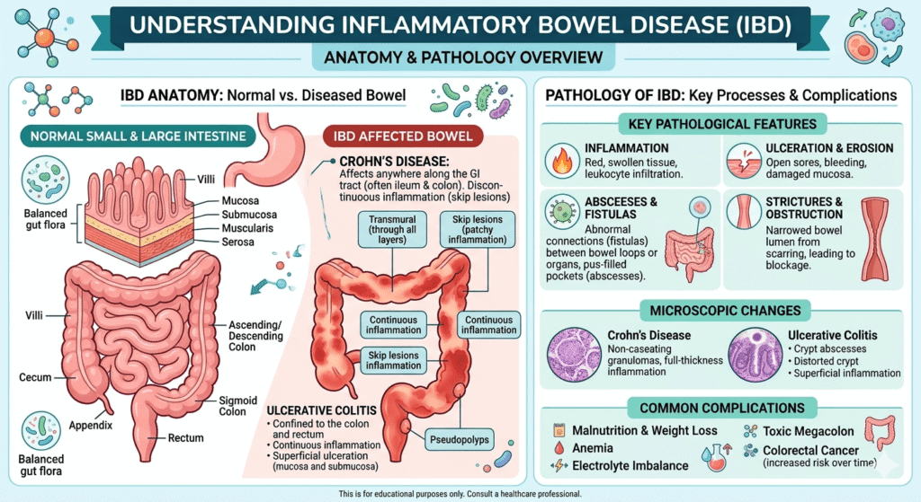
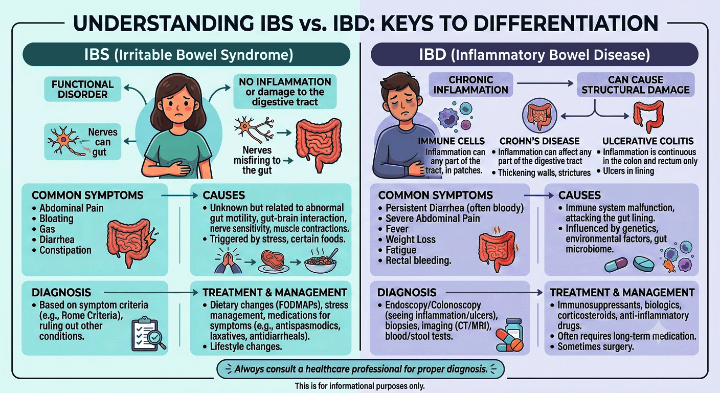
IBS vs. IBD: What Is the Difference?

IBS vs. IBD

IBS vs. IBD: What Is the Difference?

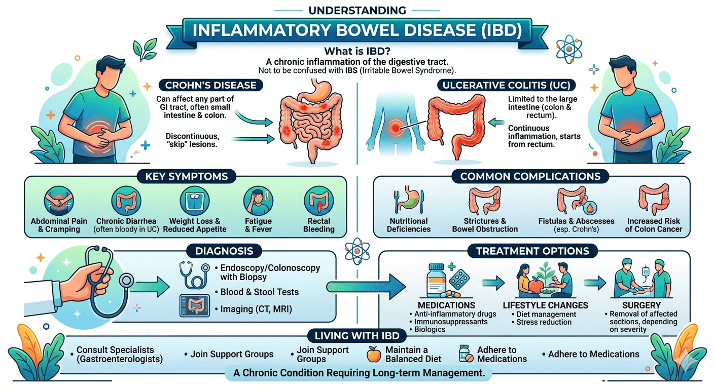
IBS (Irritable Bowel Syndrome) is a functional disorder causing chronic abdominal pain, bloating, and altered bowel habits, but no structural damage. IBD (Inflammatory Bowel Disease), including Crohn’s and ulcerative colitis, is a serious autoimmune condition causing chronic inflammation, tissue ulcers, and visible damage to the digestive tract.

Introduction

Digestive disorders affect millions of people worldwide, but many people are confused by the terms IBS and IBD. Although their names sound similar and both can cause symptoms such as abdominal pain, diarrhea, and changes in bowel habits, they are fundamentally different conditions. Irritable Bowel Syndrome (IBS) is a functional gastrointestinal disorder that impacts how the digestive tract works without causing visible inflammation or permanent damage. Inflammatory Bowel Disease (IBD), on the other hand, is a group of chronic inflammatory conditions that cause ongoing inflammation and structural damage to the gastrointestinal tract.

Understanding the difference between IBS vs. IBD is important because their causes, diagnostic approaches, treatments, and long-term health implications vary significantly. While IBS can greatly impact quality of life, IBD may lead to serious complications if left untreated. This guide explores the key differences between IBS and IBD, including their symptoms, causes, diagnosis, treatment options, and when to seek medical attention.

Understanding IBS and IBD
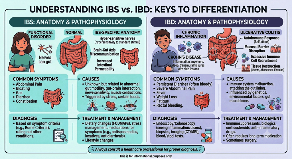
IBS vs. IBD Anatomy

What Is IBS?

Irritable Bowel Syndrome (IBS) is a functional gastrointestinal disorder. This means that the digestive tract appears structurally normal but does not function properly.

In IBS:

  • The intestines are not damaged.
  • No significant inflammation is present.
  • Symptoms arise from abnormal gut motility, altered gut-brain communication, and increased intestinal sensitivity.

IBS affects millions of people worldwide and is one of the most common reasons for gastroenterology consultations.

Common Types of IBS

  • IBS-D: Predominantly diarrhea
  • IBS-C: Predominantly constipation
  • IBS-M: Mixed diarrhea and constipation
  • IBS-U: Unclassified

What Is IBD?

Inflammatory Bowel Disease (IBD) is a group of chronic inflammatory disorders that cause actual damage to the digestive tract.

The two major forms are:

Crohn’s Disease

Can impact any part of the gastrointestinal tract, from the mouth to the anus.

Characteristics:

  • Patchy areas of inflammation (“skip lesions”)
  • Involves the entire bowel wall thickness
  • May cause fistulas and strictures

Ulcerative Colitis

Affects only the colon and rectum.

Characteristics:

  • Continuous inflammation
  • Limited primarily to the inner lining of the colon
  • Often causes bloody diarrhea (Gastrointestinal bleeding)

Unlike IBS, IBD causes visible inflammation and tissue injury.

IBS vs. IBD: Key Differences at a Glance

FeatureIBSIBD
Type of ConditionFunctional disorderInflammatory disease
Intestinal DamageNoYes
InflammationMinimal or absentSignificant
BleedingRareCommon
Cancer RiskNo increased riskIncreased risk in some patients
Weight LossUncommonCommon
FeverRareMay occur
Blood TestsUsually normalOften abnormal
Colonoscopy FindingsTypically normalInflammation visible
Life-Threatening ComplicationsRarePossible

Causes of IBS

The exact cause of IBS remains incompletely understood.

Most experts believe multiple factors contribute.

Common Causes and Risk Factors

Gut-Brain Axis Dysfunction

The gut and brain communicate continuously.

In IBS:

  • Signals become exaggerated.
  • The brain can interpret the normal, everyday movements of the gut as painful sensations.

Abnormal Intestinal Motility

Food may move:

  • Too quickly (diarrhea)
  • Too slowly (constipation)

Visceral Hypersensitivity

Visceral hypersensitivity is an increased sensitivity to sensations arising from internal organs, especially the gastrointestinal (GI) tract. People with visceral hypersensitivity may experience pain, discomfort, bloating, or urgency from normal levels of intestinal stretching or activity that would not bother most people.

Stress and Psychological Factors

Stress does not cause IBS directly but often worsens symptoms.

Common associations include:

  • Anxiety
  • Depression
  • Chronic stress

Microbiome Changes

Alterations in gut bacteria may contribute to symptoms.

Less Common Contributing Factors

  • Prior gastrointestinal infections
  • Food intolerances
  • Small intestinal bacterial overgrowth (SIBO)
  • Genetic predisposition

Causes of IBD

Unlike IBS, IBD involves a true inflammatory process.

Immune System Dysfunction

The immune system mistakenly attacks the digestive tract.

This leads to:

  • Chronic inflammation
  • Tissue injury
  • Ulcer formation

Genetic Factors

Numerous genetic variants increase susceptibility.

Having a close relative with IBD significantly raises risk.

Environmental Factors

Potential triggers include:

  • Smoking (especially Crohn’s disease)
  • Western dietary patterns
  • Certain infections
  • Urban living environments

Microbiome Alterations

Changes in intestinal bacteria may activate abnormal immune responses.

Symptoms of IBS

IBS symptoms typically fluctuate over time.

Many patients describe periods of improvement followed by flare-ups.

Typical Symptoms

Abdominal Pain

A hallmark symptom.

Pain often:

  • Improves after bowel movements
  • Varies in intensity
  • Recurs frequently

Bloating

Patients frequently describe:

  • Abdominal fullness
  • Distension
  • Excess gas

Diarrhea

Particularly in IBS-D.

May include:

  • Urgency
  • Frequent loose stools

Constipation

Common in IBS-C.

May involve:

  • Hard stools
  • Straining
  • Incomplete evacuation

Mixed Bowel Habits

Alternating constipation and diarrhea.

Symptoms Not Typical of IBS

The presence of these symptoms should prompt further evaluation:

  • Blood in stool
  • Fever
  • Significant weight loss
  • Nighttime symptoms
  • Persistent anemia

Symptoms of IBD

IBD symptoms are often more severe and progressive.

Digestive Symptoms

Chronic Diarrhea

May occur multiple times daily.

Blood in Stool

Particularly common in ulcerative colitis.

Abdominal Pain

Usually caused by active inflammation.

Urgent Bowel Movements

Patients may struggle to delay defecation.

Systemic Symptoms

Because IBD is an inflammatory disease, symptoms may affect the whole body.

Fatigue

One of the most common complaints.

Weight Loss

Results from:

  • Poor absorption
  • Reduced appetite
  • Chronic inflammation

Fever

May occur during active disease.

Extraintestinal Manifestations

IBD can impact organs beyond the gut.

Examples include:

  • Arthritis
  • Eye inflammation
  • Skin disorders
  • Liver disease

For medical students, these extraintestinal manifestations are important diagnostic clues.

How IBS and IBD Are Diagnosed

Diagnosis begins with a thorough clinical evaluation.

Medical History

Doctors assess:

  • Symptom patterns
  • Duration
  • Family history
  • Dietary triggers
  • Associated symptoms

Physical Examination

May reveal:

  • Abdominal tenderness
  • Signs of malnutrition
  • Weight loss
  • Dehydration

IBS examinations are often normal.

IBD may show objective abnormalities.

Blood Tests

Common tests include:

  • Complete Blood Count (CBC)
  • C-reactive Protein (CRP)
  • Erythrocyte Sedimentation Rate (ESR)
  • Electrolytes
  • Liver function tests
  • Lipid profile
  • RBS & HbA1c

Findings

IBS:

  • Usually normal
  • RBS & HbA1c (abnormal finding if patient is diabetic)

IBD:

May show:

  • Elevated inflammatory markers
  • Anemia
  • Nutritional deficiencies

Stool Tests

Doctors may test for:

  • Infection
  • Blood
  • Fecal calprotectin

Elevated fecal calprotectin strongly suggests intestinal inflammation and helps distinguish IBD from IBS.

Endoscopy and Colonoscopy

These are among the most essential investigations for IBD.

Doctors can:

  • Visualize inflammation
  • Obtain biopsies
  • Assess disease severity

IBS typically shows normal findings.

Imaging Studies

Ultrasound

Can evaluate:

  • Bowel wall thickness
  • Abscesses
  • Inflammation

CT Scan

Useful for:

  • Complications
  • Acute flare-ups
  • Abscess detection

MRI

Particularly valuable for:

  • Crohn’s disease
  • Fistulas
  • Pelvic involvement

Biopsy

Tissue samples obtained during colonoscopy confirm the diagnosis.

Potential Complications of IBS

IBS does not damage the intestines, but it can significantly impact quality of life.

Complications include:

  • Chronic discomfort
  • Anxiety
  • Depression
  • Social limitations
  • Work and school absenteeism

Importantly, IBS does not increase the risk of colon cancer.

Potential Complications of IBD

IBD can lead to serious complications.

Intestinal Complications

Crohn’s Disease

  • Fistulas
  • Abscesses
  • Strictures
  • Bowel obstruction

Ulcerative Colitis

  • Severe bleeding
  • Toxic megacolon
  • Colon perforation

Nutritional Problems

Patients may develop:

  • Iron deficiency
  • Vitamin B12 deficiency
  • Protein malnutrition

Cancer Risk

Long-standing IBD increases the risk of colorectal cancer, especially when inflammation is extensive and poorly controlled.

Treatment of IBS

Treatment focuses on symptom management.

Dietary Changes

Many patients benefit from:

  • Low-FODMAP diets
  • Increased fiber (selected patients)
  • Identifying trigger foods

Common triggers include:

  • Caffeine
  • Alcohol
  • Fatty foods

Stress Management

Helpful strategies include:

  • Cognitive behavioral therapy
  • Meditation
  • Regular exercise
  • Mindfulness techniques

Medications

Depending on symptoms:

  • Anti diarrheals
  • Laxatives
  • Antispasmodics
  • Certain antidepressants

Treatment of IBD

Treatment aims to control inflammation and prevent complications.

Anti-Inflammatory Medications

Examples:

  • Mesalamine
  • Sulfasalazine

Corticosteroids

Used for short-term flare control.

Examples:

  • Prednisone
  • Budesonide

Immunomodulators

Help suppress abnormal immune activity.

Examples:

  • Azathioprine
  • Methotrexate

Biologic Therapies

Modern treatments targeting specific inflammatory pathways.

Examples:

  • Anti-TNF agents
  • Integrin inhibitors
  • IL-12/23 inhibitors

Surgery

It may be necessary for:

  • Severe disease
  • Obstruction
  • Fistulas
  • Cancer prevention

Surgery can cure ulcerative colitis by removing the colon but does not cure Crohn’s disease.

Living With IBS and IBD

Receiving a diagnosis can feel overwhelming. Fortunately, both conditions can be managed effectively.

Practical Lifestyle Advice

Keep a Symptom Diary

Track:

  • Foods
  • Stress levels
  • Medications
  • Symptoms

Patterns often emerge.

Follow Medical Advice

Regular follow-up improves outcomes.

Stay Physically Active

Exercise can:

  • Improve bowel function
  • Reduce stress
  • Support overall health

Prioritize Mental Health

Digestive disorders and emotional health are closely linked.

Seeking support is a sign of strength, not weakness.

When to Seek Medical Attention

Contact a healthcare professional if you experience:

  • Blood in stool
  • Persistent weight loss
  • Fever
  • Severe abdominal pain
  • Nighttime diarrhea
  • Symptoms that worsen despite treatment

These may indicate IBD or another serious condition rather than IBS.

Final Thoughts

The differentiation between IBS and IBD remains the most crucial topic in gastroenterology. Although similar in many ways as far as symptoms and severity are concerned, there exist several differences that make each of the diseases biologically unique.

On the patient’s side, learning about the difference between the two disorders will help decrease anxiety levels and get proper medical attention. For a medical student, on the other hand, such differentiation becomes key in assessing the problem.

Most importantly, whether it is IBS or IBD that a person has, adequate management and good relations with his/her healthcare provider can truly change one’s life.

Disclaimer: The information provided in this blog post is for educational and informational purposes only and should not be considered medical advice. It is not intended to replace professional medical consultation, diagnosis, or treatment.
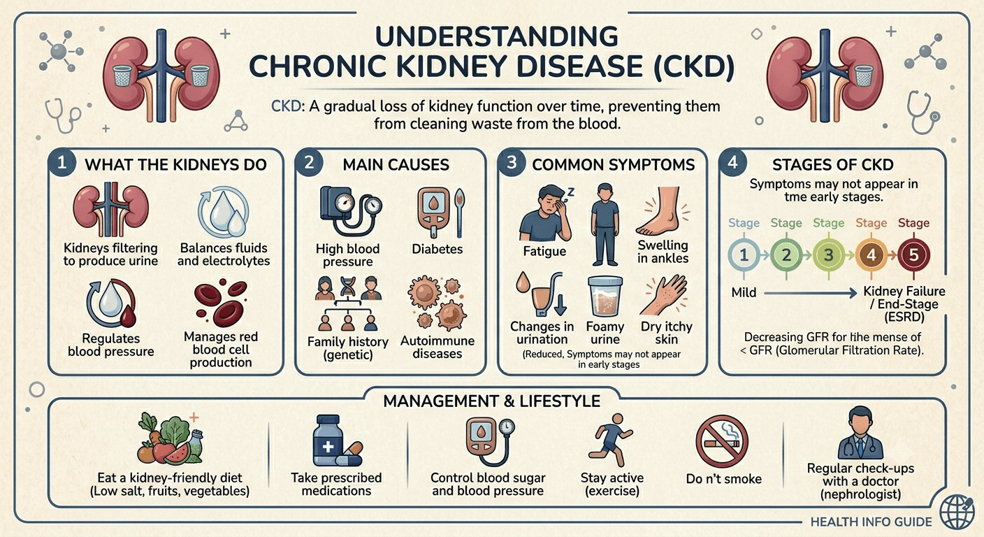
Understanding Chronic Kidney Disease (CKD): A Comprehensive Guide

Chronic Kidney Disease

Understanding Chronic Kidney Disease (CKD): A Comprehensive Guide

Here is a complete guide to Chronic Kidney Disease (CKD). Learn about symptoms, causes, early warning signs, and treatment of this condition and turn complicated clinical data into easily digestible and perfect for patients and caregivers.

Introduction

Chronic Kidney Disease is a condition whereby there is deterioration in the performance of the kidney in ridding the body of toxic substances. This is so because most cases of kidney impairment take place gradually over periods of several weeks or even years. The consequence of this is that one takes some time to figure out that one suffers from chronic kidney disease. CKD is one of the biggest health threats in the contemporary world due to the fact that millions of people suffer from this disease. Which increases the likelihood of contracting diseases such as high blood pressure, anemia, heart problems, and kidney failure.

Patients suffering from CKD are generally concerned about their future; for instance, they may be asking themselves such questions as, Will I have renal failure? How will this affect me? These are just a few of the concerns of the patients; on the other hand, medical students may be interested in getting the facts and a clear understanding of this condition.

Here is a complete guide to Chronic Kidney Disease (CKD). Learn about symptoms, causes, early warning signs, and treatment of this condition and turn complicated clinical data into easily digestible and perfect for patients and caregivers.

What Is Chronic Kidney Disease?

https://www.youtube.com/watch?v=BCb0lFZfyD0

Chronic Kidney Disease (CKD) is a long-term condition in which the kidneys gradually lose their ability to filter waste products, excess fluids, and toxins from the blood.

CKD is typically defined as:

  • Kidney damage lasting 3 months or longer, or
  • A glomerular filtration rate (GFR) below 60 mL/min/1.73 m² for at least 3 months.

The condition impacts millions of people worldwide and is a major cause of illness and premature death. Because kidney damage often develops slowly, many people do not realize they have CKD until significant loss of function has occurred.

Kidney Anatomy and Function

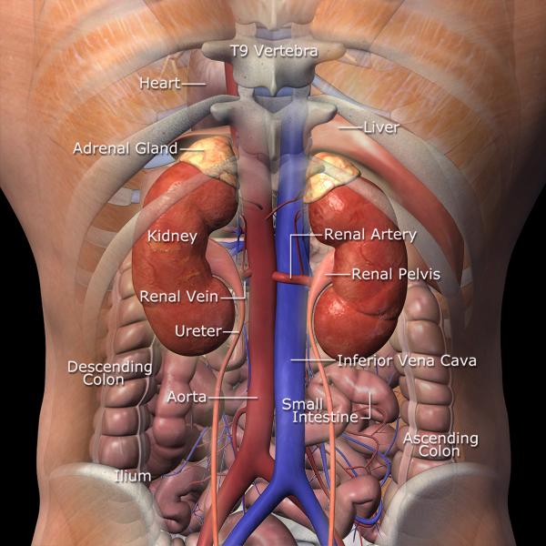
kidney anatomy1

The kidneys are two bean-shaped organs located in the back of the abdomen, on either side of the spine, just below the rib cage. Each kidney contains approximately one million filtering units called nephrons.

What Do the Kidneys Do?

The kidneys perform several essential functions:

1. Blood Filtration

They remove waste products such as:

  • Urea
  • Creatinine
  • Toxins

2. Fluid Balance

They regulate the amount of water in the body.

3. Electrolyte Regulation

They maintain healthy levels of:

  • Sodium
  • Potassium
  • Calcium
  • Phosphate

4. Blood Pressure Control

They help regulate blood pressure through the renin-angiotensin-aldosterone system (RAAS).

5. Red Blood Cell Production

They produce erythropoietin, a hormone that stimulates red blood cell formation.

6. Bone Health

They activate vitamin D, which supports calcium absorption and bone strength.

Causes of Chronic Kidney Disease

CKD is not a single disease. Instead, it is the result of many different conditions that damage the kidneys over time.

Common Causes

Diabetes Mellitus

Diabetes mellitus is the most common cause worldwide.

High blood sugar damages the tiny blood vessels in the kidneys, leading to diabetic nephropathy.

Hypertension (High Blood Pressure)

Persistently elevated blood pressure (hypertension) damages kidney blood vessels and filtering units.

Glomerulonephritis

A group of disorders involving inflammation of the kidney’s filtering structures (glomeruli).

Polycystic Kidney Disease

An inherited condition characterized by multiple kidney cysts that gradually impair kidney function.

Less Common Causes

Autoimmune Diseases

Examples include:

  • Systemic Lupus Erythematosus
  • Vasculitis
  • Goodpasture syndrome

Chronic Infections

Examples include:

Obstructive Uropathy

Blockage of urine flow due to:

Drug-Induced Kidney Damage

Long-term exposure to:

  • Certain pain medications
  • Some antibiotics
  • Chemotherapy agents

Genetic Disorders: Inherited conditions can predispose individuals to kidney dysfunction.

Risk Factors

Certain factors increase the likelihood of developing CKD:

  • Diabetes
  • High blood pressure
  • Obesity
  • Smoking
  • Cardiovascular disease
  • Family history of kidney disease
  • Older age
  • Chronic use of nephrotoxic medications

Stages of Chronic Kidney Disease

CKD is classified into five stages based primarily on GFR.

StageGFR (mL/min/1.73m²)Description
1≥90Kidney damage with normal function
260–89Mild reduction
3a45–59Mild-to-moderate reduction
3b30–44Moderate-to-severe reduction
415–29Severe reduction
5<15Kidney failure (end-stage kidney disease)

Symptoms of Chronic Kidney Disease

One of the most challenging aspects of CKD is that symptoms often develop gradually.

Early Symptoms

Many patients have no symptoms at all.

Possible early signs include:

  • Fatigue
  • Mild swelling in the ankles
  • Increased urination at night
  • Elevated blood pressure

Progressive Symptoms

As kidney function declines, patients may experience:

Fluid Retention

Leading to:

  • Swollen feet
  • Leg swelling
  • Puffiness around the eyes

Fatigue and Weakness

Typically caused by anemia and toxin buildup.

Nausea and Vomiting

Waste products accumulating in the bloodstream can irritate the digestive system.

Loss of Appetite

Patients may notice reduced interest in food and unintentional weight loss.

Itching

Known as uremic pruritus, frequently due to waste accumulation.

Muscle Cramps

Related to electrolyte imbalances.

Difficulty Concentrating

Sometimes referred to as brain fog.

Shortness of Breath

Can result from fluid overload or anemia.

How Chronic Kidney Disease Is Diagnosed

Diagnosis involves a combination of history, examination, laboratory testing, and imaging.

Medical History

Doctors assess:

  • Diabetes history
  • Hypertension history
  • Family history
  • Medication use
  • Symptoms

Physical Examination

Healthcare providers may look for:

  • High blood pressure
  • Edema (swelling)
  • Signs of fluid overload
  • Evidence of underlying systemic disease

Blood Tests

Serum Creatinine

Used to estimate kidney function.

Estimated GFR (eGFR)

Provides a measure of filtration capacity.

Electrolytes

Assess abnormalities in:

  • Potassium
  • Sodium
  • Calcium
  • Phosphate

Complete Blood Count

May reveal anemia.

Urine Tests

Urinalysis

Can detect:

  • Protein
  • Blood
  • Infection

Albumin-to-Creatinine Ratio

A key test for early kidney damage.

Imaging Studies

Ultrasound

Frequently the first imaging study performed.

It can evaluate:

  • Kidney size
  • Obstruction
  • Structural abnormalities

CT Scan

Provides more detailed anatomical information.

MRI

Useful in selected patients when further characterization is needed.

Kidney Biopsy

In certain situations, a small tissue sample is obtained to identify the exact cause of kidney disease.

A biopsy may be especially useful when:

  • The diagnosis is unclear
  • Glomerular disease is suspected
  • Treatment decisions depend on pathology findings

Complications of Chronic Kidney Disease

CKD affects far more than the kidneys alone.

Cardiovascular Disease

Heart attacks and strokes are major causes of death in CKD patients.

Anemia

Reduced erythropoietin production leads to fewer red blood cells.

Bone Disease

Abnormal calcium, phosphate, and vitamin D metabolism weakens bones.

Electrolyte Imbalances

Particularly elevated potassium levels, which can become life-threatening.

Fluid Overload

Can cause:

  • Swelling
  • High blood pressure
  • Heart failure
  • Pulmonary edema

Metabolic Acidosis

An excess of acid in the body due to impaired kidney function.

Uremia

A buildup of waste products that can impact multiple organ systems.

Treatment of Chronic Kidney Disease

A crucial principle is:

CKD treatment focuses on slowing disease progression and addressing the underlying cause.

Managing Diabetes

Careful blood sugar control helps reduce kidney damage.

Controlling Blood Pressure

Target blood pressure goals often require:

  • Lifestyle modification
  • Medications

Commonly used drugs include:

  • ACE inhibitors
  • ARBs

These medications can protect kidney function in many patients.

Dietary Management

Patients may be advised to:

  • Limit sodium intake
  • Moderate protein consumption
  • Restrict phosphorus if needed
  • Control potassium intake in advanced disease

Treating Anemia

Treatment may include:

  • Iron supplementation
  • Erythropoiesis-stimulating agents

Managing Bone and Mineral Disorders

This may involve:

  • vitamin D therapy
  • Phosphate binders
  • Dietary adjustments

Lifestyle Modifications

Important measures include:

  • Smoking cessation
  • Regular exercise
  • Weight management
  • Limiting alcohol
  • Adequate sleep

Dialysis

When kidney function becomes severely impaired, dialysis may be required.

Hemodialysis

Blood is filtered through a machine.

Peritoneal Dialysis

The lining of the abdomen serves as a natural filter.

Kidney Transplantation

For eligible patients with end-stage kidney disease, transplantation often provides the best long-term outcomes and quality of life.

Prognosis

The outlook for CKD varies widely.

Key factors influencing prognosis include:

  • Underlying cause
  • Stage at diagnosis
  • Blood pressure control
  • Diabetes management
  • Presence of cardiovascular disease
  • Adherence to treatment

Many patients with early-stage CKD can maintain stable kidney function for years or even decades with appropriate care.

Others may progress to kidney failure despite optimal treatment.

Early detection significantly improves outcomes.

Living With Chronic Kidney Disease

Practical Advice for Patients

If you have CKD, remember that a diagnosis is not necessarily a sentence to kidney failure.

Many individuals live active, fulfilling lives with the condition.

Keep Regular Medical Appointments

Routine monitoring allows doctors to detect problems early.

Take Medications Consistently

Follow prescribed treatment plans carefully.

Monitor Blood Pressure

Home blood pressure checks can be extremely valuable.

Stay Active

Regular exercise supports cardiovascular and kidney health.

Eat a Kidney-Friendly Diet

Work with a dietitian when possible.

Avoid Harmful Medications

Some over-the-counter pain medications may worsen kidney function.

Always discuss new medications with your healthcare provider.

When to Seek Medical Attention

Contact a healthcare professional promptly if you experience:

  • Sudden swelling
  • Severe shortness of breath
  • Chest pain
  • Significant reduction in urine output
  • Persistent vomiting
  • Confusion
  • Rapid weight gain from fluid retention

These symptoms may indicate worsening kidney function or complications requiring urgent care.

Key Takeaways

Chronic kidney disease is a progressive condition characterized by long-term loss of kidney function. Diabetes and hypertension remain the most common causes, but many other disorders can contribute. Early diagnosis, careful management of underlying conditions, healthy lifestyle choices, and regular medical follow-up can significantly slow disease progression and improve quality of life.

For patients, the most important message is that CKD is manageable, especially when detected early. For medical students, understanding CKD requires appreciating both the pathophysiology of nephron loss and the systemic effects that follow. In both cases, a patient-centered approach remains the cornerstone of effective care.

Splenomegaly: A Comprehensive Guide

Splenomegaly

to Causes, Symptoms, Diagnosis, and Treatment

Introduction

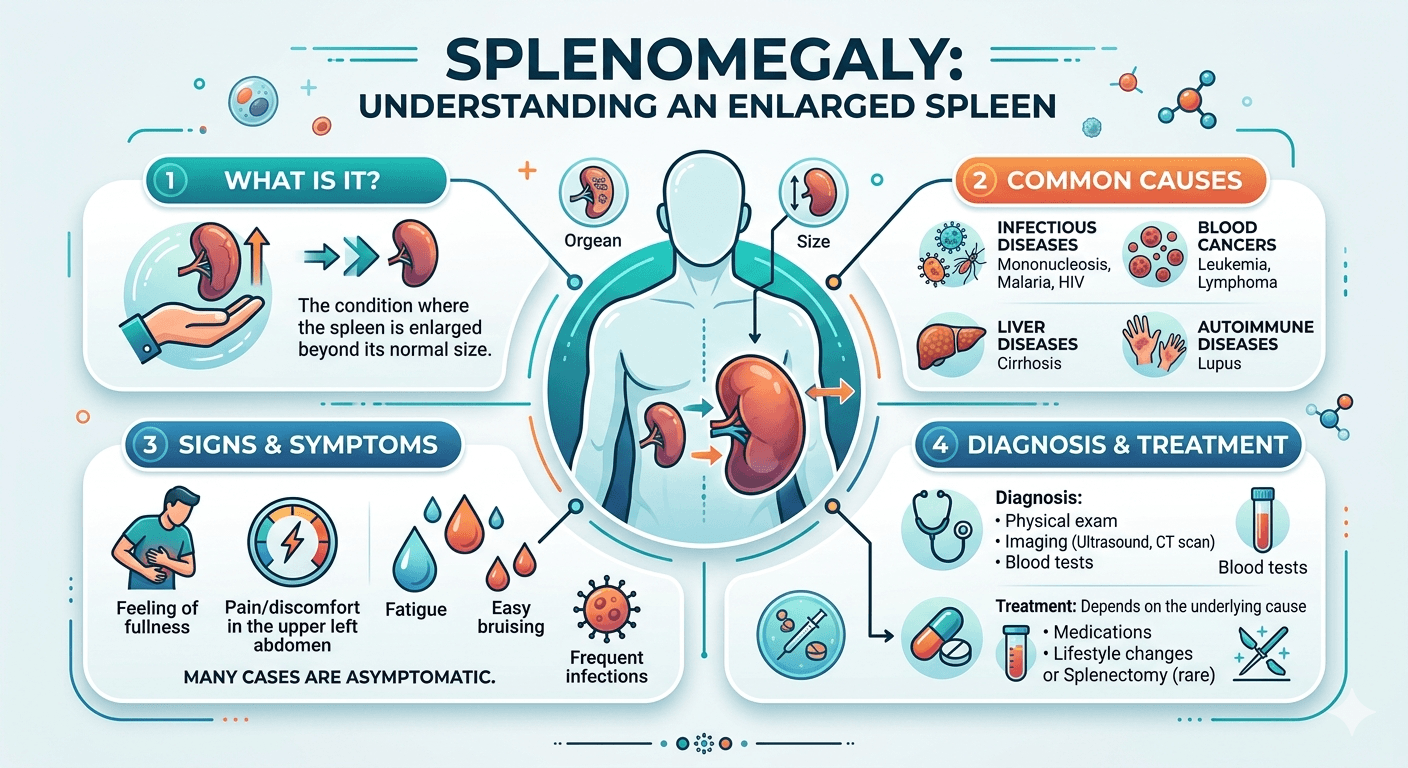
Discovering that you have an enlarged spleen or hearing the term splenomegaly for the first time can be unsettling. Patients often wonder whether it is serious, while medical students may be trying to understand the many conditions that can cause it. The good news is that splenomegaly is not a disease itself; rather, it is a sign that something else is affecting the body.

Explore splenomegaly (enlarged spleen) with this comprehensive guide covering causes, symptoms, diagnosis, treatment, complications, and warning signs that require medical attention.

This guide aims to bridge the gap between patient education and medical learning. Whether you are seeking reassurance about your health or building your clinical knowledge, understanding splenomegaly begins with understanding the remarkable organ at the center of it all: the spleen.

What Is Splenomegaly?

Splenomegaly is the medical term for an enlarged spleen.

In healthy adults, the spleen is usually about the size of a clenched fist and is typically not felt during a physical examination. When the spleen becomes larger than normal, it is described as splenomegaly.

From a clinical perspective, splenomegaly is important because it often serves as a clue to an underlying medical condition. These conditions range from temporary infections to chronic liver disease, blood disorders, autoimmune diseases, and cancers.

For patients, it is helpful to remember that an enlarged spleen is a symptom or finding not a diagnosis by itself.

Understanding the Spleen: Anatomy and Function

Where Is the Spleen Located?

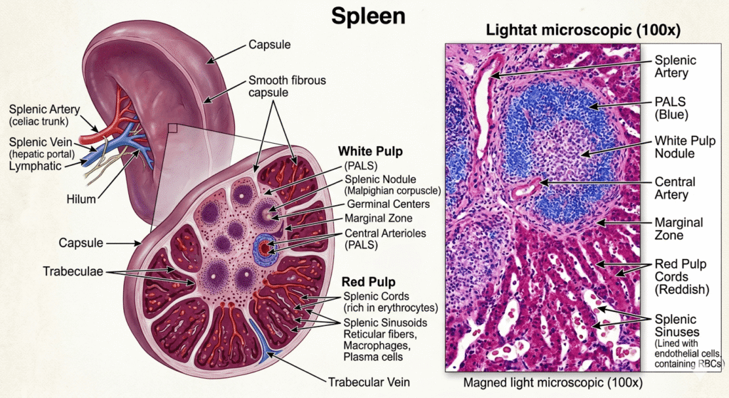
The spleen sits in the upper left part of the abdomen, tucked beneath the rib cage and behind the stomach.

Normally, the spleen is protected by the lower ribs and cannot be felt externally.

Spleen Anatomy

What Does the Spleen Do?

Although many people rarely think about the spleen, it plays several critical roles:

1. Blood Filtration

The spleen acts like a sophisticated filter, removing:

  • Old red blood cells
  • Damaged blood cells
  • Cellular debris

2. Immune Defense

The spleen helps the body fight infections by:

  • Producing immune cells
  • Filtering bacteria and viruses from the bloodstream
  • Supporting antibody production

3. Blood Storage

The spleen stores:

  • Platelets
  • White blood cells
  • Small reserves of blood

4. Fetal Blood Production

Before birth, the spleen helps produce blood cells. Certain diseases can reactivate this function later in life, contributing to enlargement.

How Does Splenomegaly Develop?

The spleen enlarges when it becomes overworked, congested, infiltrated by abnormal cells, or stimulated by immune activity.

Common mechanisms include:

  • Increased destruction of blood cells
  • Excess immune activation
  • Blood flow obstruction
  • Cancer cell infiltration
  • Infection-related inflammation

For medical students, splenomegaly is often categorized as:

  1. Congestive
  2. Infectious
  3. Hematologic
  4. Neoplastic
  5. Inflammatory/autoimmune
  6. Infiltrative

This framework helps narrow the differential diagnosis.

Causes of Splenomegaly

The causes are numerous, but they can be organized into major categories.

Infections

Infections are among the most common causes worldwide.

Viral Infections

Examples include:

  • Infectious Mononucleosis
  • COVID-19
  • Viral hepatitis
  • Cytomegalovirus (CMV)
  • HIV infection

Bacterial Infections

Examples:

  • Tuberculosis
  • Infective endocarditis
  • Typhoid fever

Parasitic Infections

Common in certain regions:

  • Malaria
  • Leishmaniasis
  • Schistosomiasis

Liver Disease and Portal Hypertension

One of the most common causes of significant splenomegaly in adults is liver disease.

Conditions include:

  • Cirrhosis
  • Chronic hepatitis
  • Portal vein thrombosis

When blood flow through the liver becomes restricted, pressure increases in the portal venous system. Blood backs up into the spleen, causing congestive splenomegaly.

Blood Disorders

Several hematologic conditions enlarge the spleen.

Hemolytic Anemias

The spleen removes damaged red blood cells. If destruction becomes excessive, the spleen enlarges.

Examples:

  • Sickle Cell Disease
  • Hereditary spherocytosis
  • Autoimmune hemolytic anemia

Myeloproliferative Disorders

Examples:

  • Polycythemia Vera
  • Primary Myelofibrosis
  • Essential thrombocythemia

These conditions often produce marked enlargement.

Cancers

Cancer-related splenomegaly usually occurs when abnormal cells infiltrate the spleen.

Examples include:

  • Leukemia
  • Lymphoma
  • Metastatic cancers

Massive splenomegaly is classically associated with certain leukemias and Myeloproliferative diseases.

Autoimmune and Inflammatory Diseases

The immune system can stimulate splenic enlargement.

Examples:

  • Systemic Lupus Erythematosus
  • Rheumatoid Arthritis
  • Felty syndrome
  • Sarcoidosis

Storage and Infiltrative Disorders

These are less common but important.

Examples:

  • Gaucher Disease
  • Amyloidosis
  • Niemann-Pick disease

Abnormal substances accumulate within the spleen, causing enlargement.

Symptoms of Splenomegaly

Many people with mild splenomegaly have no symptoms at all.

When symptoms occur, they may arise directly from the enlarged spleen or from the underlying disease.

Upper Left Abdominal Pain

Patients may experience:

  • Dull discomfort
  • Pressure sensation
  • Fullness under the ribs

Pain Radiating to the Left Shoulder

Known as Kehr’s sign, this may occur if the spleen irritates the diaphragm.

Early Satiety

A growing spleen can press against the stomach.

Patients often report:

I feel full after eating only a small amount.

Abdominal Distension

A very enlarged spleen may create a visible abdominal bulge.

Symptoms Related to the Underlying Cause

Infection-Related Symptoms

  • Fever
  • Chills
  • Fatigue
  • Night sweats

Blood Disorder Symptoms

  • Weakness
  • Shortness of breath
  • Easy bruising

Cancer-Related Symptoms

  • Weight loss
  • Persistent fatigue
  • Enlarged lymph nodes
  • Night sweats

Liver Disease Symptoms

  • Jaundice
  • Swollen abdomen
  • Leg swelling

How Is Splenomegaly Diagnosed?

Diagnosis involves determining both:

  1. Whether the spleen is enlarged
  2. Why it is enlarged

Medical History

Doctors ask about:

  • Recent infections
  • Travel history
  • Alcohol use
  • Family history
  • Weight loss
  • Fever
  • Bleeding problems

For medical students, history-taking often provides the strongest clues to etiology.

Physical Examination

During examination, the physician feels beneath the left rib cage.

A normal spleen is usually not palpable.

Findings may include:

  • Palpable spleen tip
  • Massive enlargement extending across the abdomen
  • Associated liver enlargement

Blood Tests

Blood work frequently reveals the underlying cause.

Common tests include:

TestPurpose
Complete Blood Count (CBC)Evaluates red cells, white cells, platelets
Peripheral Blood SmearExamines blood cell morphology
Liver Function TestsAssesses liver disease
Viral TestingDetects viral infections
Inflammatory MarkersEvaluates immune disorders
Coagulation StudiesAssesses clotting function

Ultrasound

First-Line Imaging Study

Ultrasound is:

  • Safe
  • Inexpensive
  • Radiation-free

It accurately measures spleen size and evaluates blood flow.

CT Scan

CT scans provide detailed images and help identify:

  • Tumors
  • Abscesses
  • Trauma
  • Portal hypertension

MRI

MRI is particularly useful when:

  • Soft tissue characterization is needed
  • Vascular abnormalities are suspected
  • Radiation exposure should be minimized

Bone Marrow Biopsy

When blood cancers or marrow disorders are suspected, a bone marrow biopsy may be necessary.

This test can diagnose:

  • Leukemia
  • Lymphoma
  • Myelofibrosis
  • Other hematologic diseases

For medical students, this is typically a key step in evaluating unexplained splenomegaly with abnormal blood counts.

Treatment of Splenomegaly

The most important principle is simple:

Treat the underlying cause, not just the enlarged spleen.

Treatment of Infections

Examples include:

  • Antibiotics for bacterial infections
  • Antiviral therapies when indicated
  • Antiparasitic medications for malaria and related diseases

Once the infection resolves, the spleen often returns to normal size.

Treatment of Liver Disease

Management may involve:

  • Avoiding alcohol
  • Treating viral hepatitis
  • Managing portal hypertension
  • Liver transplantation in advanced cases

Treatment of Blood Disorders

Depending on the condition:

  • Corticosteroids
  • Immunosuppressive medications
  • Blood transfusions
  • Targeted therapies

Treatment of Cancer

Cancer treatment may include:

  • Chemotherapy
  • Radiation therapy
  • Immunotherapy
  • Stem cell transplantation

Successful treatment often reduces spleen size.

Splenectomy (Spleen Removal)

In selected situations, removal of the spleen may be necessary.

Common indications:

  • Severe hypersplenism
  • Splenic rupture
  • Certain blood disorders
  • Persistent symptomatic enlargement

However, splenectomy increases susceptibility to certain infections.

Patients require:

  • Vaccinations
  • Preventive medical follow-up
  • Education about infection risks

Prognosis

The outlook for splenomegaly varies considerably.

Generally Favorable Prognosis

Examples:

  • Infectious mononucleosis
  • Temporary viral infections
  • Treatable bacterial infections

Variable Prognosis

Examples:

  • Cirrhosis
  • Autoimmune disorders
  • Chronic hemolytic anemia

More Serious Prognosis

Examples:

  • Advanced leukemia
  • Aggressive lymphoma
  • Severe myelofibrosis

Ultimately, prognosis depends on:

  • Underlying diagnosis
  • Severity of disease
  • Response to treatment
  • Presence of complications

Living With Splenomegaly

For patients, receiving a diagnosis of splenomegaly can feel overwhelming. Fortunately, many people live well while the underlying condition is investigated and treated.

Protect Your Spleen

Avoid activities with a high risk of abdominal trauma, including:

  • Contact sports
  • Martial arts
  • High-impact activities unless approved by your doctor

Attend Follow-Up Appointments

Regular monitoring helps track:

  • Spleen size
  • Blood counts
  • Treatment effectiveness

Know the Warning Signs

Seek urgent medical attention if you develop:

  • Severe left-sided abdominal pain
  • Fainting
  • Sudden dizziness
  • Significant bleeding
  • High fever

These symptoms may indicate a serious complication.

Maintain General Health

Helpful measures include:

  • Eating a balanced diet
  • Staying physically active as advised
  • Avoiding excessive alcohol
  • Keeping vaccinations up to date

Managing chronic illnesses carefully

Splenomegaly FAQ

Key Takeaways

For patients, the most important message is reassurance: many causes of splenomegaly are treatable, and modern diagnostic tools allow physicians to identify the underlying problem effectively. For medical students, splenomegaly remains a classic clinical finding that reflects diverse pathophysiologic processes and demands a systematic diagnostic approach. Understanding why the spleen enlarges is often the key to understanding the disease itself.

Disclaimer: The information provided in this blog post is for educational and informational purposes only and should not be considered medical advice. It is not intended to replace professional medical consultation, diagnosis, or treatment. Always seek the guidance of a qualified healthcare professional regarding any medical condition or health-related concerns.
Understanding Nephritic and Nephrotic Syndrome

Nephritic and Nephrotic syndrome

A Detailed Medical Guide

Introduction

The kidneys play a vital role in keeping the body balanced. They do this by filtering our blood, regulating fluids and electrolytes, and flushing out waste.

Understand Nephritic and Nephrotic Syndrome with this comprehensive guide covering causes, symptoms, diagnosis, treatment, and key differences. Learn how Nephritic and Nephrotic Syndrome affect kidney function and what patients and medical students need to know.

This comprehensive guide covers the causes, symptoms, diagnosis, and treatment of both syndromes Nephritic syndrome and Nephrotic syndrome.

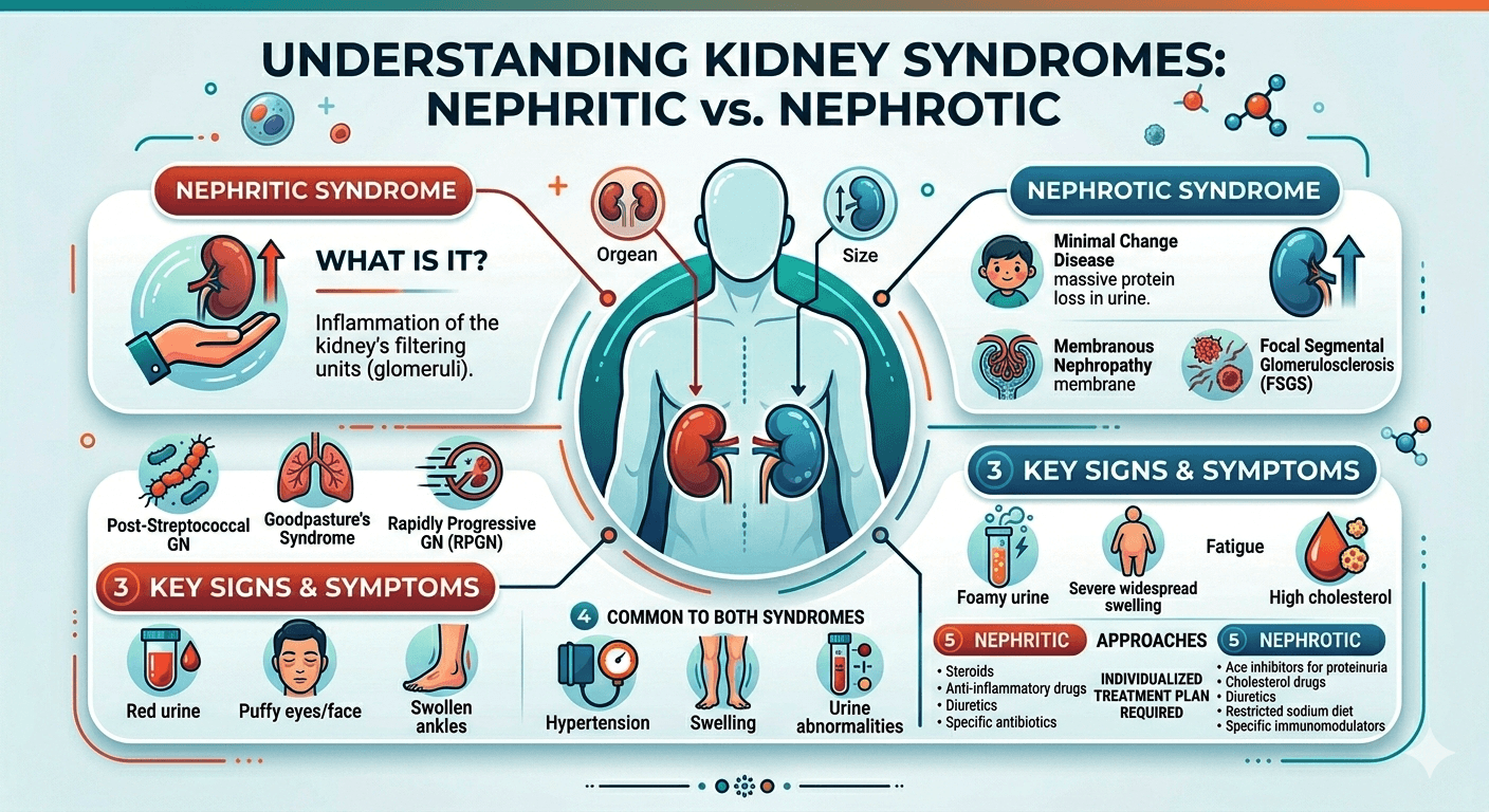
Conditions that affect the glomeruli microscopic filtration structures found in the kidney typically manifest as either nephritic syndrome or nephrotic syndrome.

While having an identical underlying structure, these two syndromes differ drastically in terms of the pathology, symptoms, laboratory findings, and management. The ability to distinguish between these two syndromes is an important skill for anyone interested in the physiology of kidney diseases.

What Is a Glomerulus?

The Glomerulus is a ball-like structure composed of capillaries that filters the blood in the kidney. It allows blood to flow out of the kidney while holding back the cells and proteins.

Glomerular filtration barrier includes the following layers:

1.         Fenestrated endothelium.

2.         Glomerular basement membrane (GBM)

3.         Slit-diaphragm podocytes.

Damage to different components of this barrier results in distinct clinical syndromes.

Nephritic Syndrome

Definition

It is a medical syndrome marked by inflammation of the glomerulus, hence disrupting the filtration process and allowing red blood cells to appear in urine.

Features of Nephritic Syndrome

  • Hematuria
  • Oliguria
  • Hypertension
  • Mild to moderate proteinuria
  • Azotemia
  • Edema

Pathophysiology of Nephritic Syndrome

Glomerulonephritis develops due to inflammation of the glomeruli, which disrupts normal kidney filtration and may lead to progressive kidney damage.

Immune complexes, antibodies, and the cellular immune system cause damage to the endothelial cells lining the glomerular capillary. There are several consequences, including:

  • Decreased glomerular filtration rate
  • Hematuria caused by leakage of the red blood cells into the urine
  • Natriuresis and water retention
  • Activating the RAAS

Consequently, there is hypertension and swelling.

Clinical Presentation of Nephritic Syndrome

1. Hematuria

The hallmark of nephritic syndrome.

The color of urine appears:

  • Cola-colored
  • Tea-colored
  • Smoky brown

Under microscopy, one can see:

  • Dysmorphic red blood cells
  • Blood casts in the urine

2.Oliguria

There is oliguria due to low GFR.

3.Hypertension

Retention of fluids increases the intravascular volume resulting in high BP(Hypertension)

4.Edema

This is usually mild and usually seen on:

  • Facial
  • Lids
  • Peri-orbital areas

5. Azotemia

High levels of serum urea and creatinine

Causes of Nephritic Syndrome

Post Streptococcal Glomerulonephritis (PSGN)

Develops after infection with a nephritogenic strain of Group A Streptococcus.

Common in children.

IgA Nephropathy (Berger Disease)

It is the most common form of primary glomerulonephritis.

Deposition of IgA in the mesangium is its hallmark.

Rapidly Progressive Glomerulonephritis (RPGN)

It is a severe type that leads to the quick progression of renal failure.

Some of its causes include:

  • Anti-GBM disease
  • ANCA Vasculitis
  • Lupus nephritis

Lupus Nephritis

An important complication of SLE.

Membranoproliferative Glomerulonephritis (MPGN)

Laboratory Abnormalities in Nephritic Syndrome

Urinalysis

Abnormal findings include:

  • Hematuria
  • RBC casts
  • Proteinuria

Blood Tests

  • Elevated creatinine
  • Elevated blood urea nitrogen (BUN)
  • Reduced complement levels (for some causes)

Kidney Biopsy

  • Hypercellularity
  • Immune complex deposition
  • Crescent formation

Nephrotic Syndrome

Description

Nephrotic syndrome is caused by increased permeability of the glomerular filtration barrier, leading to massive protein loss through the urine.

Diagnosis Criteria

  • Proteinuria of >3.5g/day
  • Hypoalbuminemia
  • Generalized edema
  • Hyperlipidemia
  • Lipiduria

Nephrotic Syndrome Pathophysiology

Damage occurs to podocytes and glomerular basement membranes.

Consequences include:

1. Massive urinary protein loss

2. Decreased plasma oncotic pressure

3. Fluid movement from capillaries to interstitial spaces

4. Edema formation

The liver responds by synthesizing more proteins and lipoproteins leading to hyperlipidemia.

Clinical Manifestations of Nephrotic Syndrome

1. Massive proteinuria

Excretion of more than 3.5g of protein daily.

2. Edema formation

Known as anasarca.(anasarca is a medical condition in which excessive fluid buildup throughout the body, causing widespread swelling of the skin and tissues.)

Sites include:

  • Face
  • Legs
  • Abdomen
  • Scrotum

3. Hypoalbuminemia

Decreased serum albumin due to excessive urinary losses.

4. Hyperlipidemia

Elevated level of cholesterol and triglycerides is common.

5. Lipiduria

Microscopy of urine reveals:

  • Oval fat bodies
  • Fatty casts

Producing the characteristic “Maltese cross” when viewed under polarized light.

Causes of Nephrotic Syndrome

Minimal Change Disease (MCD)

The most common cause in children.

Features:

• Normal light microscopy

• Podocyte foot process effacement on electron microscopy

Focal Segmental Glomerulosclerosis (FSGS)

Commonly seen in adults.

Associated with:

• HIV infection

• Obesity

• Sickle cell disease

Membranous Nephropathy

Common cause in older adults.

Associated with anti-PLA2R antibodies.

Diabetic Nephropathy

The diabetes is the commonest cause of nephrotic syndrome globally.

Amyloidosis

Extracellular deposition of amyloid proteins damages the kidneys.

Complications of Nephrotic Syndrome

Thromboembolism

Deficiency of anticoagulant proteins like ant thrombin III makes it easier to form clots.

Complications of nephrotic syndrome include:

  • Renal vein thrombosis
  • Deep vein thrombosis
  • Pulmonary embolism

Infections

Due to loss of immunoglobulins, there is increased susceptibility to infections.

Examples of such infections include:

  • Pneumonia
  • Cellulitis
  • Peritonitis

Malnutrition

Persistent protein loss causes muscle wastage.

Acute kidney injury

May occur in severe cases.

Laboratory Abnormalities in Nephrotic Syndrome

Urinalysis

Massive proteinuria

Fatty casts

Oval fat bodies

Blood Tests

Common findings include:

Test                           Result
Serum albumin         Decreased
Cholesterol               Increased
Triglycerides             Increased

Creatinine Variable

Kidney Biopsy

Required in adults to determine the underlying cause.

Difference Between Nephritic and Nephrotic Syndrome

FeatureNephritic SyndromeNephrotic Syndrome
DefinitionA glomerular disorder characterized primarily by inflammation and hematuria.A glomerular disorder characterized primarily by massive protein loss in the urine.
Primary PathologyInflammation of the glomeruli.Increased permeability of the glomerular filtration barrier.
ProteinuriaMild to moderate (<3.5 g/day).Severe (>3.5 g/day).
HematuriaProminent; urine may appear smoky, tea-colored, or cola-colored.Usually absent or microscopic if present.
Red Blood Cell CastsCommon and characteristic.Rare.
EdemaMild to moderate, often around the eyes.Severe, generalized edema (anasarca).
Blood PressureFrequently elevated (hypertension).Usually normal or mildly elevated.
Serum AlbuminSlightly reduced or normal.Markedly decreased (Hypoalbuminemia).
Serum LipidsUsually normal.Elevated cholesterol and triglycerides (hyperlipidemia).
Urine AppearanceDark, smoky, or bloody.Frothy or foamy due to excess protein.
Kidney FunctionOften reduced, with decreased glomerular filtration rate (GFR).Usually preserved initially.
Fluid RetentionPresent due to reduced kidney function.Marked due to low plasma oncotic pressure.
Risk of Blood ClotsLess common.Increased risk because of loss of anticoagulant proteins in urine.
Common CausesPost-streptococcal glomerulonephritis, IgA nephropathy, rapidly progressive glomerulonephritis, lupus nephritis.Minimal change disease, focal segmental glomerulosclerosis (FSGS), membranous nephropathy, diabetic nephropathy.
Key Clinical FeaturesHematuria, hypertension, oliguria, and mild edema.Massive proteinuria, severe edema, hypoalbuminemia, and hyperlipidemia.
Treatment FocusControlling inflammation, blood pressure, and treating the underlying cause.Reducing proteinuria, managing edema, preventing complications, and treating the underlying disease.

Diagnostic Approaches

History

The essential queries include:

  • Any recent infection?
  • Indications of autoimmune illness
  • The patient’s medication list
  • History of any hereditary condition?

Physical examination

Check for:

  • Level of blood pressure
  • The extent of edema
  • Systemic disease signs

Laboratory Tests

Suggested investigations: 

            Urinalysis
            Measurement of urine proteins
            Serum creatinine
            Serum albumin
            Kidney function test (KFT)
            Liver function test
            LDH level
            Lipid profile
            D-dimer test
            Testing of complement levels
            Anti nuclear antibody (ANA)
            Anti-neutrophil cytoplasmic antibodies (ANCA)
            Anti GBM antibodies

Imaging

  • Ultrasound (USG) whole abdomen
  • CT abdomen (as decided by physician)
  • Renal biopsy

Generally aids in making definitive diagnosis

Approach to Treatment

Approach to Nephritic Syndrome

Treat based on cause

General Approach:

  • Treatment of hypertension
  • Limited intake of fluids
  • Limitation in sodium
  • Use of diuretics

Specific approach for the particular disease may include:

  • Steroids
  • Cytotoxic therapy
  • Rituximab
  • Plasmapheresis

(Treatment entirely depends upon physician)

Approach to Nephrotic Syndrome

General Approach

  • Limit salt consumption
  • Administration of diuretics
  • ACE inhibitors
  • ARBs

Hyperlipidemia Management

Use statins when indicated

Prevention of thrombosis

Indicated in high-risk cases

Specific treatment depends upon diagnosis

Steroids, calcineurin inhibitors, rituximab, etc.

Prognosis

Prognosis is based upon cause.

Good prognosis

  • Minimal change disease
  • Post Streptococcal Glomerulonephritis

Average prognosis

  • IgA nephropathy
  • Membranous nephropathy

conclusion

Both nephritic and nephrotic syndrome are important types of kidney diseases that occur due to injury to glomeruli, but there are significant differences in their development, symptoms, lab results, and treatment modalities. First of all, nephritic syndrome is caused by inflammation of glomeruli, which produces symptoms like hematuria, hypertension, and decreased functioning of kidneys. Nephrotic syndrome, on the other hand, occurs when the permeability of glomeruli increases, resulting in heavy proteinuria, edema, Hypoalbuminemia, and hyperlipidemia.

Understanding the difference between these syndromes is key to accurate diagnostics, effective treatment, and even preventing serious complications in the form of chronic kidney disease or renal failure. The knowledge about pathogenesis and signs of nephritic and nephrotic syndromes plays an especially vital role for future medical professionals. Patients should be informed about symptoms like edema, abnormal urination, high blood pressure, or unusual tiredness. With proper care, many cases of nephritic or nephrotic syndromes can be successfully managed.

Frequently Asked Questions (FAQs)

1. What is the easiest way to differentiate nephritic and nephrotic syndrome?

Nephritic syndrome presents with hematuria and inflammation, while nephrotic syndrome presents with massive proteinuria and generalized edema.

2. Why does nephrotic syndrome cause hyperlipidemia?

The liver compensates for protein loss by increasing synthesis of lipoproteins, resulting in elevated cholesterol and triglyceride levels.

3. Are RBC casts specific for nephritic syndrome?

Yes. RBC casts strongly suggest glomerular inflammation and are a classic feature of nephritic syndrome.

4. Which syndrome causes more severe edema?

Nephrotic syndrome causes more severe edema because of significant hypoalbuminemia.

5. What is the most common nephrotic syndrome in children?

Minimal Change Disease is the most common cause in children.

6. Can nephritic syndrome progress to kidney failure?

Yes. Severe forms such as rapidly progressive glomerulonephritis can lead to acute or chronic kidney failure if untreated.

Disclaimer: The information provided in this blog post is for educational and informational purposes only and should not be considered medical advice. It is not intended to replace professional medical consultation, diagnosis, or treatment. Always seek the guidance of a qualified healthcare professional regarding any medical condition or health-related concerns. The author and publisher are not responsible for any actions taken based on the information presented in this article.
Read More

Pain in Epigastric Region

Pain in Epigastric region

What Your Belly Is Trying to Tell You

Pain in Epigastric Region: What Your Belly Is Trying to Tell You. Discover the common causes, symptoms, warning signs, diagnosis, treatment options, and when to seek medical attention for upper abdominal pain.

Introduction

Pain in the upper middle part of your abdomen can be alarming. This area, known as the epigastric region, sits just below the breastbone and above the belly button. While occasional discomfort may result from something as simple as overeating, persistent or severe pain can signal an underlying medical condition that requires attention.

Pain in Epigastric Region: What Your Belly Is Trying to Tell You. Discover the common causes, symptoms, warning signs, diagnosis, treatment options, and when to seek medical attention for upper abdominal pain.

Understanding what your body is trying to tell you can help you seek the right care at the right time. This article explores the causes, symptoms, diagnosis, treatment, and prevention of epigastric pain based on current medical knowledge.

Understanding the Epigastric Region

The abdomen is divided into several regions to help healthcare professionals identify the source of pain. The epigastric region is located in the upper central abdomen.

Several important organs are found in or near this area, including:

  • Stomach
  • Pancreas
  • Liver
  • Gallbladder
  • Duodenum (first part of the small intestine)
  • Lower esophagus
  • Major blood vessels

Because so many organs are located here, pain in this region can have numerous causes.

Why Epigastric Pain Should Not Be Ignored

Epigastric pain can range from mild discomfort to severe, debilitating pain. The sensation may be:

  • Burning
  • Aching
  • Sharp
  • Cramping
  • Gnawing
  • Pressure-like

In some cases, the pain is harmless and temporary. However, it can also indicate conditions such as ulcers, gallstones, pancreatitis, or even heart disease.

The duration, intensity, and associated symptoms often provide clues about the underlying cause.

Common Symptoms That Accompany Epigastric Pain

Pain rarely occurs alone. Many patients experience additional symptoms that help narrow down the diagnosis.

Common accompanying symptoms include:

SymptomPossible Association
HeartburnAcid reflux, GERD
NauseaGastritis, pancreatitis
VomitingUlcers, infections
BloatingDyspepsia, indigestion
Loss of appetiteGastritis, ulcers
FeverInfection, pancreatitis
Chest discomfortGERD, cardiac causes
Weight lossChronic digestive disease

Gastroesophageal Reflux Disease (GERD)

One of the most common causes of pain in the epigastric region is GERD, commonly known as acid reflux.

GERD occurs when stomach acid flows backward into the esophagus, irritating its lining.

How Acid Reflux Causes Pain

Symptoms often include:

  • Burning sensation in the upper abdomen
  • Heartburn
  • Sour taste in the mouth
  • Difficulty swallowing
  • Symptoms worsening after meals

Certain foods can trigger GERD, including:

  • Spicy foods
  • Fatty meals
  • Chocolate
  • Caffeine
  • Alcohol

Frequent reflux should be evaluated because long-term acid exposure can damage the esophagus.

Gastritis and Stomach Inflammation

Gastritis refers to inflammation of the stomach lining. It may develop suddenly or gradually over time.

Common causes include:

  • Helicobacter pylori infection
  • Excessive alcohol consumption
  • Long-term use of NSAIDs such as ibuprofen
  • Stress-related illness
  • Autoimmune conditions

Risk Factors for Gastritis

  • Helicobacter pylori (H. pylori) infection: One of the most common causes of chronic gastritis worldwide. This bacterium weakens the stomach’s protective barrier and promotes inflammation.
  • Regular use of non-steroidal anti-inflammatory drugs (NSAIDs): Medications such as aspirin, ibuprofen, and naproxen can damage the stomach lining, especially when used frequently or in high doses.
  • Excessive alcohol consumption: Alcohol irritates and erodes the stomach mucosa, increasing the risk of inflammation and bleeding.
  • Smoking: Tobacco use impairs the stomach’s natural defense mechanisms and delays healing of the gastric mucosa.
  • Severe physical stress: Major surgery, severe burns, trauma, or critical illness can lead to stress-related gastritis.
  • Autoimmune disorders: In autoimmune gastritis, the immune system mistakenly attacks the stomach lining, leading to chronic inflammation and vitamin B12 deficiency.
  • Advancing age: Older adults are at greater risk due to thinning of the stomach lining and increased likelihood of H. pylori infection or medication use.
  • Bile reflux: Backflow of bile from the small intestine into the stomach can irritate the gastric mucosa.
  • Chronic medical conditions: Diseases such as chronic kidney disease, liver disease, HIV infection, and Crohn’s disease may increase the risk of gastritis.
  • Dietary and lifestyle factors: Frequent consumption of highly spicy foods, excessive caffeine, and poor dietary habits may worsen symptoms in susceptible individuals, although they are not primary causes.

Peptic Ulcers

Peptic ulcers are open sores that develop in the stomach or upper small intestine.

These ulcers commonly result from:

  • H. pylori infection
  • Frequent NSAID use
  • Smoking
  • Excessive alcohol intake

Signs of a Stomach or Duodenal Ulcer

Symptoms may include:

  • Gnawing upper abdominal pain
  • Pain that improves or worsens after eating
  • Bloating
  • Nausea
  • Nighttime discomfort

Complications such as bleeding ulcers require immediate medical attention.

Gallbladder Disorders

The gallbladder stores bile, a digestive fluid produced by the liver.

When gallstones form, they may block bile ducts and trigger pain.

Gallstones and Upper Abdominal Pain

Typical symptoms include:

  • Sudden upper abdominal pain
  • Pain after fatty meals
  • Pain radiating to the back or right shoulder
  • Nausea and vomiting

Gallbladder pain can sometimes be mistaken for stomach-related discomfort because it occurs near the epigastric region.

Pancreatitis

The pancreas plays a vital role in digestion and blood sugar regulation.

Inflammation of the pancreas, known as pancreatitis, can cause severe epigastric pain.

Acute vs. Chronic Pancreatitis

Acute pancreatitis develops suddenly and may require hospitalization.

Chronic pancreatitis develops gradually and can cause long-term digestive problems.

Common symptoms include:

  • Severe upper abdominal pain
  • Pain radiating to the back
  • Nausea
  • Vomiting
  • Fever

Excessive alcohol use and gallstones are among the leading causes.

Heart-Related Causes of Epigastric Pain

Many people assume heart problems always cause chest pain. In reality, heart disease can sometimes present as upper abdominal discomfort.

When It Mimics a Heart Attack

Heart-related warning signs may include:

  • Pressure-like epigastric pain
  • Sweating
  • Shortness of breath
  • Dizziness
  • Pain spreading to the jaw, neck, or arm

Individuals with these symptoms should seek emergency medical care immediately.

Functional Dyspepsia

Functional dyspepsia refers to chronic indigestion without an identifiable structural cause.

Patients often report:

  • Fullness after eating
  • Bloating
  • Burning discomfort
  • Early satiety

Although not life-threatening, it can significantly affect quality of life.

Diagnosing Epigastric Pain

Doctors begin with a detailed medical history and physical examination.

Additional tests may include:

Laboratory Tests

  • Complete blood count
  • Liver function tests
  • Viral marker
  • Lipid profile
  • Pancreatic enzyme tests (S. Amylase, S. Lipase)
  • H. pylori testing
  • RBS, HbA1c

Imaging Studies

  • Ultrasound (whole abdomen)
  • CT scan
  • MRI

Endoscopy

An upper gastrointestinal endoscopy allows doctors to directly examine the esophagus, stomach, and duodenum.

This test is especially useful when ulcers, gastritis, or bleeding are suspected.

Treatment Options

Treatment depends entirely on the underlying cause.

Medications

Common medications include:

  • Antacids
  • Proton pump inhibitors
  • H2 blockers
  • Antibiotics for H. pylori infection
  • Pain management medications

Procedures

Some conditions require intervention:

  • Gallbladder surgery
  • Endoscopic ulcer treatment
  • Pancreatic procedures

Dietary Management

Many patients benefit from:

  • Smaller meals
  • Reduced fatty food intake
  • Limiting caffeine
  • Avoiding alcohol
  • Maintaining hydration

Lifestyle Changes for Prevention

Healthy habits can reduce the risk of many digestive disorders.

Consider the following strategies:

Eat Mindfully

  • Avoid overeating
  • Chew food thoroughly
  • Eat slowly

Maintain a Healthy Weight

Excess body weight increases abdominal pressure and can worsen reflux symptoms.

Avoid Smoking

Smoking damages the digestive tract and increases ulcer risk.

Manage Stress

Chronic stress may aggravate digestive symptoms and worsen functional gastrointestinal disorders.

Stay Physically Active

Regular exercise supports digestive health and overall well-being.

For additional information World Gastroenterology Organisation.

When to Seek Emergency Care

Seek immediate medical attention if epigastric pain is accompanied by:

  • Severe chest pain
  • Difficulty breathing
  • Vomiting blood
  • Black or tarry stools
  • Sudden severe abdominal pain
  • Fainting
  • High fever
  • Persistent vomiting

These symptoms may indicate a serious medical emergency.

Frequently Asked Questions

1. What is the most common cause of pain in the epigastric region?

Acid reflux, gastritis, and indigestion are among the most common causes of epigastric pain.

2. Can stress cause epigastric pain?

Yes. Stress can worsen digestive disorders and contribute to symptoms such as indigestion and stomach discomfort.

3. Is epigastric pain always related to the stomach?

No. The pancreas, gallbladder, liver, heart, and nearby organs can also cause pain in this region.

4. When should I worry about epigastric pain?

You should seek medical evaluation if the pain is severe, persistent, recurrent, or accompanied by alarming symptoms such as weight loss, vomiting blood, or chest pain.

5. Can a heart attack feel like stomach pain?

Yes. Some heart attacks present as upper abdominal discomfort, especially in older adults, women, and people with diabetes.

6. What foods should I avoid if I have recurring epigastric pain?

Common triggers include spicy foods, fatty meals, alcohol, caffeinated beverages, and acidic foods.

7. Can ulcers heal on their own?

Some ulcers may improve temporarily, but proper diagnosis and treatment are important to prevent complications.

8. Does pancreatitis always cause severe pain?

Acute pancreatitis usually causes severe pain, while chronic pancreatitis may produce persistent but less intense discomfort.

Conclusion

Pain in the epigastric region: What Your Belly Is Trying to Tell You is a question worth paying attention to because this seemingly simple symptom can have many different causes. While indigestion and acid reflux are common explanations, conditions such as ulcers, gallbladder disease, pancreatitis, and even heart disease can also be responsible.

The key is to recognize accompanying symptoms, understand potential triggers, and seek medical evaluation when pain is severe, persistent, or associated with warning signs. Early diagnosis often leads to more effective treatment and better long-term outcomes.

Disclaimer: The information provided in this blog post is for educational and informational purposes only and should not be considered medical advice. It is not intended to replace professional medical consultation, diagnosis, or treatment. Always seek the guidance of a qualified healthcare professional regarding any medical condition or health-related concerns. The author and publisher are not responsible for any actions taken based on the information presented in this article.
Early Warning Signs of Kidney Disease

Early warning signs of kidney disease

Early warning signs of kidney disease

Early warning signs of kidney disease are often overlooked. Discover key symptoms, risk factors, and why early diagnosis matters.

Introduction

The condition of kidney dysfunction is frequently referred to as a “silent disease.” This is because there is usually no symptom manifestation until a great deal of harm is done to the kidney. The function of the kidneys involves the filtration of the blood for any wastes, the regulation of fluid balance and electrolytes, controlling blood pressure, and releasing hormones that produce red blood cells and contribute to bone health.

Learn the early warning signs of kidney disease, why they occur, who is at risk, and when to seek medical attention. A comprehensive guide for patients and medical students.

Based on statistics in global health care, chronic kidney disease (CKD) is common around the world, and there are hundreds of millions of individuals affected with this ailment. It is one of the causes of disability and death.

It is important for both health care professionals and patients to recognize the earliest manifestations of kidney disease.

Understanding the Role of Healthy Kidneys

Before moving on to the discussion about the signs of kidney disease, it should be understood what normal kidneys do: 

Cleanse metabolic waste products out of the body via filtration through the kidneys;
Maintain the right water balance in the body by filtering out toxic substances through Urine.   
  Electrolyte balance control (sodium, potassium, calcium, and phosphate);
Blood pressure regulation through RAAS (renin-angiotensin-aldosterone system);
Erythropoietin production to promote red blood cells synthesis;
Vitamin D activation for bones health preservation.

With the deterioration of renal function, these functions are disturbed and cause various symptoms.

Why Early Symptoms Are Often Missed

Kidneys possess a remarkable functional reserve. A person can lose a substantial portion of kidney function before experiencing noticeable symptoms.

Early symptoms are often:

  • Mild
  • Non-specific
  • Mistaken for stress, aging, or other medical conditions

As a result, many patients remain undiagnosed until laboratory abnormalities or advanced disease become evident.

Early Warning Signs of Kidney Disease You Shouldn’t Ignore

Fatigue and Weakness

Unexplained fatigue is a classic initial symptom.

Reasons Behind It

There are several contributing factors:

            Increased accumulation of metabolic wastes (uremia)

            Anemia secondary to decreased erythropoietin production

Balancing of electrolytes

            Impaired sleep patterns

Clinical Presentation

Patients usually have complaints about:           

Diminishing energyPoor exercise tolerance
Concentration difficultiesGeneral muscle weakness

Medical students are advised to consider CKD when there is fatigue along with certain risk factors such as diabetes mellitus or hypertension.

Alterations in Urination Patterns

Alterations in urination are very common initial symptoms.

Increased Urination

Patients typically experience:

            Increase in urinary frequency, especially nighttime (nocturia)

            Large volume of urine

Decreased Urination

Sometimes, patients complain about:

            Reduced volume of urination

            Difficult voiding

Frothy Urine

When urine becomes foamy or bubbly, it is due to:

            Proteinuria

            Glomerulopathy

The loss of proteins through the kidneys is a major indicator of their damage.

Bloody Urine

There might be:

            Pinkish urine

            Reddish urine

            Cola-colored urine

Prompt medical attention should be sought for any kind of bloody urine.

Swelling of the Lower Limbs, Ankles, Face

What Causes the Swelling?

Excess water and sodium are less easily absorbed by damaged kidneys.

The result is fluid accumulation in:

Ankles Feet
 Legs Hands
 Around the eyesPeri orbital Puffiness

An indication of early protein loss through the kidneys may be morning eye puffiness.

Clinical Importance.

Those who experience persistent swelling should never be ignored, particularly when dealing with:

 Diabetes.

 Hypertension.

 Heart disease.

High Blood Pressure.

Hypertension and kidney disease are linked in both directions..

Kidney Disease Can Cause Hypertension.

Damaged kidneys may:

 Retain sodium.

 Activate the RAAS system.

 Increase vascular resistance.

Hypertension Can Damage Kidneys.

Uncontrolled blood pressure for an extended period can damage kidney blood vessels and accelerate the development of CKD.

Red Flag.

The diagnosis of underlying kidney disease in individuals with hard-to-control hypertension is necessary.

Loss of Appetite and Nausea.

During the decline of kidney function, the accumulation of toxins in the bloodstream occurs.

Symptoms Include.

  • Reduced appetite.
  • A taste of metallic odor is present in the mouth.
  • Nausea.
  • Early satiety.
  • Uremic Symptoms.
  • Advanced toxin buildup may cause:
  • Vomiting.
  • Weight loss.
  • Malnutrition.

Medical evaluation is necessary for individuals at high risk who experience mild changes in their appetite.

Difficulty Concentrating and Brain Fog.

Similarly, the kidneys and brain are closely linked by vascular and metabolic pathways.

Why It Occurs.

Possible mechanisms include:

 Anemia. Uremia.
 Sleep disturbances. Electrolyte abnormalities.
Common Complaints.Patients may describe:
 Memory problems. Poor concentration.
 Mental fatigue. Reduced work performance.

Although not commonly recognized, these symptoms can be crucial indicators.

Persistent Itching (Pruritus)

Itching may be a sign of an unanticipated illness in early kidney disease.

Underlying Mechanisms.

Factors may include:

  • Accumulation of metabolic waste.
  • Elevated phosphate levels.
  • Chronic inflammation.
  • Dry skin.

Clinical Features.

Patients often report:

  • Generalized itching.
  • Symptoms worse at night.
  • Skin rash without any obvious signs.

If you experience chronic itching, it’s important to consider systemic factors like kidney disease.

Muscle Cramps.

Why Do They Occur?

Kidney dysfunction can disrupt:

  • Calcium balance.
  • Potassium regulation.
  • Magnesium levels.

These anomalies cause more muscle cramps.

Typical Presentation.

  • Night-time leg cramps.
  • Muscle spasms.
  • Painful contractions.

Despite being common and often benign, frequent cramps may indicate an underlying kidney problem.

Shortness of Breath.

It is possible to experience shortness of breath in the early stages of kidney disease.

Contributing Factors.

Anemia.

The reduction of red blood cell count and oxygen supply is caused by a decrease in erythropoietin production.

Fluid overload.

Excess fluid can accumulate in the lungs, leading to breathing difficulties.

Warning Signs.

Seek prompt medical attention if you are experiencing breathlessness and:

  • Chest pain.
  • Significant swelling.
  • Rapid weight gain.

Unexplained Weight Changes.

Weight Gain.

Fluid retention can cause:

  • Rapid weight gain.
  • Swelling.
  • Bloating.

Weight Loss.

Progressive kidney disease may cause:.

  • Poor appetite.
  • Nausea.
  • Muscle wasting.

Consider studying sudden weight fluctuations.

Sleep Problems.

CKD is increasingly associated with sleep disturbances.?

Common Issues.

  • Insomnia.
  • Restless legs syndrome.
  • Frequent nighttime urination.
  • Obstructive sleep apnea.

Impact.

Poor sleep contributes to:

  • Fatigue.
  • Cognitive impairment.
  • Reduced quality of life.

Major Risk Factors for Kidney Disease

Patients with the following conditions should undergo regular kidney screening:

Risk FactorReason
Diabetes mellitusLeading cause of CKD
HypertensionDamages kidney blood vessels
Cardiovascular diseaseShared vascular risk factors
ObesityIncreases kidney workload
Family history of kidney diseaseGenetic predisposition
SmokingAccelerates vascular damage
Autoimmune disordersCan directly affect kidneys
Advanced ageNatural decline in kidney function

When Should Patients Seek Medical Attention?

Consult a healthcare professional if you experience:

 Persistent fatigueFoamy urine
Blood in urineSwelling of legs or face
Uncontrolled hypertensionFrequent nighttime urination
Persistent nauseaUnexplained itching

Early evaluation can prevent progression to advanced kidney disease.

Key Diagnostic Tests

Blood Tests

  • CBC
  • ESR
  • CRP
  • Lipid profile
  • Kidney function test (KFT)
  • Liver function tests
  • RBS, HbA1c
  • Estimated glomerular filtration rate (eGFR)
  • Blood urea nitrogen (BUN)
  • Electrolyte panel

Urine Tests       

  • Urinalysis
  • Urine albumin-to-creatinine ratio (ACR)
  • Protein quantification

Imaging

  •  Ultrasound (whole abdomen)
  • CT scan (selected cases)
  • MRI (when indicated)

Kidney Biopsy

Kidney biopsy done when the diagnosis remains uncertain or glomerular disease is suspected.

Prevention Strategies

Many cases of kidney disease can be delayed or prevented.

Healthy Lifestyle Measures

  • Maintain healthy blood pressure
  • Control diabetes effectively
  • Stay physically active
  • Limit excessive salt intake
  • Avoid smoking
  • Maintain a healthy weight
  • Stay adequately hydrated

Medication Safety

Avoid unnecessary use of nephrotoxic medications such as:

  • Excessive NSAIDs (e.g., ibuprofen)
  • Certain herbal supplements
  • Unregulated over-the-counter remedies

Always consult a healthcare professional before starting new medications.

Frequently Asked Questions (FAQs)

1. Can kidney disease develop without symptoms?

Yes. Early kidney disease is often asymptomatic, which is why regular screening is important for high-risk individuals.

2. Is foamy urine always a sign of kidney disease?

Not always. However, persistent foamy urine may indicate proteinuria and should be medically evaluated.

3. Can kidney disease be reversed?

Some causes of kidney injury are reversible if identified early. Chronic kidney disease is usually not fully reversible, but progression can often be slowed.

4. Who should be screened for kidney disease?

People with diabetes, hypertension, cardiovascular disease, obesity, or a family history of kidney disease should undergo regular screening.

5. Why does kidney disease cause swelling?

Damaged kidneys may retain sodium and water and lose protein through urine, leading to fluid accumulation in tissues.

6. What is the most essential test for kidney function?

The estimated glomerular filtration rate (eGFR) is one of the most important measures of kidney function and is typically calculated using serum creatinine.

7. Can young adults develop kidney disease?

Yes. Although risk increases with age, kidney disease can occur in younger individuals due to genetic disorders, autoimmune diseases, infections, or diabetes.

8. When is dialysis needed?

Dialysis is generally considered when kidney function becomes severely impaired and the kidneys can no longer adequately remove waste products and excess fluid.

Conclusion

Due to the silent nature of kidney disease, early detection is crucial. Why? Before developing severe kidney failure symptoms, which may include fatigue, changes in the way you urinate, swelling, elevated blood pressure and fever, itching, muscle cramps or difficulty sleeping. Patients can receive early medical evaluation and improved outcomes by being aware of these warning signs. Accurate diagnosis and patient care rely on medical students who understand the pathophysiology behind these symptoms.

The most effective approach is to use routine screening of high-risk individuals and appropriately manage diabetes, hypertension, and other contributing conditions to detect potential risks early. These early warning signals can assist in preserving kidney function and improving long-term health by working with patients and their healthcare providers.

Disclaimer: The information provided in this blog post is for educational and informational purposes only and should not be considered medical advice. It is not intended to replace professional medical consultation, diagnosis, or treatment.
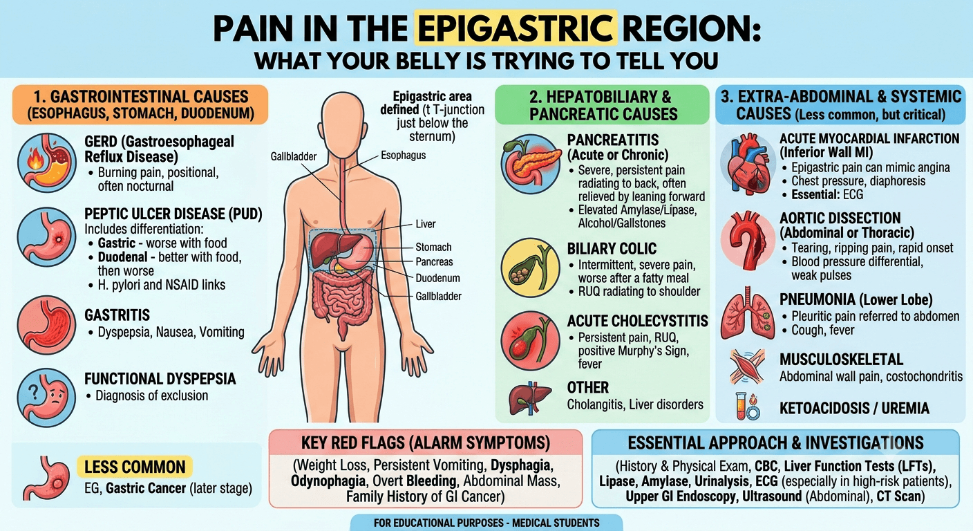
Living With Hypothyroidism

Living with hypothyroidism

How to Fix Brain Fog and Anxiety

Introduction

That’s correct, Hypothyroidism is indeed one of the most common endocrine disorders worldwide. While many people Living With Hypothyroidism recognize symptoms such as fatigue, weight gain, dry skin, and cold intolerance. The cognitive and emotional effects of hypothyroidism are often underestimated. Patients frequently describe feeling mentally foggy, forgetful, slow to process information, and unusually anxious. These symptoms can significantly affect quality of life, work performance, academic achievement, and relationships.

Living With Hypothyroidism: How to Fix Brain Fog and Anxiety a comprehensive guide for patients and medical students covering causes, diagnosis, treatment, lifestyle strategies, and the science behind thyroid-related cognitive and mood symptoms.

For patients, understanding why these symptoms occur can be reassuring. For medical students, understanding the underlying pathophysiology provides valuable insight into the complex relationship between thyroid hormones, brain function, and mental health.

This article explores the causes of brain fog and anxiety in hypothyroidism and discusses evidence-based strategies to improve cognitive function and emotional well-being.

Understanding Hypothyroidism

Hypothyroidism occurs when the thyroid gland does not produce sufficient thyroid hormones, primarily thyroxine (T4) and triiodothyronine (T3). These hormones regulate metabolism throughout the body, including the brain.

https://www.youtube.com/watch?v=JBvkueM7lpw

Common causes include:

  • Autoimmune thyroiditis (especially Hashimoto’s Thyroiditis)
  • Thyroid surgery
  • Radioactive iodine treatment
  • Certain medications
  • Congenital thyroid disorders
  • Iodine deficiency

Thyroid hormones play critical roles in:

  • Energy production
  • Neuronal signaling
  • Neurotransmitter regulation
  • Memory formation
  • Mood regulation
  • Cognitive processing speed

When hormone levels decline, brain function may be affected long before laboratory abnormalities become severe.

What Is Brain Fog?

Brain fog is not a formal medical diagnosis. Instead, it is a collection of cognitive symptoms that patients commonly report.

Typical symptoms include:

  • Difficulty concentrating
  • Forgetfulness
  • Slower thinking
  • Reduced mental clarity
  • Trouble finding words
  • Difficulty multitasking
  • Mental fatigue
  • Reduced productivity

Patients often describe it as feeling like their brain is running through mud.

For medical students, these symptoms reflect disruptions in attention, executive function, working memory, and information processing speed.

Why Does Hypothyroidism Cause Brain Fog?

1. Reduced Cerebral Metabolism

Thyroid hormones increase metabolic activity throughout the body, including the brain.

In hypothyroidism:

  • Neurons consume less energy
  • Glucose utilization decreases
  • Neural signaling slows
  • Cognitive processing becomes less efficient

Brain imaging studies have demonstrated reduced cerebral blood flow and metabolic activity in untreated hypothyroidism.

2. Neurotransmitter Changes

Thyroid hormones influence several neurotransmitter systems:

  • Serotonin
  • Dopamine
  • Norepinephrine
  • GABA

Reduced thyroid hormone levels can alter neurotransmitter production and receptor sensitivity, contributing to cognitive slowing and mood symptoms.

3. Sleep Disturbance

Many hypothyroid patients experience:

  • Excessive sleepiness
  • Poor sleep quality
  • Obstructive sleep apnea
  • Non-restorative sleep

Sleep disruption alone can significantly worsen cognitive function.

4. Inflammation and Autoimmunity

In autoimmune thyroid disease, inflammatory cytokines may impact brain function independently of thyroid hormone levels.

Some patients continue to report cognitive symptoms despite apparently normal laboratory results, suggesting that inflammation may contribute to persistent symptoms.

The Connection Between Hypothyroidism and Anxiety

Many people assume hypothyroidism causes only depression. However, anxiety is also common.

Symptoms may include:

  • Excessive worry
  • Irritability
  • Restlessness
  • Panic attacks
  • Social anxiety
  • Feeling overwhelmed

Studies suggest that thyroid dysfunction affects multiple brain regions involved in emotional regulation, including:

  • Amygdala
  • Hippocampus
  • Prefrontal cortex

These structures rely on normal thyroid hormone signaling for optimal function.

Why Anxiety Can Persist Even After Treatment

Some patients continue to experience anxiety despite achieving normal thyroid function tests.

Possible explanations include:

Residual Neurochemical Changes

The brain may require months to fully adapt after thyroid hormone levels normalize.

Coexisting Mental Health Conditions

Patients with hypothyroidism may also have:

  • Generalized anxiety disorder
  • Depression
  • Panic disorder
  • Health anxiety

These conditions often require separate treatment.

Lifestyle Factors

Persistent anxiety may be aggravated by:

  • Poor sleep
  • Chronic stress
  • Nutritional deficiencies
  • Sedentary behavior
  • Excessive caffeine consumption

The First Step: Optimize Thyroid Hormone Replacement

For most patients, effective treatment begins with appropriate thyroid hormone replacement.

The most commonly prescribed therapy is:

  • Levothyroxine

Key principles include:

  • Taking medication consistently
  • Taking it on an empty stomach
  • Avoiding interference from calcium or iron supplements
  • Monitoring thyroid function regularly

Medical students should remember that treatment targets are individualized. A laboratory value within the reference range does not always guarantee symptom resolution. Persistent symptoms warrant a comprehensive evaluation rather than automatic dose escalation

Addressing Common Nutritional Deficiencies

Several nutritional deficiencies can mimic or worsen brain fog and anxiety.

Iron Deficiency

Iron supports:

  • Oxygen transport
  • Dopamine production
  • Cognitive performance

Low ferritin levels may contribute to fatigue and poor concentration.

Vitamin B12 Deficiency

Deficiency may cause:

  • Memory problems
  • Cognitive slowing
  • Neuropathy
  • Mood disturbances

Vitamin D Deficiency

Low vitamin D levels have been associated with:

  • Depression
  • Anxiety
  • Fatigue

Selenium

Selenium participates in thyroid hormone metabolism and antioxidant defense.

Dietary sources include:

  • Brazil nuts
  • Seafood
  • Eggs

Supplementation should only be considered when clinically appropriate.

Improving Brain Fog Through Sleep Optimization

Sleep is one of the most powerful interventions for cognitive recovery.

Practical Strategies

  • Maintain a consistent sleep schedule
  • Avoid screens before bedtime
  • Limit caffeine in the afternoon
  • Keep the bedroom cool and dark
  • Evaluate for sleep apnea if symptoms suggest it

Many patients notice meaningful improvements in concentration after improving sleep quality.

Exercise: A Natural Brain Booster

Physical activity improves:

Recommended activities include:

Aerobic Exercise

Examples:

  • Walking
  • Cycling
  • Swimming
  • Jogging

Resistance Training

Strength training supports:

  • Metabolic health
  • Cognitive performance
  • Psychological well-being

Even 20–30 minutes of moderate activity most days can improve mental clarity.

Managing Anxiety Effectively

Cognitive Behavioral Therapy (CBT)

Cognitive Behavioral Therapy is among the most effective treatments for anxiety.

CBT helps patients:

  • Identify negative thought patterns
  • Reduce catastrophic thinking
  • Build coping skills
  • Improve resilience

Mindfulness and Meditation

Research suggests mindfulness practices can:

  • Reduce stress hormones
  • Improve emotional regulation
  • Enhance attention and focus

Even 10 minutes daily may provide measurable benefits.

Dietary Approaches for Better Cognitive Function

No special “thyroid diet” cures hypothyroidism, but healthy nutrition supports recovery.

Focus On

  • Lean proteins
  • Vegetables
  • Fruits
  • Whole grains
  • Healthy fats

Brain-Supportive Foods

Examples include:

  • Fatty fish
  • Nuts
  • Seeds
  • Olive oil
  • Berries

These foods provide nutrients that support neuronal function and reduce inflammation.

When Brain Fog Is Not Just Hypothyroidism

Persistent symptoms deserve further evaluation.

Possible alternative or additional causes include:

  • Sleep apnea
  • Depression
  • Anxiety disorders
  • ADHD
  • Anemia
  • Chronic fatigue syndrome
  • Medication side effects
  • Long COVID
  • Menopause
  • Vitamin deficiencies

Clinicians should avoid attributing every symptom solely to thyroid disease.

Special Considerations for Medical Students

Medical students frequently encounter patients who report symptoms despite “normal” laboratory results.

Important clinical lessons include:

Treat the Patient, Not Just the Lab Values

Laboratory normalization does not always equal symptom resolution.

Consider Comorbidities

Fatigue, cognitive dysfunction, and anxiety are multifactorial.

Avoid Premature Diagnostic Closure

Always maintain a broad differential diagnosis.

Listen to Patient Experiences

Subjective symptoms provide valuable clinical information that may not be reflected in laboratory tests alone.

Frequently Asked Questions (FAQs)

1. Can hypothyroidism cause severe brain fog?

Yes. Cognitive slowing, memory difficulties, and poor concentration are common manifestations of untreated hypothyroidism.

2. How long does it take for brain fog to improve after starting treatment?

Many patients improve within weeks, but complete recovery may take several months depending on severity and duration of disease.

3. Can anxiety occur with hypothyroidism?

Yes. Although depression is more commonly recognized, anxiety can also occur due to thyroid hormone effects on neurotransmitters and brain function.

4. Does increasing thyroid medication always fix brain fog?

No. Persistent symptoms may be related to sleep disorders, nutritional deficiencies, mental health conditions, or other medical problems.

5. Which vitamin deficiencies are commonly associated with hypothyroidism?

Iron, vitamin B12, and vitamin D deficiencies are frequently encountered and may contribute to fatigue and cognitive symptoms.

6. Is exercise safe for people with hypothyroidism?

In most cases, yes. Regular physical activity improves mood, cognition, cardiovascular health, and overall quality of life.

7. Can Hashimoto’s thyroiditis affect the brain directly?

Emerging evidence suggests that autoimmune activity and inflammatory pathways may contribute to cognitive symptoms in some individuals, though thyroid hormone deficiency remains the primary mechanism.

Conclusion

Living with hypothyroidism can be challenging, particularly when brain fog and anxiety interfere with daily life. Fortunately, these symptoms are often manageable with a comprehensive approach that includes optimized thyroid hormone replacement, adequate sleep, regular exercise, nutritional assessment, stress management, and treatment of coexisting medical or psychological conditions.

For patients, the key message is that persistent cognitive and emotional symptoms are real and deserve attention. For medical students, hypothyroidism offers an important example of how endocrine disorders can profoundly influence brain function and mental health. Understanding this connection allows clinicians to provide more holistic and effective care.

By addressing both the biochemical and lifestyle factors involved, many individuals with hypothyroidism can achieve substantial improvements in mental clarity, emotional stability, and overall quality of life.

Disclaimer: The information provided in this blog post is for educational and informational purposes only and should not be considered medical advice. It is not intended to replace professional medical consultation, diagnosis, or treatment. Always seek the guidance of a qualified healthcare professional regarding any medical condition or health-related concerns. The author and publisher are not responsible for any actions taken based on the information presented in this article.
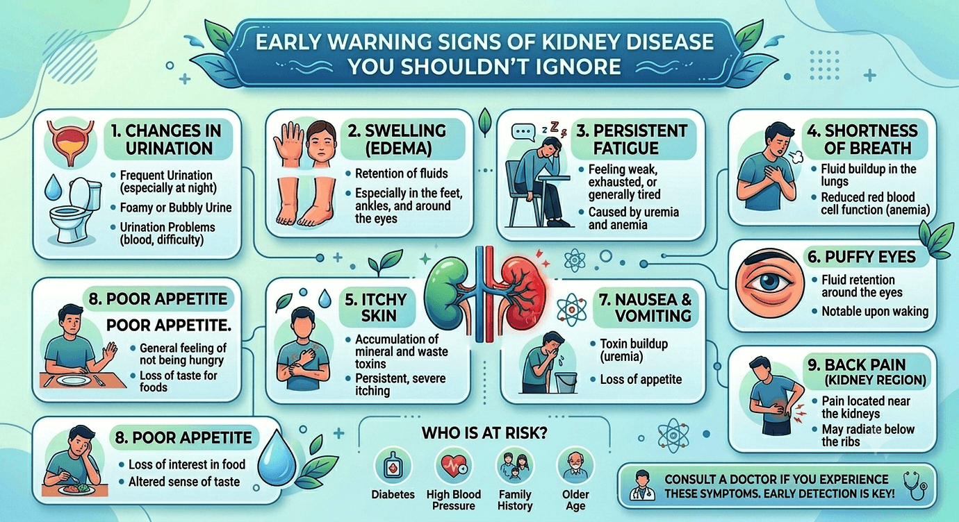
Hypertension: Symptoms, Causes, & Treatment

Hypertension

Introduction

Hypertension, commonly known as high blood pressure, is one of the most prevalent cardiovascular disorders worldwide. It is often referred to as the “silent killer” because many individuals have no symptoms until significant damage has occurred to vital organs such as the heart, brain, kidneys, and eyes.

Blood pressure represents the force exerted by circulating blood against the walls of the arteries. Persistent elevation of this pressure increases the risk of serious complications, including heart attack, stroke, heart failure, chronic kidney disease, and premature death.

This article provides a detailed overview of hypertension for both patients and medical students, covering its symptoms, causes, diagnosis, treatment, and prevention.

What Is Blood Pressure?

Blood pressure is measured using two numbers:

Systolic Blood Pressure

  • The upper number
  • Represents pressure in the arteries when the heart contracts

Diastolic Blood Pressure

  • The lower number
  • Represents pressure in the arteries when the heart relaxes between beats

Example:

120/80 mmHg

  • Systolic = 120 mmHg
  • Diastolic = 80 mmHg

Classification of Blood Pressure

According to current clinical guidelines:

CategorySystolic (mmHg)Diastolic (mmHg)
Normal<120<80
Elevated120-129<80
Stage 1 Hypertension130-13980-89
Stage 2 Hypertension≥140≥90
Hypertensive Crisis≥180≥120

A hypertensive crisis requires immediate medical attention.

Symptoms of Hypertension

Most patients with hypertension experience no symptoms, even when blood pressure reaches dangerously high levels.

When symptoms occur, they may include:

Common Symptoms

  • Persistent headaches
  • Dizziness
  • Fatigue
  • Shortness of breath
  • Blurred vision
  • Palpitations
  • Nosebleeds
  • Chest discomfort

Symptoms of Severe Hypertension

Severely elevated blood pressure may cause:

  • Severe headache
  • Confusion
  • Visual disturbances
  • Chest pain
  • Difficulty breathing
  • Neurological deficits
  • Seizures

These symptoms may indicate a hypertensive emergency requiring urgent treatment.

Causes of Hypertension

Hypertension is broadly divided into two categories.

1. Primary (Essential) Hypertension

This accounts for approximately 90–95% of cases.

No single cause is identified, but multiple factors contribute:

  • Genetic predisposition
  • Aging
  • Obesity
  • Sedentary lifestyle
  • High salt intake
  • Excess alcohol consumption
  • Smoking
  • Chronic stress
  • Insulin resistance

2. Secondary Hypertension

Secondary hypertension results from an identifiable underlying condition.

Common causes include:

Kidney Disorders

  • Chronic kidney disease
  • Glomerulonephritis
  • Polycystic kidney disease
  • Renal artery stenosis

Endocrine Disorders

  • Hyperthyroidism
  • Hypothyroidism
  • Cushing syndrome
  • Primary hyperaldosteronism
  • Pheochromocytoma

Sleep Disorders

  • Obstructive sleep apnea

Medications

  • Oral contraceptives
  • Corticosteroids
  • NSAIDs
  • Decongestants
  • Certain antidepressants

Other Causes

  • Pregnancy-related hypertension
  • Congenital vascular abnormalities
  • Recreational drug use

Risk Factors for Hypertension

Several factors increase the likelihood of developing hypertension.

Non-Modifiable Risk Factors

  • Increasing age
  • Family history
  • Male sex (younger adults)
  • Postmenopausal status in women
  • Ethnicity

Modifiable Risk Factors

  • Obesity
  • Physical inactivity
  • High-sodium diet
  • Excessive alcohol consumption
  • Smoking
  • Diabetes mellitus
  • Dyslipidemia
  • Chronic stress
  • Poor sleep quality

Complications of Hypertension

Uncontrolled hypertension can damage multiple organs.

Cardiovascular Complications

  • Coronary artery disease
  • Myocardial infarction
  • Heart failure
  • Left ventricular hypertrophy
  • Peripheral arterial disease

Neurological Complications

  • Stroke
  • Transient ischemic attack (TIA)
  • Vascular dementia

Renal Complications

  • Chronic kidney disease
  • Hypertensive nephropathy
  • End-stage renal disease

Ocular Complications

  • Hypertensive retinopathy
  • Vision loss

Vascular Complications

  • Aortic aneurysm
  • Aortic dissection

Diagnosis of Hypertension

Blood Pressure Measurement

Diagnosis requires:

  • Proper cuff size
  • Correct patient positioning
  • Multiple readings on separate occasions

Home Blood Pressure Monitoring

Provides more accurate assessment in many patients and helps detect:

  • White coat hypertension
  • Masked hypertension

Ambulatory Blood Pressure Monitoring (ABPM)

Measures blood pressure over 24 hours and is considered the gold standard for diagnosis.

Laboratory Investigations

Initial evaluation often includes:

Blood Tests

  • Complete blood count
  • Blood glucose
  • HbA1c
  • Lipid profile
  • Liver function tests
  • Kidney function tests
  • Electrolytes
  • Thyroid function tests

Urine Tests

  • Urinalysis
  • Urine albumin-to-creatinine ratio

Additional Tests

  • Electrocardiogram (ECG)
  • Echocardiography
  • Fundoscopic examination
  • Renal ultrasound (when indicated)

Treatment of Hypertension

Treatment aims to reduce blood pressure and prevent complications.

Lifestyle Modifications

Lifestyle changes are recommended for all patients.

Reduce Salt Intake

Aim for less than 5 grams of salt daily.

Maintain Healthy Weight

Even modest weight loss can significantly reduce blood pressure.

Regular Physical Activity

Recommended:

  • At least 150 minutes of moderate exercise weekly

Examples:

  • Brisk walking
  • Cycling
  • Swimming

Healthy Diet

The DASH diet emphasizes:

  • Fruits
  • Vegetables
  • Whole grains
  • Lean proteins
  • Low-fat dairy products

Limit Alcohol

Excessive alcohol intake contributes to hypertension.

Quit Smoking

Smoking greatly increases cardiovascular risk.

Stress Management

Helpful techniques include:

  • Meditation
  • Yoga
  • Relaxation exercises
  • Adequate sleep

Medications for Hypertension

Drug selection depends on age, comorbidities, and individual risk factors.

ACE Inhibitors

Examples:

  • Lisinopril
  • Enalapril

Benefits:

  • Kidney protection in diabetes
  • Cardiovascular risk reduction

Angiotensin Receptor Blockers (ARBs)

Examples:

  • Losartan
  • Valsartan

Often used when ACE inhibitors are not tolerated.

Calcium Channel Blockers

Examples:

  • Amlodipine
  • Nifedipine

Thiazide Diuretics

Examples:

  • Hydrochlorothiazide
  • Chlorthalidone

Beta Blockers

Examples:

  • Metoprolol
  • Atenolol

Particularly useful in certain cardiac conditions.

Hypertensive Emergency

A hypertensive emergency occurs when severe hypertension is accompanied by acute organ damage.

Possible manifestations include:

  • Stroke
  • Pulmonary edema
  • Acute kidney injury
  • Aortic dissection
  • Hypertensive encephalopathy

Treatment typically requires hospitalization and intravenous antihypertensive medications.

Prevention of Hypertension

Preventive strategies include:

  • Maintaining a healthy body weight
  • Exercising regularly
  • Limiting salt intake
  • Eating a balanced diet
  • Avoiding tobacco products
  • Limiting alcohol consumption
  • Managing diabetes and cholesterol
  • Regular blood pressure monitoring

Prognosis

The outlook for hypertension is generally excellent when diagnosed early and treated appropriately. Consistent lifestyle modification, adherence to prescribed medications, and regular follow-up can significantly reduce the risk of heart disease, stroke, kidney failure, and other complications.

Key Takeaways

  • Hypertension is a chronic condition characterized by persistently elevated blood pressure.
  • Most patients have no symptoms, making routine screening essential.
  • Major risk factors include obesity, high salt intake, aging, diabetes, smoking, and family history.
  • Untreated hypertension can damage the heart, brain, kidneys, eyes, and blood vessels.
  • Lifestyle modification remains the foundation of treatment.
  • Medications are often necessary to achieve blood pressure control and prevent complications.
  • Early diagnosis and long-term management dramatically improve outcomes and quality of life.

Frequently Asked Questions (FAQs)

1. Why is hypertension called the “silent killer”?
Because many people have high blood pressure for years without noticeable symptoms, while organ damage gradually occurs.

2. Can hypertension be cured?
Most cases cannot be permanently cured, but they can be effectively controlled through lifestyle changes and medications.

3. What is the best diet for hypertension?
The DASH diet, rich in fruits, vegetables, whole grains, and low-fat dairy products, is highly effective.

4. Can stress cause high blood pressure?
Chronic stress can contribute to elevated blood pressure and unhealthy lifestyle habits that worsen hypertension.

5. What happens if hypertension is left untreated?
Untreated hypertension increases the risk of heart attack, stroke, heart failure, kidney disease, vision loss, and premature death.

Disclaimer: The information provided in this blog post is for educational and informational purposes only and should not be considered medical advice. It is not intended to replace professional medical consultation, diagnosis, or treatment. Always seek the guidance of a qualified healthcare professional regarding any medical condition or health-related concerns. The author and publisher are not responsible for any actions taken based on the information presented in this article.
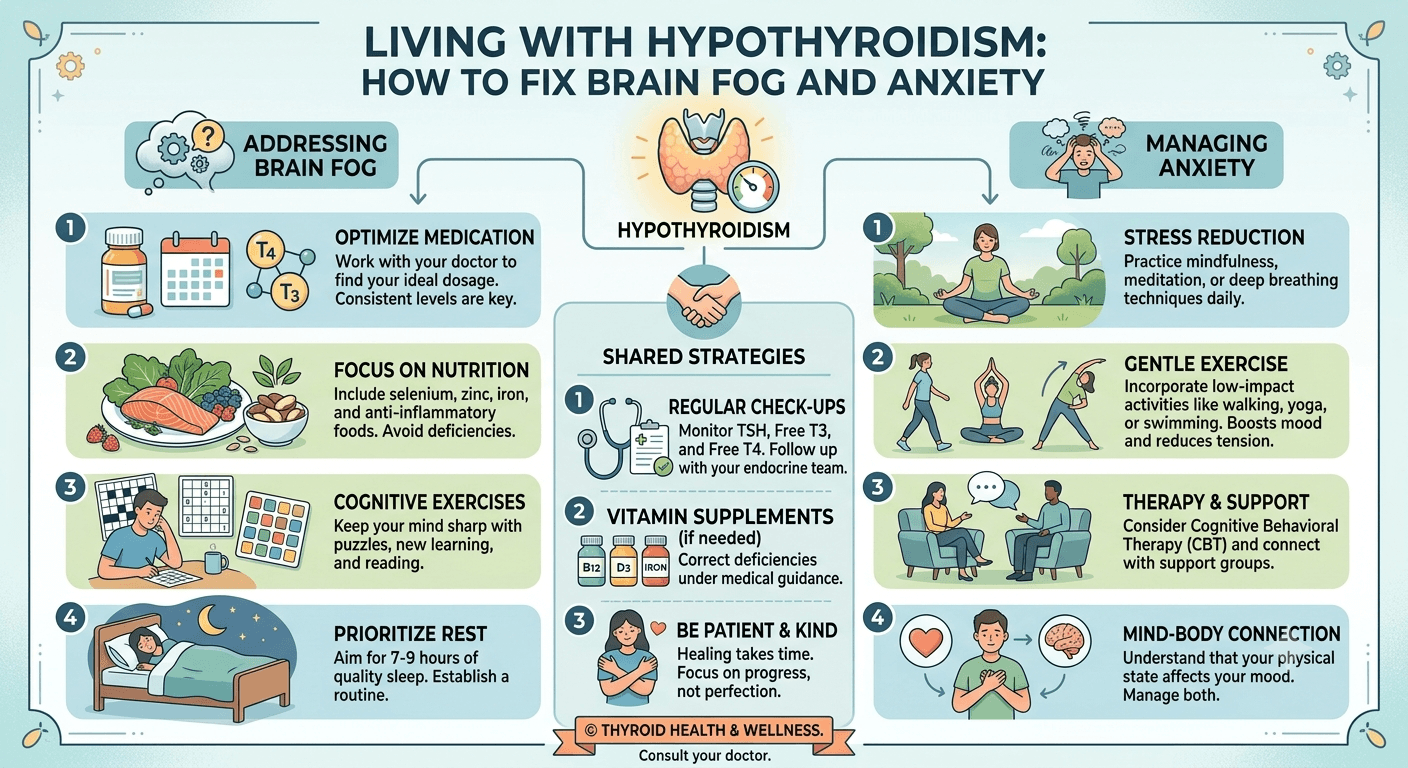
Osteoarthritis vs. Rheumatoid Arthritis

Rheumatoid Arthritis

Key Differences & Symptoms

Introduction

Arthritis is a common condition that affects millions of people worldwide, causing joint pain, stiffness, and reduced mobility. Among the many types of arthritis, Osteoarthritis (OA) and Rheumatoid Arthritis (RA) are the most frequently diagnosed. Although both conditions impact the joints and can significantly impact quality of life, they have different causes, symptoms, and treatment approaches.

Learn the key differences between Osteoarthritis (OA) and Rheumatoid Arthritis (RA), including symptoms, causes, risk factors, diagnosis, and treatment options. Discover how to recognize early warning signs and manage joint pain effectively.

What Is Osteoarthritis (OA)?

Osteoarthritis is the most common form of arthritis. It is a degenerative joint disease that occurs when the protective cartilage covering the ends of bones gradually wears away. As cartilage deteriorates, bones may rub against each other, causing pain, stiffness, and reduced joint function.

OA is often referred to as a wear-and-tear arthritis because it typically develops over time due to aging and repeated stress on the joints.

Commonly Affected Joints in OA

  • Knees
  • Hips
  • Hands
  • Lower back
  • Neck
  • Feet

What Is Rheumatoid Arthritis (RA)?

Rheumatoid arthritis is a chronic autoimmune disease in which the body’s immune system mistakenly attacks the lining of the joints (synovium). This causes inflammation that can damage cartilage, bone, and surrounding tissues.

Unlike OA, RA is a systemic disease, meaning it can affect other organs such as the lungs, heart, eyes, and blood vessels.

Commonly Affected Joints in RA

  • Fingers
  • Wrists
  • Hands
  • Feet
  • Ankles
  • Elbows
  • Knees

RA usually impacts the same joints on both sides of the body.

Key Differences Between OA and RA

FeatureOsteoarthritis (OA)Rheumatoid Arthritis (RA)
CauseWear and tear of cartilageAutoimmune inflammation
Age of OnsetUsually after age 50Can occur at any age
Joint InvolvementOften affects one side more than the otherUsually symmetrical
Morning StiffnessLess than 30 minutesMore than 30–60 minutes
InflammationMild or absentSignificant inflammation
Systemic SymptomsRareCommon
Disease ProgressionGradualCan progress rapidly
Other Organ InvolvementNoYes

Symptoms of Osteoarthritis (OA)

Osteoarthritis (OA) is a degenerative joint disease caused by the gradual breakdown of cartilage. Symptoms often develop slowly and worsen over time. Common symptoms include:

  • Joint pain that increases with activity and improves with rest
  • Morning stiffness lasting less than 30 minutes
  • Reduced range of motion and joint flexibility
  • Joint swelling or tenderness
  • A grating or cracking sensation (crepitus) during movement
  • Bony enlargement of affected joints, especially the fingers, knees, hips, and spine

Symptoms of Rheumatoid Arthritis (RA)

Rheumatoid Arthritis (RA) is a chronic autoimmune disease that causes inflammation of the joints and other tissues. Symptoms often affect multiple joints symmetrically and may include:

  • Persistent joint pain, swelling, and tenderness
  • Morning stiffness lasting more than 30–60 minutes
  • Warm, inflamed joints, especially in the hands, wrists, and feet
  • Fatigue, weakness, and reduced energy levels
  • Low-grade fever and general malaise
  • Loss of joint function and progressive deformity in advanced disease

Risk Factors for Osteoarthritis

Several factors increase the risk of developing OA:

  • Aging
  • Obesity
  • Metabolic disorders such as diabetes and dyslipidemia
  • Previous joint injuries
  • Repetitive joint stress
  • Family history
  • Joint deformities
  • Certain occupations involving heavy physical work

Risk Factors for Rheumatoid Arthritis

Risk factors for RA include:

  • Family history
  • Female sex
  • Smoking
  • Obesity
  • Environmental exposures
  • Hormonal influences
  • Certain genetic factors

Diagnosis

Diagnosing Osteoarthritis

Doctors may use:

Lab Tests: There is no specific diagnostic lab test for Osteoarthritis. However, lab tests play a massive role in the diagnostic process. Their primary job is differential diagnosis to rule out other conditions (like Rheumatoid Arthritis or gout) that mimic OA.

Medical history

Physical examination

X-rays

MRI scans in selected cases

Typical findings include cartilage loss, narrowed joint spaces, and bone spurs.

Diagnosing Rheumatoid Arthritis

Diagnosis may involve:

Physical examination

Blood tests

  • Rheumatoid Factor (RF)
  • Anti-CCP antibodies
  • ESR
  • CRP
  • Kidney Function Tests: (Chronic NSAID use can impair renal function).
  • Liver Function Tests: (To monitor liver health if the patient is using high-dose acetaminophen or certain prescription NSAIDs for pain management)
  • X-rays
  • Ultrasound
  • MRI scans

    These tests help identify inflammation and joint damage.

    Treatment of Osteoarthritis

    Treatment focuses on symptom relief and maintaining joint function.

    Lifestyle Changes

    • Weight loss
    • Regular exercise
    • Physical therapy
    • Joint protection techniques

    Medications

    • Acetaminophen
    • Nonsteroidal anti-inflammatory drugs (NSAIDs) like ibuprofen, naproxen, or meloxicam.
    • Topical pain relievers

    Advanced Treatments

    • Corticosteroid injections
    • Hyaluronic acid injections
    • Joint replacement surgery

    Treatment of Rheumatoid Arthritis

    Early treatment is essential to prevent permanent joint damage.

    Disease-Modifying Medications

    • DMARDs such as methotrexate (usually once a week)
    • Biologic agents
    • Targeted synthetic DMARDs

    Symptom Relief

    • NSAIDs like ibuprofen, naproxen, or meloxicam.
    • Corticosteroids

    Supportive Care

    • Physical therapy
    • Occupational therapy
    • Regular exercise
    • Smoking cessation

    Can OA and RA Occur Together?

    Yes. Some individuals with rheumatoid arthritis may also develop osteoarthritis, especially as they age. In such cases, diagnosis and treatment become more complex and require careful evaluation by a healthcare professional.

    When to See a Doctor

    Seek medical attention if you experience:

    • Persistent joint pain
    • Joint swelling
    • Morning stiffness lasting more than 30 minutes
    • Difficulty performing daily activities
    • Unexplained fatigue or weight loss
    • Progressive loss of joint movement

    Early diagnosis can significantly improve long-term outcomes.

    Conclusion

    Although osteoarthritis and rheumatoid arthritis both cause joint pain and stiffness, they are fundamentally different diseases. Osteoarthritis results from cartilage degeneration, whereas rheumatoid arthritis is an autoimmune condition characterized by chronic inflammation. OA typically develops gradually with age, while RA can affect people of any age and may involve multiple organs.

    Recognizing the key differences and symptoms of these conditions allows for earlier diagnosis, better treatment, and improved quality of life. If you experience persistent joint symptoms, consult a healthcare professional for proper evaluation and management.

    Frequently Asked Questions (FAQs)

    1. What is the main difference between osteoarthritis and rheumatoid arthritis?

    Osteoarthritis is caused by the breakdown of joint cartilage, while rheumatoid arthritis is an autoimmune disease that attacks joint tissues.

    2. Which condition is more painful, OA or RA?

    Both can cause significant pain. RA often causes more inflammation and systemic symptoms, while OA pain is usually related to joint use and degeneration.

    3. Can rheumatoid arthritis be cured?

    There is currently no cure for RA, but modern treatments can control symptoms and slow disease progression.

    4. Is osteoarthritis an autoimmune disease?

    No. Osteoarthritis is a degenerative joint condition that is not caused by an abnormal immune response.

    5. Does exercise help arthritis?

    Yes. Appropriate exercise can improve joint flexibility, strengthen muscles, reduce pain, and enhance overall function in both OA and RA.

    Disclaimer: The information provided in this blog post is for educational and informational purposes only and should not be considered medical advice. It is not intended to replace professional medical consultation, diagnosis, or treatment. Always seek the guidance of a qualified healthcare professional regarding any medical condition or health-related concerns. The author and publisher are not responsible for any actions taken based on the information presented in this article.
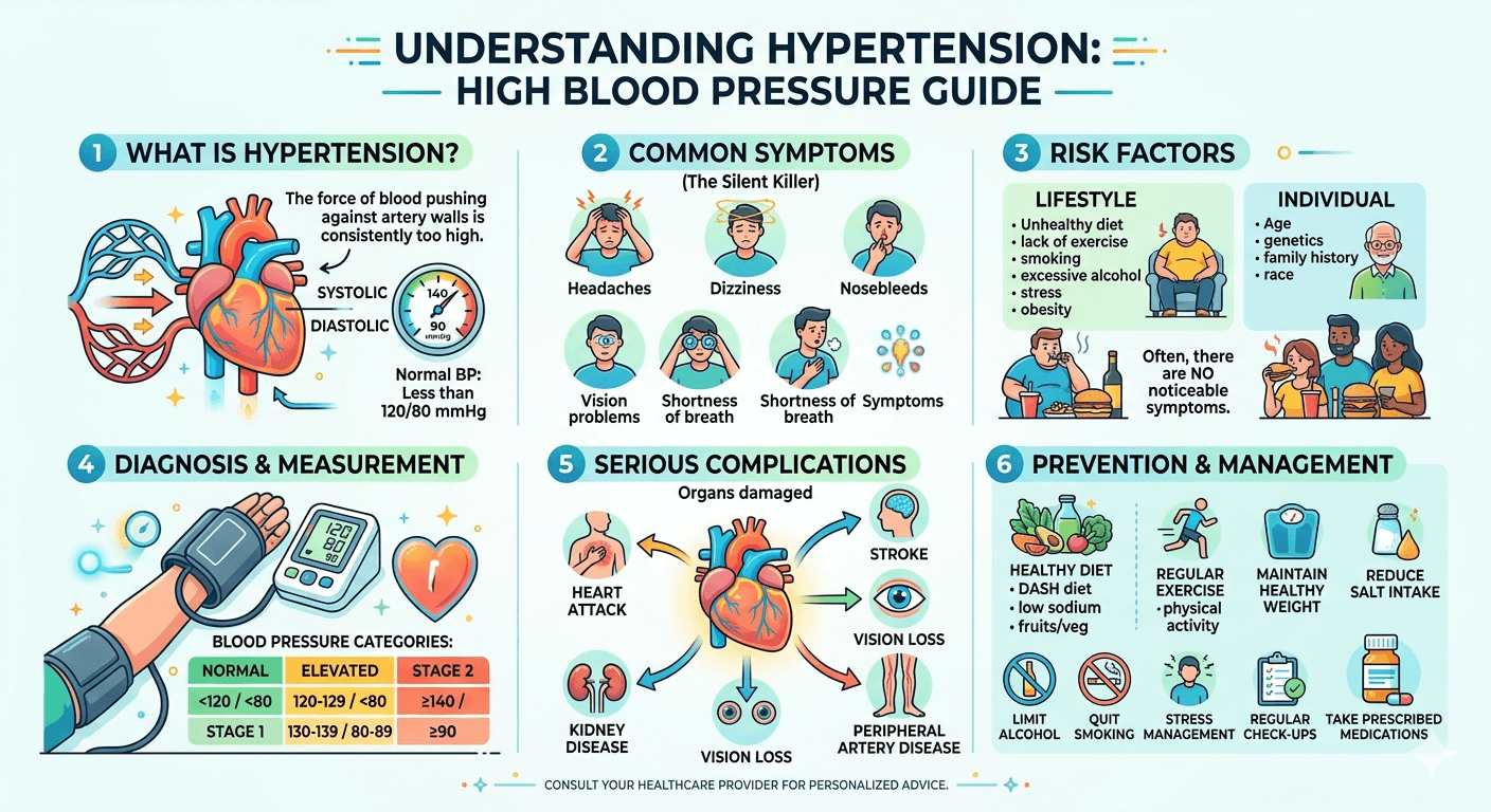
    Pyelonephritis and Cystitis: A Detailed Guide

    Pyelonephritis and cystitis

    Introduction

    Pyelonephritis and cystitis are among the most common infections affecting the urinary tract. Both conditions are forms of urinary tract infection (UTI), but they differ in their location, severity, clinical presentation, and potential complications. Understanding these disorders is important because prompt diagnosis and treatment can prevent serious outcomes such as kidney damage and sepsis.

    Learn about Pyelonephritis and cystitis, two common urinary tract infections. This detailed guide covers causes, symptoms, diagnosis, treatment options, complications, and prevention strategies for patients and medical students.

    Overview of the Urinary Tract

    The urinary system consists of:

    • Kidneys
    • Ureters
    • Urinary bladder
    • Urethra

    The kidneys filter blood and produce urine. Urine passes through the ureters into the bladder, where it is stored before being expelled through the urethra.

    Urinary tract infections may involve different parts of this system:

    • Cystitis: Infection of the urinary bladder
    • Pyelonephritis: Infection of the kidney and renal pelvis.

    What is Cystitis?

    Cystitis is inflammation of the urinary bladder, most commonly caused by bacterial infection. It is considered a lower urinary tract infection and is one of the most frequent bacterial infections encountered in clinical practice.

    Women are affected more often than men because the female urethra is shorter, allowing bacteria easier access to the bladder.

    Causes of Cystitis

    The most common causative organism is:

    • Escherichia coli (E. coli)

    Other pathogens include:

    • Klebsiella species
    • Proteus species
    • Enterococcus species
    • Staphylococcus saprophyticus

    These bacteria usually originate from the gastrointestinal tract and enter the urinary tract through the urethra.

    Risk Factors for Cystitis

    • Female sex
    • Sexual activity
    • Pregnancy
    • Menopause
    • Diabetes mellitus
    • Poor hydration
    • Urinary catheterization
    • Use of spermicides
    • Previous urinary tract infections
    • Urinary obstruction

    Symptoms of Cystitis

    Patients commonly present with:

    • Dysuria (burning or pain during urination)
    • Increased urinary frequency
    • Urinary urgency
    • Suprapubic discomfort
    • Cloudy urine
    • Foul-smelling urine
    • Hematuria (blood in urine)

    Systemic symptoms such as high fever are usually absent.

    Physical Examination Findings

    • Mild Suprapubic tenderness
    • Usually normal vital signs
    • No costovertebral angle tenderness

    What is Pyelonephritis?

    Pyelonephritis is a bacterial infection involving the kidney tissue and renal pelvis. It is an upper urinary tract infection and is generally more severe than cystitis.

    Most cases develop when bacteria spread upward from an infected bladder into one or both kidneys.

    Types of Pyelonephritis

    Acute Pyelonephritis

    A sudden bacterial infection characterized by fever, flank pain, and urinary symptoms. It may become life-threatening if not treated promptly.

    Chronic Pyelonephritis

    A recurrent or persistent kidney infection leading to progressive renal scarring and deterioration of kidney function. It is less common and often associated with urinary tract abnormalities.

    Pathophysiology

    The infection typically follows an ascending route:

    1. Bacteria enter the urethra.
    2. The bladder becomes infected (cystitis).
    3. Organisms ascend through the ureters.
    4. One or both kidneys become infected (pyelonephritis).

    Less commonly, infection may spread to the kidneys through the bloodstream.

    Risk Factors for Pyelonephritis

    • Untreated bladder infection
    • Kidney stones
    • Vesicoureteral reflux
    • Enlarged prostate
    • Pregnancy
    • Diabetes mellitus
    • Urinary tract obstruction
    • Immunosuppression
    • Urinary retention
    • Long-term catheter use

    These conditions facilitate bacterial growth or impair urinary drainage.

    Clinical Features of Pyelonephritis

    Symptoms

    Patients often present with:

    • High fever
    • Chills and rigors
    • Flank pain
    • Back pain
    • Nausea and vomiting
    • Dysuria
    • Urinary urgency
    • Increased urinary frequency
    • Malaise
    • Fatigue

    The presence of fever and flank pain strongly suggests kidney involvement.

    Physical Examination

    Typical findings include:

    • Fever
    • Tachycardia
    • Costovertebral angle tenderness
    • Signs of dehydration
    • Occasionally hypotension in severe infection

    Comparison Between Cystitis and Pyelonephritis

    FeatureCystitisPyelonephritis
    Site of infectionBladderKidney
    Type of UTILower UTIUpper UTI
    FeverRareCommon
    Flank painAbsentPresent
    Nausea/VomitingRareCommon
    SeverityMildModerate to severe
    HospitalizationRarely neededSometimes required
    Risk of sepsisLowHigher

    Diagnosis

    Medical History

    Important questions include:

    • Duration of symptoms
    • Presence of fever
    • Flank pain
    • Previous UTIs
    • Pregnancy status
    • Diabetes history
    • Urinary obstruction symptoms

    Urinalysis

    Urinalysis often demonstrates:

    • Pyuria (white blood cells)
    • Bacteriuria
    • Positive leukocyte esterase
    • Positive nitrites
    • Microscopic hematuria

    Urinalysis is one of the most useful initial investigations.

    Urine Culture

    Urine culture identifies the causative organism and determines antibiotic sensitivity.

    It is especially important in:

    • Pyelonephritis
    • Recurrent infections
    • Complicated UTIs
    • Pregnancy

    Blood Tests

    Particularly useful in pyelonephritis:

    • Complete blood count (CBC)
    • Elevated white blood cell count
    • Blood cultures in severe infection
    • Urine culture & sensitivity test
    • Renal function tests
    • RBS, HbA1c
    • Lipid Profile

    Imaging Studies

    Imaging is generally unnecessary in uncomplicated cystitis.

    USG whole abdomen

    Imaging may be indicated when:

    • Symptoms persist despite treatment
    • Obstruction is suspected
    • Recurrent infections occur
    • Renal abscess is suspected

    Common modalities include:

    • Ultrasonography
    • CT scan
    • MRI

    Treatment of Cystitis

    Antibiotics

    Commonly prescribed antibiotics include:

    • Nitrofurantoin
    • Trimethoprim-sulfamethoxazole
    • Fosfomycin
    • Selected cephalosporins

    Choice depends on local resistance patterns and patient-specific factors.

    Supportive Measures

    • Increase fluid intake
    • Complete the antibiotic course
    • Avoid bladder irritants
    • Maintain good hygiene

    Treatment of Pyelonephritis

    Pyelonephritis requires prompt treatment because complications can develop rapidly.

    Outpatient Management

    Suitable for stable patients who can tolerate oral medications.

    Management includes:

    • Oral antibiotics
    • Adequate hydration
    • Analgesics and antipyretics

    Hospital Admission

    Hospitalization may be necessary for:

    • Severe infection
    • Sepsis
    • Persistent vomiting
    • Pregnancy
    • Urinary obstruction
    • Immunocompromised patients

    Intravenous Antibiotics

    Examples include:

    • Ceftriaxone
    • Piperacillin-tazobactam
    • Fluoroquinolones when appropriate
    • Carbapenems for resistant organisms

    Therapy should be adjusted according to culture results.

    Complications

    Complications of Cystitis

    • Recurrent urinary tract infections
    • Ascending infection causing pyelonephritis
    • Hemorrhagic cystitis (rare)

    Complications of Pyelonephritis

    • Renal abscess
    • Kidney scarring
    • Chronic kidney disease
    • Acute kidney injury
    • Sepsis
    • Septic shock
    • Kidney failure

    Untreated Pyelonephritis can become life-threatening.

    Pyelonephritis During Pregnancy

    Pregnant women are at increased risk because hormonal and anatomical changes promote urinary stasis.

    Potential complications include:

    • Maternal sepsis
    • Preterm labor
    • Low birth weight infants
    • Increased maternal morbidity

    Screening and treatment of asymptomatic bacteriuria during pregnancy help reduce risk.

    Prevention

    Preventive measures include:

    • Drinking adequate water
    • Avoiding delayed urination
    • Proper perineal hygiene
    • Urinating after sexual intercourse
    • Controlling diabetes
    • Avoiding unnecessary catheter use
    • Completing prescribed antibiotics

    When Should Patients Seek Urgent Medical Care?

    Immediate medical attention is required if a patient develops:

    • High fever
    • Severe flank pain
    • Persistent vomiting
    • Blood in urine
    • Pregnancy with urinary symptoms
    • Confusion
    • Difficulty breathing
    • Symptoms of sepsis

    These findings may indicate a severe kidney infection requiring urgent treatment.

    Key Learning Points for Medical Students

    • Cystitis is a lower urinary tract infection involving the bladder.
    • Pyelonephritis is an upper urinary tract infection involving the kidneys.
    • E. coli is the most common pathogen.
    • Fever, chills, and flank pain strongly suggest pyelonephritis.
    • Urinalysis and urine culture remain the cornerstone of diagnosis.
    • Imaging is reserved for complicated or refractory cases.
    • Early antibiotic therapy prevents renal damage and sepsis.
    • Recurrent infections warrant investigation for structural abnormalities.

    Conclusion

    Cystitis and pyelonephritis are common but clinically important urinary tract infections. Cystitis usually remains confined to the bladder and is often uncomplicated, whereas pyelonephritis affects the kidneys and may result in serious complications if not treated promptly. Recognizing the symptoms, identifying risk factors, performing appropriate investigations, and initiating timely antimicrobial therapy are essential for optimal patient outcomes. Early diagnosis and treatment significantly reduce the risk of kidney damage, recurrent infection, and sepsis.

    FAQ 1: What is the difference between cystitis and Pyelonephritis?

    Cystitis is an infection of the urinary bladder and is classified as a lower urinary tract infection. It commonly causes burning during urination, urinary frequency, and urgency. Pyelonephritis is a more serious infection that affects one or both kidneys and is considered an upper urinary tract infection. In addition to urinary symptoms, it often causes fever, chills, flank pain, nausea, and vomiting.

    FAQ 2: Can a bladder infection (cystitis) turn into a kidney infection (Pyelonephritis)?

    Yes. If cystitis is left untreated or inadequately treated, bacteria can travel from the bladder through the ureters to the kidneys, leading to Pyelonephritis. Early diagnosis and appropriate antibiotic treatment are important to prevent this complication.

    FAQ 3: What are the warning signs that a urinary tract infection has reached the kidneys?

    Symptoms suggesting kidney involvement include:

    • High fever
    • Chills or shaking
    • Pain in the back or side (flank pain)
    • Nausea and vomiting
    • General weakness or fatigue

    These symptoms require prompt medical evaluation because kidney infections can become serious if not treated quickly.

    FAQ 4: How are Pyelonephritis and cystitis diagnosed?

    Diagnosis typically involves:

    • Medical history and physical examination
    • Urinalysis to detect white blood cells, bacteria, and blood
    • Urine culture to identify the causative organism
    • Blood tests in severe cases
    • Imaging studies such as ultrasound or CT scan when complications or urinary tract obstruction are suspected

    FAQ 5: How can I reduce my risk of developing urinary tract infections?

    You can help prevent cystitis and pyelonephritis by:

    • Drinking plenty of water
    • Not delaying urination
    • Practicing good personal hygiene
    • Urinating after sexual intercourse
    • Managing diabetes effectively
    • Avoiding unnecessary urinary catheter use
    • Completing prescribed antibiotic treatments
    • Seeking medical attention for recurrent urinary symptoms

    These preventive measures can significantly reduce the risk of recurrent urinary tract and kidney infections.

    Disclaimer: The information provided in this blog post is for educational and informational purposes only and should not be considered medical advice. It is not intended to replace professional medical consultation, diagnosis, or treatment. Always seek the guidance of a qualified healthcare professional regarding any medical condition or health-related concerns. The author and publisher are not responsible for any actions taken based on the information presented in this article.
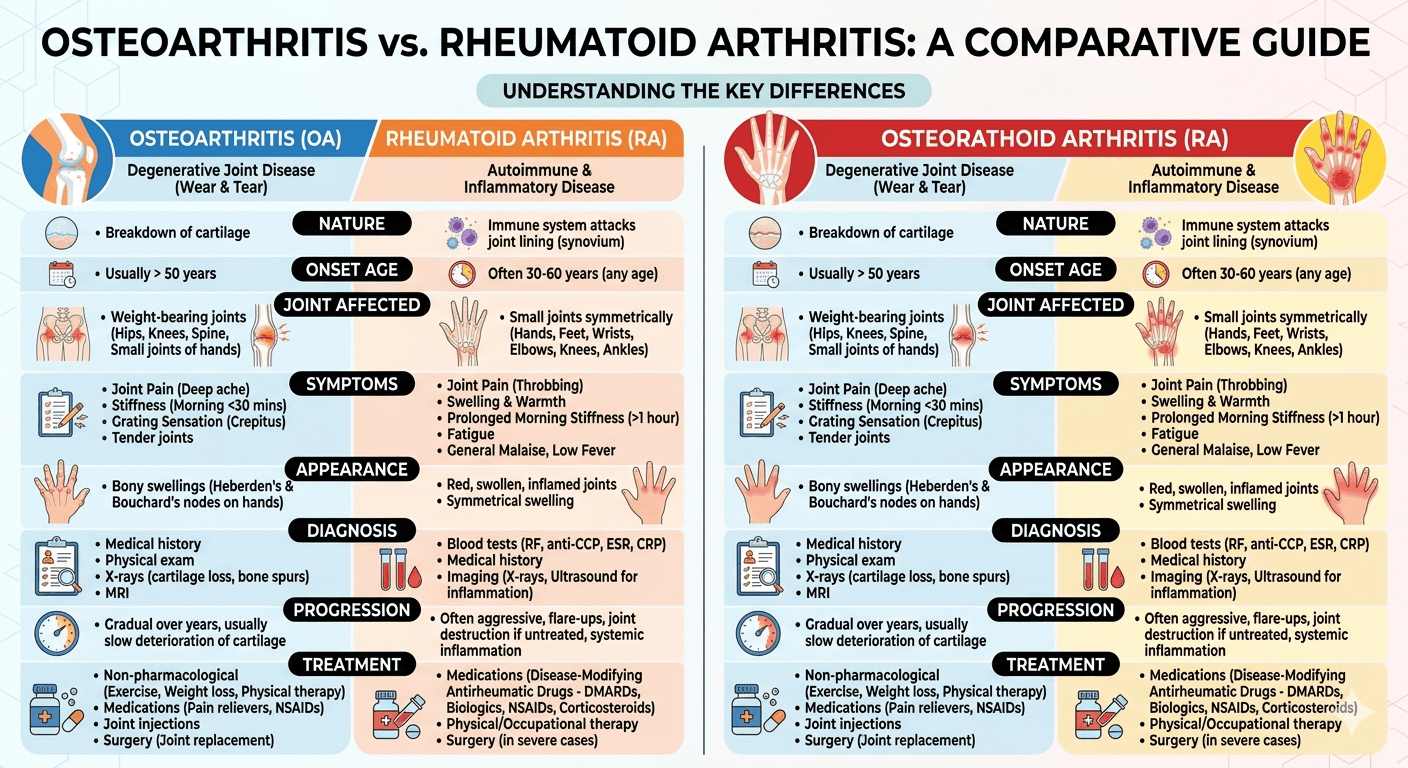
    Benign Prostatic Hyperplasia (BPH): Warning Signs You Should Never Ignore

    Benign Prostatic Hyperplasia

    And Effective Treatments That Can Improve Your Life

    Introduction

    Benign Prostatic Hyperplasia (BPH), commonly referred to as an enlarged prostate, is among the most frequent urinary disorders seen in older men. The condition occurs when the prostate gland gradually increases in size and compresses the urethra, leading to lower urinary tract symptoms (LUTS). Although BPH is a non-cancerous condition, it can greatly reduce quality of life and may result in serious complications if not properly managed.

    Learn everything about Benign Prostatic Hyperplasia (BPH), including causes, symptoms, diagnosis, treatment options, lifestyle changes, and prevention tips in this complete guide for patients and caregivers.

    BPH becomes increasingly common after the age of 50. Its progression is usually slow, with symptoms ranging from mild urinary discomfort to severe urinary obstruction. A solid understanding of prostate anatomy, disease mechanisms, clinical features, diagnosis, treatment options, and possible complications is valuable for both patients and medical students.

    Anatomy and Role of the Prostate

    The prostate is a small gland situated beneath the bladder and anterior to the rectum. It surrounds the first segment of the urethra, called the prostatic urethra.

    That’s correct. In healthy young men, the prostate gland typically weighs around 20 grams. Its main role is to produce prostatic secretions that form part of semen and help support sperm survival.

    Prostate Anatomy

    Prostate Zones

    The prostate contains several anatomical regions:

    • Peripheral Zone: Most prostate cancers develop here.
    • Transitional Zone: The primary site where BPH occurs.
    • Central Zone: Encircles the ejaculatory ducts.
    • Anterior Fibromuscular Stroma: Mainly composed of muscle tissue.

    With aging, both stromal and epithelial cells in the transitional zone undergo hyperplasia, causing prostate enlargement.

    What Is BPH?

    https://www.youtube.com/watch?v=WfOTIf8QEm0

    Benign Prostatic Hyperplasia refers to the non-malignant increase in the number of smooth muscle and epithelial cells within the prostate gland. This enlargement may partially obstruct urinary flow.

    The term includes:

    • Benign: Non-cancerous
    • Hyperplasia: Increase in cell quantity

    Although BPH is different from prostate cancer, both diseases can exist together in elderly men.

    Epidemiology

    BPH is closely linked to advancing age.

    Age Distribution

    • Rare before 40 years of age
    • Frequently develops after age 50
    • Seen in nearly half of men by age 60
    • Histological evidence is present in 80–90% of men by age 80

    Not every man with prostate enlargement develops symptoms.

    Risk Factors

    Several factors contribute to the development of BPH:

    1. Aging

    The most significant contributing factor.

    2. Hormonal Influences

    Testosterone is converted into dihydrotestosterone (DHT) by the enzyme 5-alpha reductase. DHT promotes prostate growth.

    3. Family History

    Men with affected family members may experience earlier onset.

    4. Obesity and Metabolic Syndrome

    Conditions such as diabetes, hypertension, obesity, and abnormal lipid levels are associated with higher risk.

    5. Lack of Physical Activity

    Sedentary habits may worsen disease progression.

    6. Cardiovascular Disease

    Impaired vascular health may contribute to prostate enlargement.

    Pathophysiology

    BPH develops through both mechanical and functional mechanisms.

    Static Component

    An enlarged prostate physically compresses the urethra, increasing resistance to urinary flow.

    Dynamic Component

    Increased smooth muscle tone mediated by alpha-adrenergic receptors further narrows the urethra.

    Bladder Adaptations

    Initially, the bladder compensates by increasing muscular contractions. Over time, chronic obstruction may lead to:

    • Bladder wall thickening
    • Reduced compliance
    • Urinary retention
    • Detrusor dysfunction

    Clinical Symptoms of BPH

    The symptoms are collectively known as Lower Urinary Tract Symptoms (LUTS) and are categorized as obstructive or irritative.

    Obstructive Symptoms

    • Weak urinary stream
    • Difficulty initiating urination
    • Intermittent urine flow
    • Straining during urination
    • Feeling of incomplete bladder emptying
    • Post-void dribbling
    • Urinary retention

    Irritative Symptoms

    • Increased urinary frequency
    • Urgency
    • Nocturia
    • Urge incontinence

    International Prostate Symptom Score (IPSS)

    The IPSS is commonly used to evaluate symptom severity.

    Severity Levels

    • Mild: 0–7
    • Moderate: 8–19
    • Severe: 20–35

    This scoring system helps determine appropriate management.

    Complications of Untreated BPH

    If left untreated, BPH can result in significant complications:

    1. Acute urinary retention
    2. Chronic urinary retention
    3. Recurrent urinary tract infections
    4. Bladder stone formation
    5. Hematuria
    6. Hydronephrosis
    7. Renal impairment or failure

    Diagnosis

    Diagnosis combines history-taking, physical examination, laboratory testing, and imaging.

    Clinical History

    Important information includes:

    • Severity and duration of symptoms
    • Frequency of Nocturia
    • Episodes of urinary retention
    • Presence of hematuria
    • Medication use
    • Neurological symptoms

    Physical Examination

    General Examination

    Assessment of hydration, neurological status, and bladder distension.

    Abdominal Examination

    A palpable bladder may indicate retention.

    Digital Rectal Examination (DRE)

    Typical BPH findings include:

    • Enlarged gland
    • Smooth surface
    • Firm consistency
    • Non-tender prostate

    Hard nodules may raise suspicion for prostate cancer.

    Laboratory and Imaging Studies

    Urinalysis

    CBC,

    Lipid Profile

    RBS, HbA1c

    Serum Creatinine

    Assesses kidney function Test (KFT)

    Prostate-Specific Antigen (PSA)

    PSA levels may rise in:

    • BPH
    • Prostatitis
    • Prostate cancer

    Imaging and Additional Tests

    • Ultrasound for prostate size and residual urine
    • Post-void residual volume measurement
    • Uroflowmetry to assess urinary flow
    • Cystoscopy in selected patients

    Differential Diagnosis

    Conditions resembling BPH include:

    • Prostate cancer
    • Urinary tract infection
    • Bladder cancer
    • Urethral stricture
    • Neurogenic bladder
    • Overactive bladder
    • Chronic prostatitis

    Management of BPH

    Conservative Measures

    Mild symptoms may improve with lifestyle adjustments.

    Lifestyle Recommendations

    • Reduce evening fluid intake
    • Limit caffeine and alcohol
    • Practice scheduled voiding
    • Avoid decongestants
    • Maintain healthy weight and exercise regularly

    Medical Therapy

    Alpha-1 Adrenergic Blockers

    Examples:

    • Tamsulosin
    • Alfuzosin
    • Silodosin
    • Doxazosin

    These medications relax smooth muscle in the prostate and bladder neck, improving urinary flow.

    Common Side Effects

    • Dizziness
    • Orthostatic hypotension
    • Retrograde ejaculation

    5-Alpha Reductase Inhibitors

    Examples:

    • Finasteride
    • Dutasteride

    These drugs reduce DHT production and gradually shrink prostate size.

    Benefits

    • Lower risk of retention
    • Reduced need for surgery

    Side Effects

    • Reduced libido
    • Erectile dysfunction
    • Lower ejaculate volume

    Most effective in men with larger prostates.

    Combination Therapy

    Alpha blockers and 5-alpha reductase inhibitors may be combined for moderate to severe symptoms.

    Phosphodiesterase-5 Inhibitors

    Tadalafil may help men with both BPH and erectile dysfunction.

    Surgical Treatment

    Surgery is considered when:

    • Medications fail
    • Recurrent urinary retention occurs
    • Kidney function declines
    • Recurrent infections develop
    • Bladder stones are present

    Common Procedures

    Transurethral Resection of the Prostate (TURP)

    Considered the traditional gold-standard surgery.

    Advantages

    • Excellent symptom relief
    • Improved urinary flow

    Possible Complications

    • Bleeding
    • Infection
    • Retrograde ejaculation
    • Urethral stricture
    • TUR syndrome

    Laser Procedures

    Examples:

    • HoLEP
    • GreenLight laser therapy

    Benefits include less bleeding and shorter hospitalization.

    Open or Simple Prostatectomy

    Reserved for extremely enlarged prostates.

    Minimally Invasive Therapies

    Examples:

    • UroLift
    • Rezūm therapy
    • Prostatic artery embolization

    These may better preserve sexual function in selected patients.

    Acute Urinary Retention

    This is a medical emergency.

    Symptoms

    • Severe lower abdominal pain
    • Inability to urinate
    • Distended bladder

    Treatment

    • Immediate catheterization
    • Alpha blocker therapy
    • Further evaluation for definitive management

    BPH Versus Prostate Cancer

    FeatureBPHProstate Cancer
    NatureBenignMalignant
    Main LocationTransitional zonePeripheral zone
    DRE FindingsSmooth enlargementHard nodules
    MetastasisAbsentPossible

    Prevention and Risk Reduction

    Although BPH cannot always be prevented, healthy habits may slow progression.

    Helpful Measures

    • Maintain ideal body weight
    • Exercise routinely
    • Control diabetes and hypertension
    • Eat fruits and vegetables
    • Avoid smoking
    • Limit alcohol use

    Prognosis

    The outlook for BPH is generally excellent with appropriate treatment. Many men experience major improvement in urinary symptoms and quality of life through medications or surgery. Delayed care, however, can increase the risk of bladder and kidney complications.

    Important Facts

    • BPH mainly affects the transitional zone.
    • DHT has a central role in disease development.
    • Symptoms are divided into obstructive and irritative LUTS.
    • DRE and IPSS are important assessment tools.
    • PSA is useful but not specific for cancer.
    • Alpha blockers provide rapid relief.
    • 5-alpha reductase inhibitors shrink prostate tissue.
    • TURP remains a major surgical option.
    • Acute urinary retention is an emergency.

    When to Seek Medical Care

    Patients should seek evaluation if they experience:

    • Difficulty urinating
    • Weak stream
    • Blood in urine
    • Frequent nighttime urination
    • Painful urination
    • Sudden inability to urinate
    • Recurrent urinary infections

    Early treatment helps prevent complications and improves long-term outcomes.

    Conclusion

    Benign Prostatic Hyperplasia is a very common condition in aging men and a major cause of lower urinary tract symptoms. While non-cancerous, it can substantially affect daily living and may lead to urinary retention, infections, bladder dysfunction, and kidney damage if untreated.

    Modern advances in diagnosis and therapy have significantly improved outcomes. Treatment options range from lifestyle modifications and medications to minimally invasive procedures and surgery. A thorough understanding of BPH is important for both patients and medical trainees.

    With proper medical management and regular follow-up, most men with BPH can achieve effective symptom control and maintain a high quality of life.

    Disclaimer: The information provided in this blog post is for educational and informational purposes only and should not be considered medical advice. It is not intended to replace professional medical consultation, diagnosis, or treatment. Always seek the guidance of a qualified healthcare professional regarding any medical condition or health-related concerns. The author and publisher are not responsible for any actions taken based on the information presented in this article.
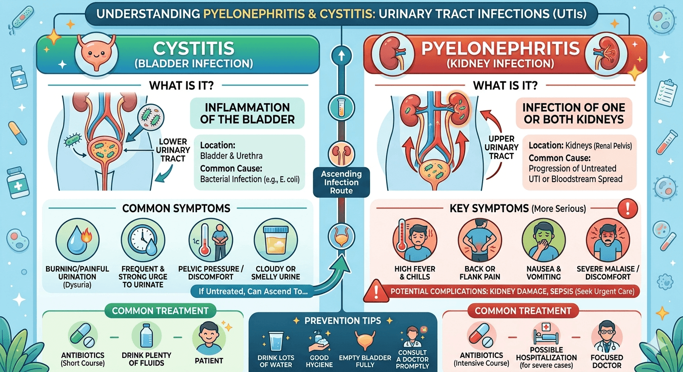
    Hidden Causes of Chronic Abdominal Pain

    Hidden Causes of Chronic Abdominal Pain

    And Digestive Problems

    Introduction

    Chronic abdominal pain and ongoing digestive problems are among the leading causes of medical consultations worldwide. Many people experience symptoms such as bloating, stomach discomfort, constipation, diarrhea, nausea, indigestion, or unexplained weight loss for months or even years without identifying the true cause. While some conditions are mild and manageable, others may indicate serious disorders involving the digestive tract, liver, pancreas, kidneys, immune system, hormones, or even cancer.

    Discover the hidden causes of chronic abdominal pain and digestive problems, including IBS, ulcers, gallbladder disease, food intolerances, pancreatitis, and more. Learn warning signs, diagnosis, and treatment options.

    For patients, learning about the hidden causes of persistent digestive symptoms can encourage earlier medical evaluation and treatment. For medical students, chronic abdominal pain is an important clinical topic that requires to be detailed history-taking, physical examination, and a structured diagnostic approach.

    This article explores the lesser-known causes of chronic abdominal pain and digestive disorders, along with their symptoms, diagnostic methods, treatment options, and warning signs that should not be overlooked.

    What Is Chronic Abdominal Pain?

    Chronic abdominal pain generally refers to pain that continues for at least three months. It may occur constantly or come and go, affecting different regions of the abdomen.

    The abdominal cavity contains several important organs, including:

    • Stomach
    • Small intestine
    • Large intestine (colon)
    • Liver
    • Gallbladder
    • Pancreas
    • Kidneys
    • Urinary tract
    • Blood vessels
    • Reproductive organs

    Because these organs share nerve pathways, pinpointing the exact source of abdominal pain can sometimes be difficult.

    Common digestive symptoms associated with chronic abdominal pain include:

    • Bloating
    • Excessive gas
    • Nausea
    • Vomiting
    • Heartburn
    • Diarrhea
    • Constipation
    • Reduced appetite
    • Weight loss
    • Feeling full quickly
    • Fatigue
    • Blood in stool
    • Difficulty swallowing

    Persistent digestive complaints should always be properly evaluated, as they may indicate structural, inflammatory, infectious, metabolic, or functional disorders.

    Functional Gastrointestinal Disorders

    One of the most frequent hidden causes of chronic abdominal pain is functional gastrointestinal disorders, where symptoms are present even though no clear structural abnormality is found.

    Irritable Bowel Syndrome (IBS)

    Irritable Bowel Syndrome is a common condition marked by:

    • Recurrent abdominal discomfort
    • Bloating
    • Changes in bowel habits
    • Constipation
    • Diarrhea
    • Mucus in stool

    Symptoms often become worse with stress, specific foods, or hormonal fluctuations.

    Types of IBS

    • IBS with constipation (IBS-C)
    • IBS with diarrhea (IBS-D)
    • Mixed IBS (IBS-M)

    Possible Causes

    • Abnormal intestinal movement
    • Increased gut sensitivity
    • Dysfunction of the gut-brain connection
    • Altered gut bacteria
    • Psychological stress

    Diagnosis and Management

    IBS is usually diagnosed after ruling out more serious conditions. Treatment may involve:

    • Dietary adjustments
    • Stress management
    • Fiber supplements
    • Probiotics
    • Antispasmodic medications
    • Psychological therapy when appropriate

    Food Intolerances and Hidden Dietary Triggers

    Many individuals with chronic digestive symptoms unknowingly suffer from food intolerances.

    Lactose Intolerance

    Lactose intolerance develops when the body does not produce enough lactase enzyme to digest dairy products.

    Symptoms

    • Bloating
    • Gas
    • Abdominal cramping
    • Diarrhea
    • Nausea

    Symptoms typically occur after consuming milk or dairy products.

    Gluten Sensitivity and Celiac Disease

    Celiac disease is an autoimmune disorder triggered by gluten consumption.

    Symptoms

    • Persistent diarrhea
    • Weight loss
    • Iron deficiency anemia
    • Abdominal pain
    • Bloating
    • Fatigue
    • Malnutrition

    If untreated, celiac disease can damage the small intestine and lead to long-term complications.

    Diagnosis

    • Tissue transglutaminase antibody testing
    • Endoscopy with intestinal biopsy

    Treatment

    A lifelong gluten-free diet is essential.

    Fructose and FODMAP Intolerance

    Certain carbohydrates can ferment in the intestines and trigger:

    • Bloating
    • Gas
    • Diarrhea
    • Cramping

    Low-FODMAP diets may benefit selected patients.

    Inflammatory Bowel Disease (IBD)

    Inflammatory Bowel Disease includes:

    • Crohn’s disease
    • Ulcerative colitis

    These conditions involve chronic inflammation of the gastrointestinal tract.

    Common Symptoms

    • Persistent abdominal pain
    • Chronic diarrhea
    • Blood in stool
    • Weight loss
    • Fatigue
    • Fever
    • Malnutrition

    Crohn’s Disease

    Crohn’s disease can affect any portion of the digestive tract from mouth to anus.

    Possible Complications

    • Intestinal narrowing
    • Fistulas
    • Abscesses
    • Malabsorption

    Ulcerative Colitis

    Ulcerative colitis mainly affects the colon and rectum.

    Symptoms

    • Bloody diarrhea
    • Urgent bowel movements
    • Rectal bleeding
    • Abdominal cramps

    Diagnosis and Treatment

    Diagnosis may include colonoscopy, biopsy, imaging, and stool tests. Treatment options include:

    • Anti-inflammatory medications
    • Immunosuppressants
    • Biologic therapies
    • Surgery in severe cases

    Chronic Gastritis and Peptic Ulcers

    Chronic Gastritis

    Gastritis refers to inflammation of the stomach lining and may result from:

    • Helicobacter pylori infection
    • Long-term NSAID use
    • Alcohol consumption
    • Autoimmune disease
    • Stress-related injury

    Symptoms

    • Upper abdominal discomfort
    • Nausea
    • Bloating
    • Indigestion
    • Early fullness

    Peptic Ulcer Disease

    Peptic ulcers develop in the stomach or duodenum.

    Warning Signs

    • Burning stomach pain
    • Vomiting blood
    • Black stools
    • Anemia
    • Weight loss

    Diagnosis and Treatment

    Diagnosis often involves endoscopy and H. pylori testing. Treatment may include:

    • Proton pump inhibitors
    • Antibiotics
    • Avoiding NSAIDs

    Gallbladder Disorders

    Gallbladder problems are often mistaken for indigestion or acidity.

    Gallstones

    Gallstones can temporarily block bile flow.

    Symptoms

    • Right upper abdominal pain
    • Pain after fatty meals
    • Nausea
    • Vomiting
    • Bloating

    Pain may spread to the back or right shoulder.

    Chronic Cholecystitis

    Repeated gallbladder inflammation can lead to ongoing digestive discomfort.

    Diagnosis and Treatment

    Doctors may use ultrasound, liver function tests, or CT scans. Treatment often involves dietary modifications or gallbladder removal surgery.

    Pancreatic Disorders

    The pancreas plays an essential role in digestion and blood sugar regulation.

    Chronic Pancreatitis

    This condition causes long-term pancreatic inflammation and scarring.

    Causes

    • Alcohol use
    • Gallstones
    • Genetic disorders
    • Autoimmune diseases
    • High triglyceride levels

    Symptoms

    • Upper abdominal pain radiating to the back
    • Fatty stools
    • Weight loss
    • Malabsorption
    • Diabetes

    Complications

    • Pancreatic insufficiency
    • Nutritional deficiencies
    • Chronic pain

    Pancreatic Cancer

    Pancreatic cancer may initially present with subtle digestive symptoms.

    Warning Signs

    • Persistent abdominal pain
    • Unexplained weight loss
    • Jaundice
    • Appetite loss
    • Newly diagnosed diabetes

    Liver Diseases and Digestive Symptoms

    Several chronic liver disorders can contribute to digestive complaints.

    Fatty Liver Disease

    Nonalcoholic fatty liver disease is increasingly common due to obesity and diabetes.

    Symptoms

    • Right upper abdominal discomfort
    • Fatigue
    • Weakness
    • Mild digestive symptoms

    Chronic Hepatitis

    Chronic liver inflammation may cause:

    • Abdominal pain
    • Nausea
    • Appetite loss
    • Jaundice
    • Enlarged liver

    Cirrhosis

    Advanced liver disease may lead to:

    • Fluid accumulation in the abdomen
    • Digestive problems
    • Gastrointestinal bleeding
    • Malnutrition

    Small Intestinal Bacterial Overgrowth (SIBO)

    SIBO occurs when excessive bacteria grow in the small intestine.

    Symptoms

    • Bloating
    • Gas
    • Diarrhea
    • Abdominal discomfort
    • Malabsorption
    • Vitamin deficiencies

    Risk Factors

    • Diabetes
    • Intestinal surgery
    • Motility disorders
    • Chronic pancreatitis

    Treatment

    Management may include antibiotics, nutritional support, and treatment of underlying conditions.

    Chronic Infections

    Some infections can produce long-term digestive symptoms.

    Parasitic Infections

    Possible symptoms include:

    • Chronic diarrhea
    • Weight loss
    • Abdominal cramps
    • Malnutrition

    Abdominal Tuberculosis

    Tuberculosis may affect the intestines or abdominal lymph nodes.

    Symptoms

    • Chronic abdominal pain
    • Fever
    • Weight loss
    • Altered bowel habits

    Gynecological Causes in Women

    Certain reproductive disorders may mimic gastrointestinal disease.

    Endometriosis

    This condition occurs when tissue similar to the uterine lining grows outside the uterus.

    Symptoms

    • Chronic pelvic pain
    • Painful periods
    • Pain during intercourse
    • Bloating
    • Bowel-related symptoms

    Ovarian Disorders

    Ovarian cysts or tumors may cause:

    • Abdominal fullness
    • Pelvic pain
    • Digestive discomfort
    • Early satiety

    Kidney and Urinary Tract Causes

    Kidney disorders may also present as abdominal discomfort.

    Kidney Stones

    Symptoms may include:

    • Flank pain
    • Lower abdominal pain
    • Nausea
    • Vomiting
    • Blood in urine

    Chronic Kidney Disease

    Chronic kidney disease may lead to:

    • Nausea
    • Poor appetite
    • Vomiting
    • Fatigue
    • Abdominal discomfort

    Hormonal and Metabolic Causes

    Diabetes Mellitus

    Diabetes can damage the nerves controlling digestion.

    Diabetic Gastroparesis

    This condition slows stomach emptying and may cause:

    • Nausea
    • Vomiting
    • Bloating
    • Early fullness
    • Abdominal discomfort

    Thyroid Disorders

    • Hypothyroidism may lead to constipation and bloating
    • Hyperthyroidism may cause diarrhea

    Psychological Factors and the Gut-Brain Connection

    Mental health significantly affects digestive function.

    Conditions linked to digestive symptoms include:

    • Anxiety
    • Depression
    • Chronic stress
    • Panic disorders

    Stress may worsen:

    • IBS
    • Acid reflux
    • Functional dyspepsia
    • Appetite changes

    The gut-brain axis plays a major role in chronic digestive symptoms.

    Gastrointestinal Cancers

    Persistent digestive complaints should never be ignored because some cancers develop gradually.

    Colorectal Cancer

    Possible symptoms include:

    • Changes in bowel habits
    • Blood in stool
    • Weight loss
    • Anemia
    • Chronic abdominal pain

    Stomach Cancer

    Early symptoms may include:

    • Indigestion
    • Feeling full quickly
    • Weight loss
    • Nausea

    Ovarian Cancer

    Ovarian cancer may also present with bloating and abdominal discomfort.

    Warning Signs Requiring Immediate Medical Attention

    The following symptoms may indicate serious disease:

    • Blood in stool
    • Vomiting blood
    • Black tarry stools
    • Significant weight loss
    • Persistent vomiting
    • Difficulty swallowing
    • Ongoing fever
    • Night sweats
    • Severe anemia
    • Jaundice
    • New digestive symptoms in older adults
    • Family history of gastrointestinal cancer

    Prompt medical evaluation is essential when these symptoms occur.

    Diagnostic Evaluation

    A thorough and systematic assessment is necessary.

    Medical History

    Doctors commonly ask about:

    • Pain location and duration
    • Relationship to meals
    • Bowel habits
    • Weight changes
    • Medication use
    • Diet
    • Family history
    • Psychological stress

    Physical Examination

    The abdomen is checked for:

    • Tenderness
    • Masses
    • Enlarged organs
    • Fluid buildup
    • Signs of malnutrition

    Laboratory Tests

    Common investigations include:

    • Complete blood count
    • Liver function tests
    • Kidney function tests
    • Blood sugar testing
    • Inflammatory markers
    • Thyroid function tests
    • Stool analysis
    • Urine analysis

    Imaging and Procedures

    Depending on symptoms, tests may include:

    • Ultrasound
    • CT scan
    • MRI
    • Endoscopy
    • Colonoscopy
    • Breath testing
    • Biopsy

    Treatment Approaches

    Treatment depends on the underlying condition.

    Lifestyle and Dietary Changes

    • Avoid trigger foods
    • Eat balanced meals
    • Stay hydrated
    • Reduce alcohol intake
    • Quit smoking
    • Exercise regularly
    • Manage stress

    Medications

    Treatment may involve:

    • Acid-reducing drugs
    • Antibiotics
    • Anti-inflammatory medications
    • Probiotics
    • Antispasmodics
    • Digestive enzyme supplements
    • Immunosuppressants

    Surgery

    Some conditions may require surgery, including:

    • Gallstones
    • Certain cancers
    • Severe inflammatory bowel disease
    • Intestinal obstruction

    Prevention and Long-Term Digestive Health

    Healthy habits can reduce the risk of chronic digestive disorders.

    Preventive Measures

    • Eat fiber-rich foods
    • Limit processed foods
    • Maintain a healthy weight
    • Exercise consistently
    • Avoid unnecessary NSAID use
    • Limit alcohol intake
    • Practice food safety
    • Undergo recommended cancer screenings

    Seeking medical care early often helps prevent complications.

    Conclusion

    Chronic abdominal pain and digestive problems can arise from many hidden causes, including functional disorders, food intolerances, inflammatory diseases, infections, metabolic conditions, and cancers. Since symptoms often overlap, accurate diagnosis requires careful evaluation and systematic clinical reasoning.

    Persistent digestive symptoms should never be ignored, especially when accompanied by weight loss, bleeding, anemia, fever, or severe pain. Early diagnosis and treatment improve outcomes and help prevent complications.

    For medical students, chronic abdominal pain demonstrates the importance of detailed history-taking, broad clinical thinking, and understanding the complex interactions between digestive, metabolic, infectious, hormonal, and psychological factors.

    Timely medical evaluation, healthy lifestyle choices, and evidence-based care remain essential for maintaining digestive health and improving quality of life.

    Hidden Signs of Gastrointestinal Bleeding

    Gastrointestinal bleeding

    You Should Never Ignore

    Introduction

    Gastrointestinal (GI) bleeding refers to bleeding that occurs anywhere within the digestive system, extending from the mouth to the rectum. The severity can vary widely, ranging from mild, slow blood loss to severe and life-threatening hemorrhage. While some individuals experience obvious symptoms such as vomiting blood or black stools, others develop subtle signs that often go unnoticed until serious complications arise.

    This article discusses the concealed symptoms, causes, risk factors, diagnostic approaches, treatment methods, and prevention of gastrointestinal bleeding.

    Identifying these less obvious symptoms early is essential for patients and healthcare students alike. Prompt diagnosis can reduce the risk of complications including severe anemia, shock, organ damage, and even death.

    Understanding Gastrointestinal Bleeding

    GI bleeding is not considered a disease on its own; instead, it is a symptom of another disorder affecting the digestive tract.

    The digestive system includes:

    • Mouth
    • Esophagus
    • Stomach
    • Small intestine
    • Large intestine (colon)
    • Rectum
    • Anus

    Types of GI Bleeding

    Upper Gastrointestinal Bleeding

    Occurs above the ligament of Treitz and commonly affects:

    • Esophagus
    • Stomach
    • Duodenum

    Lower Gastrointestinal Bleeding

    Occurs below the ligament of Treitz and may involve:

    • Small intestine
    • Colon
    • Rectum
    • Anus

    Bleeding may also be categorized as:

    • Acute: sudden and severe
    • Chronic: slow and ongoing
    • Occult: hidden blood not visible in stool

    Why GI Bleeding Is Serious

    Many people believe gastrointestinal bleeding always produces dramatic symptoms. However, slow chronic bleeding can continue unnoticed for weeks or even months.

    Unrecognized blood loss may lead to:

    • Iron deficiency anemia
    • Persistent fatigue
    • Reduced oxygen supply to tissues
    • Increased strain on the heart
    • Dizziness and falls
    • Circulatory instability
    • Hypovolemic shock in severe cases

    In some situations, GI bleeding may be the earliest sign of conditions such as stomach ulcers, inflammatory bowel disease, or gastrointestinal cancers.

    Hidden Signs of Gastrointestinal Bleeding

    1. Persistent Fatigue and Weakness

    One of the most common but overlooked signs of chronic GI bleeding is ongoing fatigue.

    Gradual blood loss lowers hemoglobin levels, reducing oxygen delivery throughout the body. Patients may experience:

    • Constant tiredness
    • Lack of energy
    • Difficulty focusing
    • Reduced ability to exercise
    • General weakness

    Iron deficiency anemia in adults without a clear explanation should raise concern for occult GI bleeding.

    2. Pale Skin and Pale Eyelids

    Long-term blood loss may cause anemia, resulting in:

    • Pale skin
    • Pale nail beds
    • Pale inner eyelids (conjunctival pallor)

    Because this develops slowly, many patients fail to notice it.

    3. Shortness of Breath With Mild Activity

    Lower hemoglobin levels reduce the blood’s oxygen-carrying ability.

    Symptoms may include:

    • Breathlessness while walking
    • Difficulty climbing stairs
    • Fast heartbeat during exertion
    • Poor stamina

    Severe anemia may even trigger chest discomfort in elderly patients or those with heart disease.

    4. Dizziness or Fainting

    Blood loss decreases circulating blood volume and oxygen supply.

    Patients may develop:

    • Lightheadedness
    • Dizziness when standing
    • Near-fainting episodes
    • Syncope (fainting)

    Dizziness accompanied by black stools or vomiting blood requires urgent medical attention.

    5. Black, Tarry Stools (Melena)

    Melena is a classic symptom of upper GI bleeding.

    The stool may appear:

    • Black
    • Sticky
    • Tarry
    • Foul-smelling

    This occurs because blood becomes digested while traveling through the digestive tract.

    6. Hidden Blood in Stool

    Occult bleeding involves blood that is not visible to the naked eye.

    Patients may have:

    • Normal-looking stool
    • Chronic anemia
    • Fatigue without obvious bleeding

    Tests such as FOBT or FIT can identify hidden blood in stool.

    Possible causes include:

    • Colon polyps
    • Colorectal cancer
    • Peptic ulcers
    • Inflammatory bowel disease
    • Angiodysplasia

    7. Vomiting Blood (Hematemesis)

    Hematemesis is a dangerous sign of upper GI bleeding.

    Vomited material may appear:

    • Bright red
    • Dark red
    • Similar to coffee grounds

    Coffee-ground vomit usually indicates partially digested blood.

    Common causes include:

    • Peptic ulcer disease
    • Esophageal varices
    • Gastritis
    • Mallory-Weiss tears

    Immediate medical care is necessary.

    8. Abdominal Pain or Discomfort

    Some bleeding disorders also cause abdominal symptoms such as:

    • Burning upper abdominal pain
    • Cramping
    • Bloating
    • Tenderness
    • Persistent discomfort after eating

    These symptoms may suggest:

    9. Unexplained Weight Loss

    Weight loss combined with GI bleeding may indicate a serious underlying illness.

    Potential causes include:

    • Stomach cancer
    • Colorectal cancer
    • Chronic inflammatory bowel disease
    • Malabsorption disorders

    10. Changes in Bowel Habits

    Lower GI bleeding may occur alongside altered bowel patterns.

    Warning signs include:

    • Persistent constipation
    • Chronic diarrhea
    • Narrow stools
    • Increased urgency
    • Feeling of incomplete bowel emptying

    These symptoms may occur in:

    • Colorectal cancer
    • Inflammatory bowel disease
    • Diverticular disease
    • Hemorrhoids

    11. Rapid Heartbeat or Palpitations

    To compensate for blood loss, the body increases heart rate.

    Patients may notice:

    • Heart palpitations
    • Racing pulse
    • Anxiety-like feelings
    • Weak pulse

    Severe blood loss may progress to shock.

    12. Rectal Bleeding

    Visible rectal bleeding may appear:

    • Bright red
    • Maroon-colored
    • Mixed with stool
    • On toilet paper

    Although hemorrhoids are common, rectal bleeding should always be evaluated.

    Common Causes of GI Bleeding

    Frequent causes include:

    • Peptic ulcer disease
    • Gastritis
    • Esophageal varices
    • Hemorrhoids
    • Diverticular disease
    • Inflammatory bowel disease
    • Gastrointestinal cancers
    • Angiodysplasia

    Risk Factors

    Factors that increase risk include:

    • NSAID use
    • Aspirin and blood thinners
    • Liver disease
    • Heavy alcohol use
    • Smoking
    • Older age
    • Previous GI disorders

    Diagnosis

    Doctors may use:

    • Medical history and physical examination
    • Complete blood count (CBC)
    • Stool testing (FOBT/FIT)
    • Liver function tests
    • Upper endoscopy (EGD)
    • Colonoscopy
    • Capsule endoscopy
    • CT angiography

    Treatment

    Treatment depends on the source and severity of bleeding.

    Emergency Care

    Severe cases may require:

    • IV fluids
    • Blood transfusion
    • Oxygen therapy
    • Monitoring of vital signs

    Medications

    Common treatments include:

    • Proton pump inhibitors (PPIs)
    • Antibiotics for H. pylori
    • Vasoconstrictor therapy such as octreotide

    Endoscopic Treatment

    Endoscopy may control bleeding through:

    • Injection therapy
    • Thermal coagulation
    • Hemoclip placement
    • Variceal band ligation

    Surgery

    Surgery may be necessary if:

    • Bleeding cannot be controlled
    • Cancer is present
    • Perforation develops
    • Recurrent severe bleeding occurs

    Complications of Untreated GI Bleeding

    Ignoring GI bleeding may lead to:

    • Severe anemia
    • Hypovolemic shock
    • Organ failure
    • Cardiac complications
    • Malnutrition
    • Death

    When Immediate Medical Attention Is Needed

    Seek emergency care for:

    • Vomiting blood
    • Black tarry stools
    • Severe rectal bleeding
    • Fainting
    • Rapid heartbeat
    • Chest pain
    • Severe weakness
    • Shortness of breath
    • Confusion

    Prevention Strategies

    Helpful preventive measures include:

    • Avoiding excessive NSAID use
    • Limiting alcohol intake
    • Quitting smoking
    • Eating a balanced diet rich in fiber and iron
    • Undergoing recommended cancer screening
    • Investigating unexplained anemia promptly

    Important Learning Points

    • GI bleeding can remain hidden and symptom-free
    • Iron deficiency anemia in adults requires investigation
    • Melena usually indicates upper GI bleeding
    • Hematochezia commonly suggests lower GI bleeding
    • Hemodynamic instability signals major blood loss
    • Endoscopy is both diagnostic and therapeutic
    • NSAIDs are a major cause of peptic ulcer bleeding

    Conclusion

    Hidden gastrointestinal bleeding can gradually harm health before severe symptoms become obvious. Fatigue, anemia, dizziness, black stools, unexplained weight loss, and bowel habit changes should never be ignored.

    Early medical assessment can prevent serious complications and improve outcomes. For medical students, understanding the many presentations of GI bleeding is essential for timely diagnosis and treatment.

    Recognizing these warning signs early may save lives, particularly when bleeding is linked to ulcers, inflammatory bowel disease, or gastrointestinal cancers. Anyone experiencing symptoms suggestive of GI bleeding should seek prompt medical evaluation.

    Disclaimer: The information provided in this blog post is for educational and informational purposes only and should not be considered medical advice. It is not intended to replace professional medical consultation, diagnosis, or treatment. Always seek the guidance of a qualified healthcare professional regarding any medical condition or health-related concerns. The author and publisher are not responsible for any actions taken based on the information presented in this article.
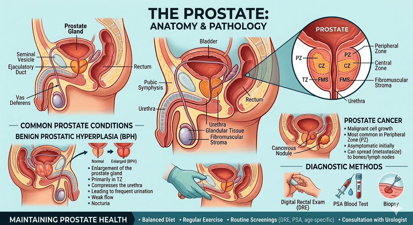
    Hidden Signs of Hypothyroidism

    hypothyroidism

    You Should Never Ignore

    Introduction

    Hypothyroidism is among the most widespread endocrine conditions globally, but many individuals remain unaware they have it because symptoms often appear slowly and can seem unrelated. This disorder develops when the thyroid gland cannot produce sufficient thyroid hormones to support the body’s metabolic needs. The primary hormones involved thyroxine (T4) and triiodothyronine (T3) help regulate metabolism, energy levels, temperature, heart activity, digestion, brain performance, and numerous other body functions.

    This guide explains hidden signs of hypothyroidism, along with its causes, underlying mechanisms, risk factors, diagnostic approaches, complications, and treatment options.

    Since thyroid hormones impact nearly every organ system, hypothyroidism may cause a broad range of subtle symptoms that are frequently mistaken for aging, stress, depression, vitamin deficiencies, or other illnesses. Detecting these warning signs early is crucial because untreated hypothyroidism can eventually result in infertility, heart disease, nerve damage, severe depression, or even Myxedema coma.

    Understanding the Thyroid Gland

    The thyroid is a butterfly-shaped endocrine gland located at the front of the neck beneath the larynx. Its main role is producing two essential hormones:

    • Thyroxine (T4)
    • Triiodothyronine (T3)

    Hormone production is controlled through the hypothalamic-pituitary-thyroid (HPT) axis involving:

    1. The hypothalamus
    2. The pituitary gland
    3. The thyroid gland

    The pituitary releases thyroid-stimulating hormone (TSH), which signals the thyroid to produce T3 and T4. When thyroid hormone levels drop, TSH levels rise in an effort to stimulate the gland.

    What Is Hypothyroidism?

    Hypothyroidism occurs when the body does not produce enough thyroid hormones. It can be divided into several categories:

    Primary Hypothyroidism

    This form originates directly in the thyroid gland. Common causes include:

    • Hashimoto’s thyroiditis
    • Iodine deficiency
    • Thyroid removal surgery
    • Radioactive iodine treatment
    • Certain medications
    • Congenital thyroid abnormalities

    Secondary Hypothyroidism

    This type results from pituitary gland dysfunction, leading to reduced TSH production.

    Tertiary Hypothyroidism

    A rare condition caused by hypothalamic dysfunction.

    How Hypothyroidism Affects the Body

    Thyroid hormones help regulate metabolism and cellular energy production. In hypothyroidism:

    • Metabolism slows down
    • Protein synthesis decreases
    • Heat generation declines
    • Fat metabolism becomes impaired
    • Digestive movement slows
    • Nervous system activity decreases

    Because these processes affect almost every organ system, symptoms can vary widely.

    Hidden Signs of Hypothyroidism

    1. Ongoing Fatigue Despite Rest

    Persistent exhaustion is one of the earliest and most common symptoms. Individuals may:

    • Feel drained after routine tasks
    • Wake up tired even after sleeping well
    • Have low endurance
    • Need frequent daytime naps

          Low thyroid hormone levels reduce energy production at the cellular level.

    2. Unexplained Weight Gain

    Even mild thyroid dysfunction can lead to gradual weight increase. Common complaints include:

    • Trouble losing weight
    • Weight gain despite normal eating habits
    • Puffiness and fluid retention

       A slower metabolism reduces calorie burning, while fluid retention contributes to             swelling

    3. Sensitivity to Cold

    People with hypothyroidism often feel cold more easily than others. Signs include:

    • Cold hands and feet
    • Poor tolerance to cold weather
    • Frequent need for extra layers

          Reduced thyroid hormone lowers heat production in the body.

    4. Dry Skin and Fragile Hair

    Changes involving the skin and hair are frequently overlooked. Symptoms may include:

    • Rough or dry skin
    • Cracked heels
    • Hair thinning or loss
    • Brittle nails
    • Loss of eyebrow hair

     Reduced oil gland activity and impaired protein production contribute to these problems.

    5. Puffy Face and Swelling

    Facial swelling, especially around the eyes, may indicate hypothyroidism. Patients may develop:

    • Puffy eyelids
    • Facial swelling
    • Enlarged tongue
    • Hoarse voice

      Fluid-like substances accumulate in tissues, producing non-pitting swelling called Myxedema

    6. Constipation

    Persistent constipation can be an early sign. Symptoms include:

    • Infrequent bowel movements
    • Hard stools
    • Abdominal bloating

       Thyroid hormone deficiency slows intestinal movement.

    7. Depression and Mood Changes

    Hypothyroidism commonly affects emotional health. Individuals may experience:

    • Depression
    • Irritability
    • Anxiety
    • Lack of motivation
    • Emotional numbness

         Thyroid hormones help regulate neurotransmitters and brain metabolism.

    8. Memory Issues and Brain Fog

    Cognitive symptoms are often missed. Patients may report:

    • Difficulty concentrating
    • Forgetfulness
    • Slower thinking
    • Trouble learning new information

         Reduced brain metabolism and blood flow may impair cognitive performance.

    9. Menstrual and Fertility Problems

    Women with hypothyroidism may develop:

    • Heavy periods
    • Irregular menstrual cycles
    • Difficulty conceiving
    • Recurrent miscarriages

         Thyroid hormones influence ovulation and reproductive hormone balance.

    10. High Cholesterol Levels

    Some individuals discover hypothyroidism during routine cholesterol testing. Findings may include:

    • Elevated LDL cholesterol
    • Increased triglycerides

         Thyroid dysfunction interferes with normal fat metabolism.

    11. Slower Heart Rate

    A low resting pulse may occur, leading to:

    • Reduced exercise tolerance
    • Fatigue during activity
    • Dizziness

       Thyroid hormones normally help maintain heart rate and cardiac output.

    12. Muscle Weakness and Joint Pain

    Musculoskeletal symptoms are common and may include:

    • Muscle cramps
    • Stiffness
    • Joint discomfort
    • Weakness
    • Delayed reflexes

        Abnormal muscle metabolism and tissue swelling contribute to these symptoms.

    13. Hoarseness

    A gradually deepening or hoarse voice may develop due to:

    • Swelling of the vocal cords
    • Enlarged tongue
    • Tissue edema

    14. Carpal Tunnel Syndrome

    Hypothyroidism can place pressure on nerves, causing:

    • Tingling
    • Numbness
    • Wrist pain
    • Weak grip strength

    15. Sleep Problems

    Despite constant fatigue, individuals may still experience:

    • Poor sleep quality
    • Excessive sleepiness
    • Obstructive sleep apnea

            Swelling of tissues and weakened respiratory muscles may contribute.

    16. Reduced Sweating

    Patients may notice:

    • Dry body skin
    • Minimal perspiration
    • Occasional heat intolerance
    • This occurs because gland activity decreases.

    17. Hearing Difficulties

    Untreated hypothyroidism may contribute to:

    • Hearing loss
    • Ear fullness
    • Balance problems

    18. Delayed Reflexes

    One classic clinical sign is slow relaxation of deep tendon reflexes, particularly in the ankle.

    Common Causes of Hypothyroidism

    Hashimoto’s Thyroiditis

    The most common cause in regions with adequate iodine intake. The immune system gradually damages thyroid tissue.

    Iodine Deficiency

    Still a major cause worldwide.

    Thyroid Surgery

    Removing part or all of the thyroid reduces hormone production.

    Radioactive Iodine Therapy

    Often used to treat hyperthyroidism or thyroid cancer.

    Medications

    Some drugs interfere with thyroid function, including:

    • Lithium
    • Amiodarone
    • Interferon-alpha
    • Certain immunotherapy medications

    Congenital Hypothyroidism

    Present from birth due to an underdeveloped or absent thyroid gland.

    Pituitary Disorders

    Pituitary tumors or injury may reduce TSH production.

    Risk Factors

    Higher-risk groups include:

    • Women
    • Adults over 60
    • Individuals with autoimmune disease
    • People with a family history of thyroid disorders
    • Patients with prior thyroid surgery
    • Those exposed to radiation
    • Pregnant or postpartum women
    • Individuals with iodine deficiency

    Diagnosis of Hypothyroidism

    Clinical Examination

    Doctors evaluate symptoms and perform a physical exam. Common findings include:

    • Dry skin
    • Slow heart rate
    • Weight gain
    • Delayed reflexes
    • Puffy face

    Laboratory Tests

    Thyroid Profile (T3, T4, TSH, FREET3, FREET4)

    Thyroid Antibodies

    Anti-TPO antibodies help diagnose Hashimoto’s thyroiditis.

    Additional Evaluations

    Depending on symptoms, physicians may order:

    • Lipid profile
    • Complete blood count
    • Liver tests
    • Electrolyte studies
    • ECG

    Subclinical Hypothyroidism

    This condition involves:

    • Elevated TSH
    • Normal free T4

    Patients may still experience mild symptoms. Treatment decisions depend on TSH levels, age, pregnancy status, symptom severity, and cardiovascular risk.

    Complications of Untreated Hypothyroidism

    Heart Disease

    Potential complications include:

    • High cholesterol
    • Hypertension
    • Heart failure
    • Atherosclerosis

    Infertility

    Hormonal imbalance may impair ovulation.

    Peripheral Neuropathy

    Chronic nerve dysfunction can damage peripheral nerves.

    Mental Health Disorders

    Severe depression and cognitive decline may develop.

    Myxedema Coma

    A rare but life-threatening emergency involving:

    • Severe hypothermia
    • Confusion
    • Respiratory depression
    • Low blood pressure

    Immediate intensive care is required.

    Treatment of Hypothyroidism

    Levothyroxine

    Synthetic T4 is the standard treatment.

    Treatment Goals

    • Normalize TSH levels
    • Relieve symptoms
    • Restore metabolism
    • Prevent complications

    Medication Tips

    Patients should:

    • Take Levothyroxine on an empty stomach
    • Use it at the same time daily
    • Avoid calcium or iron supplements within four hours

    Monitoring

    TSH levels are generally checked:

    • 6–8 weeks after dose changes
    • Every 6–12 months once stable

    Dose requirements may change because of aging, pregnancy, weight changes, or medication interactions.

    Lifestyle and Dietary Considerations

    Adequate Iodine Intake

    Healthy sources include:

    • Iodized salt
    • Dairy products
    • Seafood
    • Eggs

    Excess iodine should also be avoided.

    Selenium and Zinc

    These nutrients support thyroid health. Sources include:

    • Nuts
    • Seeds
    • Legumes
    • Meat

    Exercise

    Regular physical activity can:

    • Improve energy levels
    • Support metabolism
    • Maintain healthy weight
    • Enhance mood

    Stress Management

    Helpful strategies include:

    • Meditation
    • Yoga
    • Proper sleep
    • Counseling when needed

    Hypothyroidism in Special Populations

    Pregnancy

    Untreated hypothyroidism may increase the risk of:

    • Miscarriage
    • Preeclampsia
    • Premature delivery
    • Developmental issues in the baby

    Close monitoring is essential during pregnancy.

    Older Adults

    Symptoms may resemble aging or dementia, including:

    • Memory loss
    • Weakness
    • Depression
    • Falls

    Children and Adolescents

    Hypothyroidism may cause:

    • Growth delays
    • Poor academic performance
    • Delayed puberty

    Early diagnosis is extremely important.

    When to Seek Medical Attention

    Consult a healthcare provider if you experience:

    • Persistent fatigue
    • Weight gain without explanation
    • Cold intolerance
    • Hair loss
    • Constipation
    • Depression
    • Menstrual changes
    • Swelling of the face or neck

    Emergency care is necessary for:

    • Severe lethargy
    • Confusion
    • Difficulty breathing
    • Hypothermia
    • Loss of consciousness

    Key Clinical Points

    • Hypothyroidism affects nearly every organ system.
    • Symptoms often appear slowly and may seem unrelated.
    • Elevated TSH is the hallmark of primary hypothyroidism.
    • Hashimoto’s thyroiditis is the leading cause in developed countries.
    • Delayed reflex relaxation is a classic sign.
    • High cholesterol may result from thyroid dysfunction.
    • Thyroid testing should be considered in unexplained fatigue or depression.
    • Levothyroxine remains the standard therapy.
    • Myxedema coma is a medical emergency.

    Prognosis

    With timely diagnosis and proper treatment, the outlook for hypothyroidism is generally excellent. Most patients experience:

    • Better energy levels
    • Improved mood
    • Normalized metabolism
    • Enhanced quality of life

    Delayed treatment, however, can lead to lasting complications.

    Conclusion

    Hypothyroidism is a common but often overlooked endocrine disorder because its symptoms may develop gradually and appear unrelated. Hidden signs such as fatigue, depression, constipation, hair loss, menstrual irregularities, high cholesterol, memory problems, and sensitivity to cold should not be ignored.

    Early diagnosis and proper management are essential to prevent serious complications and improve overall well-being. With effective treatment, healthy lifestyle habits, and regular monitoring, most individuals with hypothyroidism can live healthy and productive lives.

    Disclaimer: The information provided in this blog post is for educational and informational purposes only and should not be considered medical advice. It is not intended to replace professional medical consultation, diagnosis, or treatment. Always seek the guidance of a qualified healthcare professional regarding any medical condition or health-related concerns. The author and publisher are not responsible for any actions taken based on the information presented in this article.
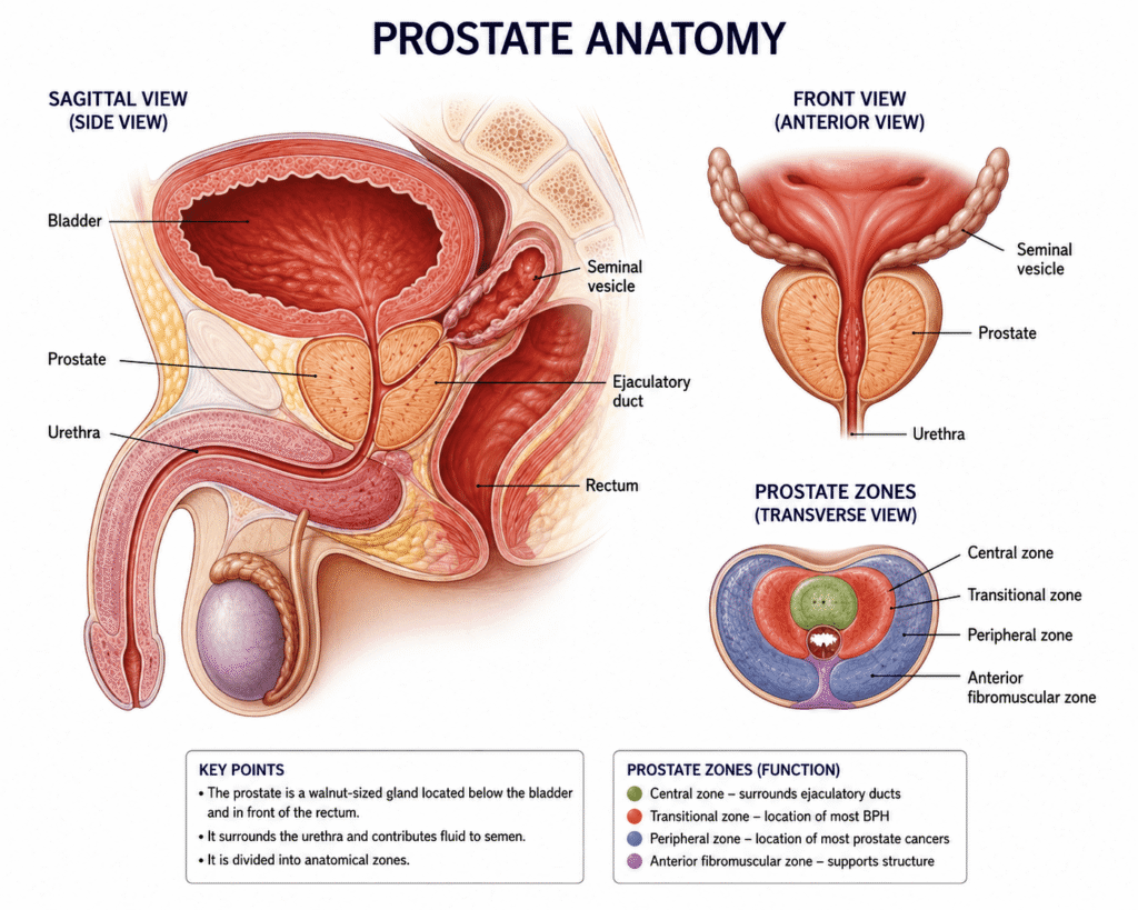
    Kidney and Urinary Disorders in Adults  

    Kidney and urinary disorders

    Early Detection and management

    Introduction

    Kidney and urinary disorders are widespread medical conditions affecting millions of adults globally. The kidneys and urinary tract play a crucial role in eliminating waste, balancing fluids, regulating blood pressure, and maintaining overall health. Damage or disease affecting any part of this system can seriously impact a person’s well-being and daily life.

    Learn the early warning signs, symptoms, causes, diagnosis, and treatment options for kidney and urinary disorders in adults. Discover practical management tips to protect kidney health and prevent serious complications.

    Many kidney and urinary conditions progress gradually and often remain unnoticed during the early stages. Therefore, timely diagnosis and proper treatment are essential to reduce the risk of complications such as chronic kidney disease, kidney failure, repeated infections, and heart-related conditions.

    This article provides an overview of kidney and urinary disorders in adults, discussing their causes, symptoms, risk factors, diagnosis, treatment options, prevention strategies, and long-term management.

    Overview of the Kidney and Urinary System

    The urinary system includes:

    • Kidneys
    • Ureters
    • Bladder
    • Urethra

    Role of the Kidneys

    The kidneys are two bean-shaped organs positioned near the spine. Their primary responsibilities include:

    • Filtering waste from the bloodstream
    • Producing urine
    • Maintaining electrolyte balance
    • Regulating blood pressure
    • Managing body fluid levels
    • Supporting red blood cell production through erythropoietin
    • Activating vitamin D to maintain bone health

    Role of the Urinary Tract

    The urinary tract is responsible for carrying and storing urine before it exits the body.

    • Ureters transport urine from the kidneys to the bladder.
    • The bladder stores urine temporarily.
    • The urethra expels urine from the body.

    Any problem affecting these organs can result in kidney or urinary disorders.

    Common Kidney and Urinary Conditions in Adults

    Chronic Kidney Disease (CKD)

    Chronic kidney disease is a progressive condition where kidney function slowly deteriorates over time.

    Common Causes

    • Diabetes
    • High blood pressure
    • Glomerulonephritis
    • Polycystic kidney disease
    • Recurring urinary tract infections
    • Kidney stones
    • Prolonged use of certain medications

    Symptoms

    • Tiredness
    • Swelling in the legs and feet
    • Reduced appetite
    • Nausea
    • Frequent urination
    • Foamy urine
    • Elevated blood pressure
    • Muscle cramps
    • Shortness of breath

    Acute Kidney Injury (AKI)

    Acute kidney injury refers to a sudden loss of kidney function that develops within hours or days.

    Causes

    • Severe dehydration
    • Sepsis
    • Significant blood loss
    • Certain drugs
    • Urinary blockage
    • Heart failure

    Symptoms

    • Reduced urine production
    • Swelling
    • Confusion
    • Fatigue
    • Nausea and vomiting
    • Chest pain in severe situations

    AKI is considered a medical emergency and requires immediate treatment.

    Urinary Tract Infection (UTI)

    UTIs occur when bacteria enter and infect the urinary tract.

    Types

    • Urethritis
    • Cystitis (bladder infection)
    • Pyelonephritis (kidney infection)

    Symptoms

    • Burning sensation during urination
    • Frequent urge to urinate
    • Cloudy or foul-smelling urine
    • Pelvic discomfort
    • Fever and chills in kidney infections

    Women are more likely to develop UTIs than men.

     Kidney Stones

    Kidney stones are solid mineral deposits that form inside the kidneys.

    Risk Factors

    • Inadequate water intake
    • High-salt diet
    • Obesity
    • Family history
    • Certain metabolic disorders

    Symptoms

    • Intense flank pain
    • Blood in the urine
    • Nausea and vomiting
    • Painful urination
    • Frequent urination

     Glomerulonephritis

    Glomerulonephritis involves inflammation of the glomeruli, the kidney’s filtering structures.

    Causes

    • Autoimmune conditions
    • Infections
    • Vasculitis
    • Diabetes

    Symptoms

    • Blood in urine
    • Protein in urine
    • Swelling
    • High blood pressure
    • Declining kidney function

     Polycystic Kidney Disease (PKD)

    PKD is an inherited disorder marked by multiple fluid-filled cysts in the kidneys.

    Symptoms

    • High blood pressure
    • Back pain
    • Enlarged kidneys
    • Blood in urine
    • Kidney failure in advanced stages

     Urinary Incontinence

    Urinary incontinence is the inability to fully control bladder function.

    Types

    • Stress incontinence
    • Urge incontinence
    • Overflow incontinence
    • Functional incontinence

    Risk Factors

    • Aging
    • Pregnancy and childbirth
    • Obesity
    • Neurological diseases
    • Enlarged prostate in men

     Benign Prostatic Hyperplasia (BPH)

    BPH is a noncancerous enlargement of the prostate gland commonly seen in older men, affecting urine flow.

    Symptoms

    • Weak urine stream
    • Frequent urination
    • Difficulty starting urination
    • Nighttime urination
    • Incomplete bladder emptying

    Risk Factors for Kidney and Urinary Disorders

    Several conditions and lifestyle habits increase the likelihood of developing kidney and urinary diseases.

    Major Risk Factors

    • Diabetes
    • Hypertension
    • Obesity
    • Smoking
    • Aging
    • Family history
    • Poor hydration
    • Excessive salt intake
    • Frequent infections
    • Long-term painkiller use
    • Autoimmune disorders
    • Sedentary lifestyle

    Early Symptoms and Warning Signs

    Many kidney diseases remain silent until they become advanced, making early recognition essential.

    Common Warning Signs

    • Changes in urination
    • Blood in urine
    • Foamy urine
    • Burning sensation while urinating
    • Swelling in the face or legs
    • Persistent fatigue
    • High blood pressure
    • Lower back or flank pain
    • Nausea
    • Difficulty concentrating
    • Unexplained itching

    Persistent symptoms should always be assessed by a healthcare provider.

    Diagnosing Kidney and Urinary Diseases

    Doctors use multiple methods to diagnose urinary and kidney disorders.

    Medical History and Physical Examination

    Healthcare providers may ask about:

    • Symptoms
    • Family medical history
    • Medication use
    • Fluid intake
    • Existing health conditions

    Laboratory Testing

    Blood Tests

    • Serum creatinine
    • Blood urea nitrogen (BUN)
    • Electrolyte levels
    • Estimated glomerular filtration rate (eGFR)

    Urine Tests

    • Urinalysis
    • Urine culture
    • Proteinuria testing
    • Microalbumin testing

    Imaging Tests

    • Ultrasound
    • CT scan
    • MRI
    • Intravenous pyelogram

    Specialized Procedures

    • Cystoscopy
    • Kidney biopsy
    • Urodynamic studies

    Stages of Chronic Kidney Disease

    CKD is categorized into five stages according to eGFR levels.

    • Stage 1: Normal kidney function with signs of kidney damage
    • Stage 2: Mild decline in function
    • Stage 3: Moderate decline
    • Stage 4: Severe reduction in kidney function
    • Stage 5: Kidney failure requiring dialysis or transplantation

    Treatment and Management

    Treatment depends on the type and severity of the condition.

    Lifestyle Changes

    Healthy Eating

    Patients are often encouraged to:

    • Lower salt intake
    • Reduce processed foods
    • Limit protein when necessary
    • Avoid excess sugar
    • Eat more fruits and vegetables
    • Maintain a healthy weight

    Proper Hydration

    Adequate water intake supports kidney health and lowers the risk of infections and stones.

    Physical Activity

    Regular exercise helps manage:

    • Blood pressure
    • Diabetes
    • Weight
    • Heart health

    Smoking Cessation

    Smoking damages blood vessels and accelerates kidney damage.

    Limiting Alcohol

    Excess alcohol can worsen dehydration and blood pressure problems.

    Medical Treatments

    Medications

    Depending on the condition, treatment may include:

    • Antibiotics
    • Blood pressure medications
    • Diuretics
    • Diabetes medications
    • Pain relievers
    • Drugs that reduce protein loss in urine

    ACE inhibitors and ARBs are commonly prescribed in CKD because they help preserve kidney function.

    Kidney Stone Management

    Treatment approaches may involve:

    • Increasing fluid intake
    • Pain control medications
    • Medical expulsive therapy
    • Lithotripsy
    • Ureteroscopy
    • Surgery for severe cases

    Dialysis

    Patients with advanced kidney failure may require dialysis.

    Types of Dialysis

    Hemodialysis

    Blood is filtered using a dialysis machine.

    Peritoneal Dialysis

    The abdominal lining is used to remove waste products.

    Kidney Transplantation

    Eligible patients with end-stage kidney disease may benefit from kidney transplantation, which can improve survival and quality of life.

    Prevention of Kidney and Urinary Diseases

    Many kidney-related conditions can be prevented or delayed through healthy habits.

    Prevention Strategies

    • Control blood sugar levels
    • Maintain healthy blood pressure
    • Stay hydrated
    • Avoid smoking
    • Exercise regularly
    • Reduce salt consumption
    • Limit unnecessary painkiller use
    • Treat infections promptly
    • Maintain a healthy weight
    • Schedule regular medical checkups

    Diet for Kidney Health

    Beneficial Foods

    • Fresh fruits
    • Vegetables
    • Whole grains
    • Healthy fats
    • Lean protein
    • Adequate water intake

    Foods to Limit

    • Processed foods
    • High-sodium foods
    • Sugary beverages
    • Excess red meat
    • Excess phosphorus and potassium in advanced CKD

    Diet plans should be customized based on the stage of kidney disease.

    Possible Complications

    Untreated kidney and urinary disorders may result in:

    • Kidney failure
    • Severe infections
    • Hypertension
    • Cardiovascular disease
    • Electrolyte imbalances
    • Anemia
    • Bone disorders
    • Fluid overload

    Medical attention is necessary if you experience:

    • Blood in urine
    • Persistent swelling
    • Severe flank pain
    • Fever with urinary symptoms
    • Reduced urine output
    • Difficulty urinating
    • Ongoing high blood pressure
    • Recurrent UTIs
    • Sudden fluid-related weight gain

    Emergency care is needed for symptoms such as chest pain, confusion, inability to urinate, or severe infection.

    Prognosis

    The outcome depends on:

    • Type of disease
    • Stage at diagnosis
    • Underlying health conditions
    • Adherence to treatment
    • Lifestyle modifications

    Early diagnosis and proper management can slow disease progression and improve long-term health outcomes.

    Emotional and Mental Health Impact

    Living with kidney disease may affect mental well-being. Patients may experience:

    • Anxiety
    • Depression
    • Stress
    • Sleep disturbances
    • Fear of dialysis or transplantation

    Support from family members, healthcare professionals, counselors, and support groups can be highly beneficial.

    Importance of Regular Screening

    Individuals at higher risk should undergo routine kidney evaluations.

    High-Risk Groups

    • People with diabetes
    • Individuals with hypertension
    • Older adults
    • People with obesity
    • Those with a family history of kidney disease

    Routine screening may include blood pressure monitoring, urine tests, and kidney function testing.

    Conclusion

    Kidney and urinary diseases are serious yet manageable health conditions affecting many adults worldwide. Early detection is essential for preventing complications and preserving kidney function.

    Recognizing symptoms, reducing risk factors, maintaining healthy habits, and obtaining timely medical care can greatly improve outcomes. Modern diagnostic tools and treatments have enabled many individuals with kidney or urinary disorders to lead healthier and longer lives.

    Anyone experiencing urinary abnormalities, swelling, fatigue, or other concerning symptoms should seek medical evaluation and appropriate treatment promptly.

    Disclaimer: The information provided in this blog post is for educational and informational purposes only and should not be considered medical advice. It is not intended to replace professional medical consultation, diagnosis, or treatment. Always seek the guidance of a qualified healthcare professional regarding any medical condition or health-related concerns.
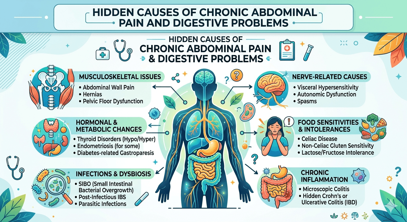
    PCOS Symptoms Every Woman Should Never Ignore

    PCOS

    introduction

    Polycystic Ovary Syndrome (PCOS) is one of the most common hormonal disorders affecting women of reproductive age. It involves a combination of hormonal imbalances, metabolic issues, and problems with ovarian function. Globally, PCOS affects approximately 6–13% of women in their reproductive years, although many cases remain undiagnosed because symptoms can differ greatly from person to person.

    PCOS is not only related to reproductive health. It is also associated with serious metabolic and psychological complications, including insulin resistance, infertility, type 2 diabetes, cardiovascular disease, and mental health disorders such as anxiety and depression.

    This article explains the pathophysiology, causes, symptoms, diagnosis, and treatment strategies for PCOS in a simplified and evidence-based format.

    Understanding the Pathophysiology of PCOS

    PCOS develops due to several interacting factors, including disruptions in hormone regulation, insulin metabolism, and ovarian activity.

    1. Hormonal Imbalance

    Normally, the hypothalamus and pituitary gland regulate ovarian function through the release of hormones. The hypothalamus secretes gonadotropin-releasing hormone (GnRH), which stimulates the pituitary gland to produce:

    • Luteinizing hormone (LH)
    • Follicle-stimulating hormone (FSH)

    In women with PCOS:

    • LH levels are often elevated
    • FSH levels may remain normal or decrease

    This imbalance overstimulates ovarian theca cells, causing excess androgen production, including testosterone. High androgen levels interfere with normal follicle growth and ovulation.

    2. Insulin Resistance

    A large number of women with PCOS experience insulin resistance, where body cells fail to respond properly to insulin. As a result:

    • The pancreas produces excess insulin
    • High insulin levels stimulate increased androgen production
    • Elevated androgens further worsen hormonal imbalance

    This creates a cycle that reinforces both insulin resistance and androgen excess.

    Insulin resistance also increases the likelihood of:

    • Type 2 diabetes
    • Obesity
    • Metabolic syndrome

    3. Ovarian Dysfunction

    Hormonal disturbances affect normal ovarian function In PCOS:

    • Follicles begin developing but do not fully mature
    • Ovulation becomes irregular or may stop completely

    These immature follicles collect around the ovaries and appear as small cyst-like structures on ultrasound imaging. Despite the name “polycystic ovary,” these are actually underdeveloped follicles rather than true cysts.

    4. Chronic Inflammation

    Research suggests that women with PCOS commonly experience low-grade chronic inflammation. Inflammatory processes may contribute to:

    • Increased androgen production
    • Greater insulin resistance

    This further worsens both reproductive and metabolic complications linked to PCOS

    Causes and Risk Factors

    The exact cause of PCOS remains unclear, but several contributing factors have been identified.

    1. Genetics

    Family history plays a significant role. Women with close relatives affected by PCOS are more likely to develop the condition.

    Genes associated with:

    • Insulin regulation
    • Hormone production
    • Ovarian function

    may contribute to susceptibility.

    2. Metabolic Factors

    Insulin resistance is considered one of the major metabolic contributors to PCOS. Risk increases in women with:

    • Obesity
    • Sedentary lifestyle
    • Poor dietary habits
    • Metabolic syndrome

    However, lean women can also develop PCOS, showing that weight alone is not the only factor involved.

    3. Hormonal Disturbances

    Excess androgen production is central to the condition and negatively affects:

    • Ovulation
    • Menstrual regularity
    • Follicular development

    4. Environmental and Lifestyle Influences

    Certain environmental and lifestyle factors may increase the risk of PCOS, including:

    • High-glycemic diets
    • Chronic stress
    • Lack of exercise
    • Exposure to endocrine-disrupting chemicals

    Symptoms of PCOS

    Symptoms vary significantly among women and can range from mild to severe.

    1. Irregular Menstrual Cycles

    Women may experience:

    • Oligomenorrhea (infrequent periods)
    • Amenorrhea (absence of periods)

    Ovulation may occur inconsistently or not at all.

    2. Symptoms of Excess Androgens

    Common androgen-related symptoms include:

    Hirsutism

    • Excess facial hair
    • Hair growth on the chest, abdomen, or back

    Acne

    • Persistent hormonal acne
    • Acne concentrated around the jawline and chin

    Androgenic Alopecia

    • Hair thinning
    • Male-pattern hair loss

    3. Polycystic Ovaries

    Ultrasound examination may reveal enlarged ovaries containing numerous small follicles. However, not every woman with polycystic ovaries has PCOS.

    4. Weight Gain

    Many women with PCOS struggle with:

    • Increased abdominal fat
    • Difficulty losing weight

    5. Fertility Challenges

    Irregular ovulation can make conception difficult. With appropriate treatment and symptom control, many women can successfully become pregnant.

    6. Metabolic Complications

    PCOS increases the risk of:

    • Insulin resistance
    • Type 2 diabetes
    • High cholesterol
    • Hypertension
    • Metabolic syndrome

    7. Mental Health Concerns

    Women with PCOS may also experience:

    • Depression
    • Anxiety
    • Low self-esteem
    • Body image concerns

    Hormonal imbalances and visible symptoms often contribute to emotional distress.

    Diagnosing PCOS

    According to the Rotterdam Criteria, PCOS is diagnosed when at least two of the following are present:

    1. Irregular or absent ovulation
    2. Clinical or laboratory evidence of elevated androgen levels
    3. Multiple ovarian follicles seen on ultrasound

    Doctors must also rule out other conditions with similar symptoms, such as:

    • Thyroid disorders
    • Hyperprolactinemia
    • Congenital adrenal hyperplasia

    Treatment Approaches for PCOS

    Although there is no permanent cure for PCOS, symptoms can often be managed successfully through lifestyle changes and medical treatment.

    1. Lifestyle Modifications

    Lifestyle changes are considered the first-line treatment, especially for women with insulin resistance or obesity.

    Nutrition

    Recommended dietary approaches include:

    • Low-glycemic foods
    • Whole grains
    • Lean proteins
    • Fiber-rich vegetables

    Exercise

    Beneficial forms of physical activity include:

    • Aerobic exercise such as walking or jogging
    • Resistance training such as weightlifting

    Research shows that losing even 5–10% of body weight can improve:

    • Ovulation
    • Insulin sensitivity
    • Menstrual regularity

    2. Medications

    Hormonal Contraceptives

    Birth control pills may help:

    • Regulate menstrual cycles
    • Lower androgen levels
    • Improve acne and excessive hair growth

    Insulin-Sensitizing Medications

    Metformin is commonly prescribed to improve insulin response and may:

    • Promote ovulation
    • Reduce androgen levels
    • Improve metabolic health

    Ovulation-Inducing Medications

    Women trying to conceive may benefit from:

    • Letrozole
    • Clomiphene citrate

    These medications help stimulate ovulation.

    Anti-Androgen Drugs

    Medications such as spironolactone may reduce symptoms like acne and unwanted hair growth. These are usually prescribed alongside contraception because they may affect pregnancy.

    3. Fertility Treatments

    If standard ovulation treatments are unsuccessful, additional fertility options include:

    • Gonadotropin injections
    • In vitro fertilization (IVF)

    These therapies can improve the chances of pregnancy.

    4. Cosmetic Treatments

    Some women use cosmetic procedures to manage physical symptoms, including:

    • Laser hair removal
    • Electrolysis
    • Dermatological acne treatments

    While these methods do not treat the root cause, they can improve confidence and quality of life.

    Long-Term Health Risks

    Without proper management, PCOS may increase the risk of:

    • Type 2 diabetes
    • Cardiovascular disease
    • Endometrial cancer
    • Infertility
    • Sleep apnea

    Early diagnosis and long-term lifestyle management can greatly reduce these complications.

    Conclusion

    Polycystic Ovary Syndrome (PCOS) is a widespread endocrine and metabolic disorder affecting millions of women globally. It develops through a combination of hormonal imbalance, insulin resistance, genetic factors, and ovarian dysfunction. Although there is currently no cure, effective symptom management through lifestyle improvements, medications, and fertility treatments can significantly enhance reproductive and metabolic health. Early awareness, diagnosis, and proper care are essential for improving long-term outcomes and overall well-being.

    Disclaimer: The information provided in this blog post is for educational and informational purposes only and should not be considered medical advice. It is not intended to replace professional medical consultation, diagnosis, or treatment. Always seek the guidance of a qualified healthcare professional regarding any medical condition or health-related concerns. The author and publisher are not responsible for any actions taken based on the information presented in this article.
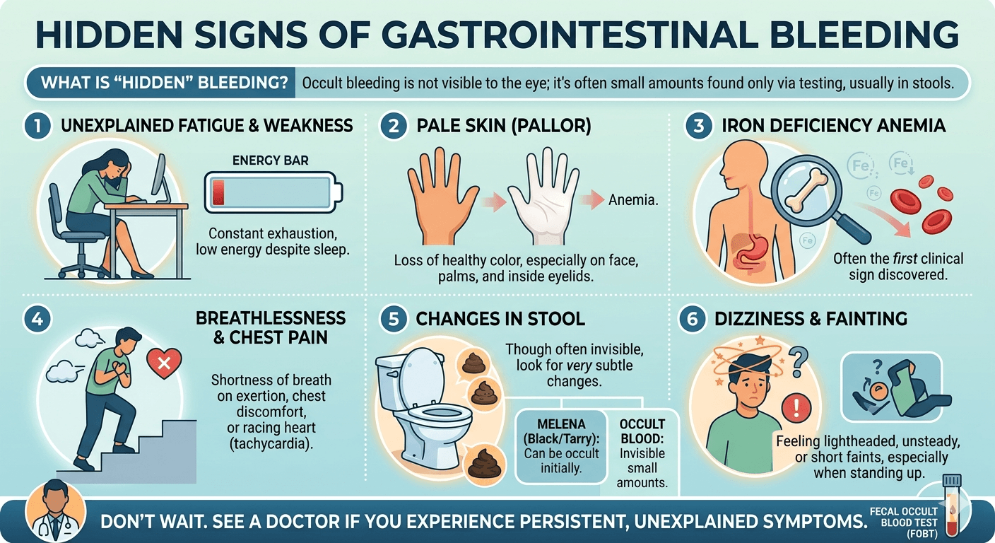
    Acute Kidney Injury

    Acute Kidney Injury

    Complete Guide for Patients and Caregivers

      Introduction

    Acute Kidney Injury (AKI), formerly referred to as acute renal failure, is a rapid decline in kidney function that develops within hours or days. This condition reduces the kidneys’ ability to remove waste products, regulate electrolytes, maintain fluid balance, and preserve acid-base equilibrium. Consequently, substances such as urea and creatinine accumulate in the bloodstream, while sodium, potassium, and fluid imbalances may develop.

    Learn everything about Acute Kidney Injury (AKI), including causes, symptoms, diagnosis, treatment, diet, and recovery tips in this complete guide for patients and caregivers.

    AKI is frequently encountered in hospitalized patients, particularly those in intensive care units. The severity can vary from mild, reversible impairment to life-threatening kidney failure requiring dialysis. Prompt diagnosis and treatment are crucial because untreated AKI may result in severe complications such as pulmonary edema, hyperkalemia, metabolic acidosis, and multiple organ dysfunction.

    The kidneys are essential for filtering blood, producing urine, controlling blood pressure, and stimulating red blood cell production. Therefore, sudden impairment of kidney function can negatively affect nearly every body system.

    Pathophysiology of Acute Kidney Injury

    AKI develops when there is a sudden decrease in the glomerular filtration rate (GFR), causing retention of waste products and disturbances in fluid and electrolyte regulation. AKI is divided into three primary categories according to its underlying mechanism:

    1. Pre renal AKI

    Pre renal AKI occurs when blood flow to the kidneys is reduced without direct kidney tissue damage. Decreased renal perfusion lowers filtration pressure.

    Common causes include:

    • Dehydration
    • Hemorrhage
    • Severe hypotension
    • Heart failure
    • Shock
    • Sepsis

    In response to reduced blood flow, the kidneys activate compensatory systems such as the renin-angiotensin-aldosterone system (RAAS) to retain sodium and water. Persistent hypoperfusion can eventually cause ischemic injury to renal tubules.

    2. Intrinsic (Intra renal) AKI

    Intrinsic AKI results from direct injury to kidney structures including tubules, glomeruli, interstitium, or blood vessels.

    Frequent causes include:

    • Acute tubular necrosis (ATN)
    • Glomerulonephritis
    • Acute interstitial nephritis
    • Nephrotoxic medications
    • Contrast-induced nephropathy

    In ATN, toxins or ischemia damage tubular epithelial cells, leading to tubular obstruction, impaired filtration, and inflammation.

    3. Post renal AKI

    Post renal AKI develops due to obstruction of urine flow anywhere in the urinary tract.

    Common causes include:

    • Kidney stones
    • Enlarged prostate
    • Bladder tumors
    • Urethral obstruction

    Urinary blockage raises pressure inside the kidneys, decreasing glomerular filtration and potentially causing progressive kidney injury if not treated.

    Causes of Acute Kidney Injury

    Pre renal Causes

    • Severe dehydration
    • Vomiting and diarrhea
    • Blood loss
    • Burns
    • Septic shock
    • Heart failure
    • Liver failure
    • Excessive use of diuretics
    • Low blood pressure

    Intrinsic Renal Causes

    • Acute tubular necrosis
    • Glomerulonephritis
    • Acute interstitial nephritis
    • Vasculitis
    • Hemolytic uremic syndrome
    • Rhabdomyolysis
    • Nephrotoxic drugs
    • Imaging contrast dyes

    Post renal Causes

    • Kidney stones
    • Benign prostatic hyperplasia
    • Urinary tract obstruction
    • Bladder cancer
    • Blood clots in the urinary tract
    • Neurogenic bladder

    Risk Factors

    Several conditions increase the likelihood of AKI, including:

    • Advanced age
    • Diabetes mellitus
    • Hypertension
    • Chronic kidney disease
    • Severe infections
    • Major surgical procedures
    • ICU admission
    • Use of nephrotoxic medications
    • Dehydration
    • Heart disease
    • Liver disease

    Signs and Symptoms

    Early Manifestations

    • Reduced urine output (oliguria)
    • Fatigue
    • Weakness
    • Loss of appetite
    • Nausea and vomiting
    • Swelling in the legs and feet
    • Mild confusion

    Progressive Manifestations

    • Shortness of breath caused by fluid overload
    • Chest discomfort
    • Severe hypertension
    • Muscle cramps
    • Persistent vomiting
    • Generalized edema
    • Drowsiness
    • Altered mental state

    Severe Complications

    • Hyperkalemia leading to cardiac arrhythmias
    • Pulmonary edema
    • Metabolic acidosis
    • Uremic encephalopathy
    • Seizures
    • Coma

    Some individuals may initially show no symptoms, and AKI may only be identified through laboratory testing.

    Diagnosis of Acute Kidney Injury

    Medical History and Physical Examination

    The physician evaluates:

    • Fluid balance
    • Medication use
    • Toxin exposure
    • Recent infections
    • Surgical history
    • Urine output

    Physical findings may include dehydration, edema, hypotension, or signs of systemic illness.

    Laboratory Tests

    Kidney function test (KFT)

    HbA1c (for diabetic patients)

    CBC

    ESR, CRP

    LFT with viral markers

    Electrolyte Assessment

    Abnormal levels of potassium, sodium, calcium, phosphate, and bicarbonate may indicate renal dysfunction.

    Urinalysis

    Urine examination may reveal:

    • Proteinuria
    • Hematuria
    • Urinary casts
    • Infection
    • Abnormal urine concentration

    Imaging Studies

    Ultrasound

    Used to identify urinary obstruction, hydronephrosis, or structural abnormalities.

    CT Scan

    Helpful in detecting stones or masses.

    Kidney Biopsy

    Performed in selected patients when the exact cause of intrinsic kidney disease remains uncertain.

    Staging of AKI

    AKI is commonly staged according to KDIGO criteria using serum creatinine levels and urine output.

    Stage 1

    • Mild rise in creatinine
    • Slight decrease in urine output

    Stage 2

    • Moderate increase in creatinine
    • More noticeable reduction in urine production

    Stage 3

    • Severe renal dysfunction
    • Markedly elevated creatinine
    • Possible need for dialysis

    Complications

    Untreated or severe AKI may lead to:

    • Hyperkalemia
    • Pulmonary edema
    • Hypertension
    • Metabolic acidosis
    • Fluid overload
    • Uremia
    • Cardiac arrhythmias
    • Chronic kidney disease
    • End-stage renal disease
    • Multi-organ failure

    Management of Acute Kidney Injury

    1. Treat the Underlying Cause

    Pre renal AKI

    • Intravenous fluid administration
    • Shock correction
    • Blood transfusion when necessary
    • Sepsis management with antibiotics.

    Intrinsic AKI

    • Discontinuation of nephrotoxic medications
    • Treatment of infections
    • Immunosuppressive therapy for Glomerulonephritis when indicated

    Post renal AKI

    • Relief of urinary obstruction
    • Catheter insertion
    • Surgical management
    • Removal of stones

    2. Fluid Management

    Maintaining proper fluid balance is essential.

    In dehydration:

    • IV normal saline may be given.

    In fluid overload:

    • Fluid restriction
    • Diuretics such as furosemide

    Excessive hydration should be avoided because it may worsen pulmonary edema.

    3. Electrolyte Management

    Hyperkalemia Treatment

    • Calcium gluconate
    • Insulin with glucose
    • Sodium bicarbonate
    • Potassium-binding agents
    • Dialysis in severe situations

    Metabolic Acidosis

    • Sodium bicarbonate therapy in selected patients

    4. Medication Adjustment

    Many medications require dose modification in AKI because impaired kidneys cannot effectively eliminate drugs.

    Drugs to avoid include:

    • NSAIDs
    • Certain antibiotics
    • Contrast agents
    • Aminoglycosides

    5. Renal Replacement Therapy (Dialysis)

    Dialysis may be necessary in severe AKI when complications cannot be controlled medically.

    Indications for Dialysis

    • Severe Hyperkalemia
    • Pulmonary edema
    • Severe metabolic acidosis
    • Uremic symptoms
    • Persistent fluid overload

    Diet Plan for AKI

    Nutrition is important for recovery and complication prevention. Diet should be tailored according to:

    • Severity of AKI
    • Electrolyte levels
    • Fluid balance
    • Dialysis status

    Dietary Goals

    • Maintain adequate nutrition
    • Prevent electrolyte disturbances
    • Reduce kidney workload
    • Avoid fluid overload

    Recommended Foods

    Low-Potassium Foods

    • Apples
    • Grapes
    • Berries
    • Cabbage
    • Cauliflower
    • White rice
    • Pasta

    Moderate Protein Sources

    • Egg whites
    • Skinless chicken
    • Fish
    • Tofu

    Low-Sodium Foods

    • Fresh vegetables
    • Homemade meals
    • Unsalted foods

    Adequate Calorie Sources

    • Rice
    • Bread
    • Healthy fats
    • Low-potassium fruits

    Foods to Avoid

    High-Potassium Foods

    • Bananas
    • Oranges
    • Potatoes
    • Tomatoes
    • Spinach
    • Coconut water

    High-Sodium Foods

    • Processed foods
    • Pickles
    • Chips
    • Fast food
    • Canned soups

    Excess Protein

    High protein intake may increase nitrogen waste accumulation.

    Excess Fluids

    Patients with reduced urine output may require fluid restriction.

    Prevention

    Preventive measures include:

    • Maintaining adequate hydration
    • Avoiding unnecessary nephrotoxic medications
    • Proper control of diabetes and hypertension
    • Monitoring kidney function in hospitalized patients
    • Prompt treatment of infections
    • Limiting contrast dye exposure when possible
    • Early detection of urinary obstruction

    Prognosis

    The prognosis of AKI depends on:

    • The underlying cause
    • Severity of injury
    • Patient age
    • Associated comorbidities
    • Timeliness of treatment

    Mild cases may completely recover with appropriate therapy, while severe AKI can progress to chronic kidney disease or permanent renal damage. Mortality is significantly higher among critically ill patients.

    Conclusion

    Acute Kidney Injury is a serious condition characterized by the rapid deterioration of kidney function. It may arise from decreased renal blood flow, direct kidney injury, or urinary tract obstruction. AKI can result in dangerous complications such as electrolyte imbalances, fluid overload, metabolic acidosis, and multi-organ failure.

    Early recognition and prompt treatment are essential for improving outcomes and minimizing complications. Management focuses on correcting the underlying cause, maintaining fluid and electrolyte balance, avoiding nephrotoxic substances, and initiating dialysis when necessary.

    Proper nutrition, hydration, and close medical monitoring are vital for recovery. Increased awareness, preventive strategies, and timely intervention can significantly reduce the burden of AKI and improve patient outcomes.

    Disclaimer: The information provided in this blog post is for educational and informational purposes only and should not be considered medical advice. It is not intended to replace professional medical consultation, diagnosis, or treatment.
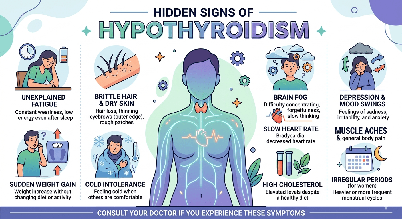
    Right Upper Quadrant Pain:

    right upper quadrant pain

    Causes, Symptoms, and When to Seek Medical Help

    Introduction

    Right upper quadrant (RUQ) pain refers to discomfort felt on the upper right side of the abdomen beneath the ribs. This area contains several vital organs responsible for digestion, metabolism, filtration, and circulation. The pain can vary from mild, short-term discomfort to intense pain that may require immediate medical care.

    Learn about right upper quadrant pain, including its causes, symptoms, diagnosis, treatment options, and when to seek medical attention for RUQ pain.

    The type, intensity, and duration of the pain often help identify its underlying cause. Some conditions, such as indigestion or muscle strain, are relatively minor, while others including gallbladder disorders, liver disease, kidney infections, or appendicitis may be serious. Recognizing related symptoms and possible causes is important for prompt diagnosis and treatment.

    Organs Associated with Right Upper Quadrant Pain

    Several organs are located within or near the RUQ, and pain may arise directly from them or from nearby structures.

    1. Liver

    The liver is the body’s largest internal organ and plays a major role in detoxification, metabolism, bile formation, and nutrient storage. Conditions such as liver inflammation, enlargement, infection, or tumors may trigger RUQ pain.

    2. Gallbladder

    The gallbladder stores bile produced by the liver and releases it during digestion. Gallstones, inflammation, or blockage of bile flow commonly lead to severe RUQ discomfort.

    3. Bile Ducts

    These channels carry bile from the liver and gallbladder to the intestine. Infections or blockages in these ducts may result in pain, fever, and jaundice.

    4. Right Kidney

    Part of the right kidney lies near the RUQ. Kidney infections, stones, or inflammation can cause pain that spreads toward the flank or back.

    5. Pancreas

    Although mainly central in location, the head of the pancreas extends toward the right side. Pancreatic inflammation may produce pain in the upper abdomen and RUQ.

    6. Intestines

    The upper portions of the intestines can also contribute to RUQ pain due to inflammation, infection, gas buildup, or digestive disorders.

    7. Right Lung and Pleura

    Conditions affecting the lower right lung or pleural lining, such as pneumonia or pleuritis, may create pain felt in the RUQ.

    8. Muscles and Ribs

    Injuries or strain involving muscles, ribs, or the chest wall can mimic pain originating from internal organs.

    Mechanisms Behind Right Upper Quadrant Pain

    The development of RUQ pain depends on the affected organ and the disease process involved.

    Inflammation

    Inflammatory conditions stimulate pain receptors and cause tissue swelling. Disorders such as hepatitis, pancreatitis, cholecystitis, and kidney infections commonly produce pain through inflammation.

    Obstruction

    Blockage within ducts or hollow organs raises internal pressure and stretches tissues, resulting in discomfort. Gallstones obstructing the bile duct are a common example.

    Infection

    Viral, bacterial, or parasitic infections trigger immune responses that cause tissue injury, swelling, fever, and pain.

    Ischemia

    Reduced blood supply deprives tissues of oxygen and nutrients, causing damage and pain.

    Muscle Spasm

    Spasms in smooth muscles of the intestines or bile ducts may cause intermittent cramping sensations.

    Capsule Stretching

    Organs such as the liver and kidneys are enclosed in fibrous coverings. Enlargement or swelling stretches these capsules and leads to discomfort.

    Common Causes of Right Upper Quadrant Pain

    1. Gallstones (Cholelithiasis)

    Gallstones are hardened deposits inside the gallbladder that may block bile flow and cause sudden, severe pain known as biliary colic.

    Symptoms

    • Sharp RUQ pain after eating fatty foods
    • Nausea and vomiting
    • Pain spreading to the right shoulder or back

    2. Acute Cholecystitis

    This refers to inflammation of the gallbladder, usually caused by gallstone blockage.

    Symptoms

    • Continuous severe RUQ pain
    • Fever
    • Tenderness over the gallbladder
    • Nausea and vomiting

    3. Hepatitis

    Hepatitis is liver inflammation caused by viruses, alcohol, medications, or autoimmune conditions.

    Symptoms

    • RUQ discomfort
    • Fatigue
    • Reduced appetite
    • Jaundice
    • Dark urine

    4. Fatty Liver Disease

    Excess fat buildup in the liver may enlarge the organ and produce discomfort.

    Risk Factors

    • Obesity
    • Diabetes
    • High cholesterol
    • Heavy alcohol use

    5. Liver Abscess

    A liver abscess is a pus-filled infection within the liver.

    Symptoms

    • Fever and chills
    • Severe RUQ pain
    • Weight loss
    • Weakness

    6. Pancreatitis

    Inflammation of the pancreas may cause upper abdominal pain extending toward the RUQ.

    Causes

    • Gallstones
    • Alcohol misuse
    • Elevated triglycerides

    7. Peptic Ulcer Disease

    Ulcers in the stomach or duodenum can produce pain that resembles RUQ discomfort.

    8. Kidney Stones

    Stones in the right kidney may cause flank pain radiating to the RUQ.

    Symptoms

    • Severe colicky pain
    • Blood in urine
    • Pain during urination

    9. Kidney Infection (Pyelonephritis)

    A bacterial kidney infection can lead to fever, flank pain, and RUQ discomfort.

    10. Pneumonia

    Infection of the lower right lung may irritate the diaphragm and cause referred abdominal pain.

    11. Appendicitis

    Although usually associated with lower abdominal pain, appendicitis may initially present with upper abdominal discomfort.

    12. Musculoskeletal Causes

    Muscle strain, rib fractures, or chest wall inflammation can also produce localized RUQ pain.

    Symptoms Associated with Right Upper Quadrant Pain

    Symptoms differ depending on the underlying condition.

    Common Symptoms

    • Sharp or dull pain in the RUQ
    • Burning or cramping sensation
    • Pain radiating to the shoulder or back
    • Nausea and vomiting
    • Fever
    • Bloating
    • Loss of appetite
    • Fatigue
    • Abdominal tenderness

    Warning Signs Requiring Immediate Attention

    • Jaundice
    • Persistent vomiting
    • High fever
    • Breathing difficulty
    • Black stools
    • Severe abdominal stiffness
    • Fainting or dizziness
    • Confusion
    • Low BP (Hypotension)
    • Rebound tenderness.

    These symptoms may indicate a serious medical emergency.

    Diagnosis of Right Upper Quadrant Pain

    Proper diagnosis requires a detailed medical history and physical examination.

    Medical History

    Doctors typically ask about:

    • Duration and nature of pain
    • Relationship to meals
    • Associated symptoms
    • Alcohol use
    • Medication history
    • Previous illnesses

    Physical Examination

    The physician may assess:

    • Tenderness
    • Enlarged organs
    • Fever
    • Jaundice
    • Abdominal guarding

    Laboratory Tests

    Blood Tests

    • Complete blood count (CBC)
    • Liver function tests (LFTs)
    • Kidney function tests
    • Lipid Profile
    • Pancreatic enzyme levels (S Amylase, S Lipase)
    • Inflammatory markers
    • Blood sugar (RBS)

    Urine Analysis

    Helps identify kidney infections or blood in the urine.

    Imaging Studies

    Ultrasound

    Often the first imaging test used to evaluate gallstones, liver conditions, and bile duct disorders.

    CT Scan

    Provides detailed images of abdominal organs.

    MRI or MRCP

    Useful for examining bile ducts and liver structures.

    Chest X-ray

    May reveal pneumonia or lung-related causes.

    Endoscopy

    Used to detect ulcers or gastrointestinal diseases.

    When to Seek Medical Care

    Medical attention is recommended if RUQ pain:

    • Persists for several hours
    • Becomes severe or progressively worse
    • Occurs with fever
    • Causes jaundice
    • Is associated with vomiting
    • Occurs alongside chest pain or breathing problems
    • Leads to weakness or fainting
    • Involves blood in vomit or stool

    Sudden, intense abdominal pain requires immediate emergency evaluation.

    Treatment of Right Upper Quadrant Pain

    Treatment depends on the underlying cause.

    1. Medications

    Pain Relievers

    Used to control mild to moderate pain.

    Antibiotics

    Prescribed for bacterial infections such as kidney infections or cholecystitis.

    Antiviral Medications

    Helpful in certain viral hepatitis infections.

    Antacids and Proton Pump Inhibitors

    Used for ulcers and acid reflux conditions.

    Antispasmodics

    Help reduce spasms in the bile ducts or intestines.

    2. Dietary Changes

    • Avoid fried and fatty foods
    • Limit alcohol consumption
    • Eat smaller, frequent meals
    • Stay well hydrated
    • Increase fruits and vegetables intake

    3. Surgical Treatment

    Cholecystectomy

    Removal of the gallbladder for gallstones or gallbladder inflammation.

    Drainage of Liver Abscess

    Performed when pus accumulates inside the liver.

    Surgery for Appendicitis or Obstruction

    Needed in emergency situations.

    4. Management of Liver Disorders

    • Weight loss for fatty liver disease
    • Avoiding alcohol
    • Treating hepatitis
    • Regular liver monitoring

    5. Treatment of Kidney Conditions

    • Adequate hydration
    • Antibiotics for infections
    • Removal of kidney stones if required

    Prevention and Precautions

    Many causes of RUQ pain can be reduced through healthy lifestyle habits.

    Healthy Diet

    • Limit oily and processed foods
    • Eat balanced, fiber-rich meals
    • Reduce excess sugar intake

    Maintain a Healthy Weight

    Obesity increases the likelihood of gallstones and fatty liver disease.

    Avoid Excess Alcohol

    Alcohol can damage both the liver and pancreas.

    Stay Hydrated

    Proper hydration supports digestive and kidney health.

    Exercise Regularly

    Physical activity improves metabolism and overall organ function.

    Vaccination

    Vaccines for hepatitis A and B help prevent viral hepatitis.

    Safe Medication Practices

    Avoid unnecessary medications that may harm the liver.

    Routine Medical Checkups

    Early detection of liver disease, gallstones, or metabolic disorders lowers the risk of complications.

    Conclusion

    Right upper quadrant pain is a common symptom with many possible causes involving the liver, gallbladder, kidneys, pancreas, lungs, intestines, or musculoskeletal structures. While some conditions are mild, others can become life-threatening if not treated promptly.

    Recognizing associated symptoms, warning signs, and risk factors is essential for early diagnosis and proper treatment. Healthy lifestyle choices, balanced nutrition, preventive care, and timely medical evaluation all play an important role in minimizing complications and improving overall health outcomes.

    Anyone experiencing persistent or severe RUQ pain should seek professional medical care for accurate diagnosis and treatment.

    Disclaimer: The information provided in this blog post is for educational and informational purposes only and should not be considered medical advice. It is not intended to replace professional medical consultation, diagnosis, or treatment.
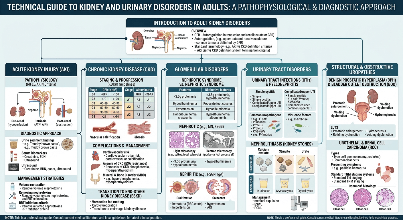
    Pancreatitis: A Complete Medical Guide For Patients And Caregivers

    Understanding Pancreatitis acute and chronic

    Introduction

    Pancreatitis is a condition marked by inflammation of the pancreas, which may occur suddenly as acute pancreatitis or develop gradually as chronic pancreatitis. The pancreas, located behind the stomach, plays a vital role in both digestion and blood sugar regulation. It produces enzymes that aid in digesting fats, proteins, and carbohydrates, while also releasing hormones like insulin and glucagon to control glucose levels.

    This guide explains pancreatitis in detail, including its causes, symptoms, diagnostic methods, treatment approaches, possible complications, and recovery recommendations.

    Pancreatitis develops when digestive enzymes become activated within the pancreas rather than in the small intestine. As a result, the pancreas begins to digest itself, leading to inflammation and tissue damage. The severity may vary from mild inflammation that improves with supportive care to severe disease involving organ failure and systemic complications.

    Acute pancreatitis appears suddenly and often resolves with proper treatment, while chronic pancreatitis causes ongoing inflammation that permanently damages pancreatic tissue and gradually impairs pancreatic function.

    Understanding the disease process, warning signs, causes, and treatment strategies is essential for early diagnosis and prevention of serious complications.

    How Pancreatitis Develops

    The primary problem in pancreatitis is the early activation of digestive enzymes inside pancreatic tissue. Normally, these enzymes remain inactive until they reach the duodenum. In pancreatitis, this protective system fails.

    Enzyme Activation and Self-Digestion

    Activation of trypsin within pancreatic acinar cells triggers a chain reaction of enzyme activation. These enzymes begin damaging pancreatic tissue, leading to:

    • Cellular destruction
    • Fat necrosis
    • Bleeding
    • Swelling
    • Inflammation

    This destructive process is known as pancreatic autodigestion.

    Inflammatory Reaction

    When pancreatic injury occurs, inflammatory chemicals such as cytokines and chemokines are released. These attract immune cells and worsen tissue damage. Severe inflammation can spread throughout the body and lead to systemic inflammatory response syndrome (SIRS).

    Impaired Blood Circulation

    Inflammation increases vascular permeability and swelling, reducing blood flow within the pancreas. Poor circulation may eventually cause tissue death and pancreatic necrosis.

    Systemic Complications

    In severe pancreatitis, inflammatory substances may enter the bloodstream and affect other organs, potentially causing:

    • Acute respiratory distress syndrome (ARDS)
    • Acute kidney injury
    • Shock
    • Sepsis
    • Multi-organ failure

    Chronic Pancreatitis

    Repeated inflammatory episodes can lead to fibrosis, calcification, and permanent destruction of pancreatic tissue. Over time, both digestive and hormonal functions decline, resulting in malabsorption and diabetes.

    Causes of Pancreatitis

    Pancreatitis may develop from several underlying conditions. The causes often differ between acute and chronic disease.

    Common Causes of Acute Pancreatitis

    Gallstones

    Gallstones are one of the most frequent causes. Small stones can block the pancreatic duct or ampulla of Vater, triggering enzyme activation and inflammation.

    Alcohol Use

    Heavy alcohol consumption damages pancreatic cells and thickens pancreatic secretions, contributing to duct blockage and inflammation.

    Hypertriglyceridemia

    Very high triglyceride levels may trigger pancreatitis due to the production of toxic free fatty acids.

    Medications

    Some medications linked to pancreatitis include:

    • Azathioprine
    • Valproic acid
    • Thiazide diuretics
    • Corticosteroids
    • Didanosine

    Infections

    Certain viral infections, including mumps, hepatitis viruses, and cytomegalovirus, can inflame the pancreas.

    Trauma

    Abdominal injuries or surgical procedures may damage pancreatic tissue.

    ERCP Procedures

    Endoscopic Retrograde Cholangiopancreatography (ERCP) is a recognized cause of procedure-related pancreatitis.

    Autoimmune Disorders

    Autoimmune pancreatitis occurs when the immune system attacks pancreatic tissue.

    Metabolic Conditions

    Additional causes include:

    • Hypercalcemia
    • Genetic abnormalities
    • Obesity

    Idiopathic Cases

    In some patients, no specific cause can be identified.

    Causes of Chronic Pancreatitis

    Common causes include:

    • Long-term alcohol abuse
    • Smoking
    • Genetic disorders
    • Autoimmune pancreatitis
    • Repeated acute pancreatitis attacks
    • Obstructive pancreatic duct disease

    Symptoms and Signs of Pancreatitis

    Clinical manifestations vary depending on the severity and duration of inflammation.

    Symptoms of Acute Pancreatitis

    Abdominal Pain

    Severe upper abdominal pain is the most characteristic symptom. The pain often:

    • Radiates to the back
    • Starts suddenly
    • Becomes worse after meals
    • Improves slightly when leaning forward

    Nausea and Vomiting

    Persistent nausea and repeated vomiting are frequently present.

    Fever

    Inflammation or infection may lead to elevated body temperature.

    Abdominal Tenderness

    The abdomen may become swollen and painful on examination.

    Loss of Appetite

    Eating often worsens pain, causing patients to avoid food.

    Tachycardia and Low Blood Pressure

    Severe disease may cause dehydration and circulatory collapse.

    Severe Features

    Serious pancreatitis may present with:

    • Breathing difficulty
    • Cyanosis
    • Confusion
    • Jaundice
    • Multi-organ dysfunction

    Features of Chronic Pancreatitis

    Persistent or Recurrent Pain

    Pain may continue for long periods or occur in repeated episodes.

    Malabsorption

    Reduced digestive enzyme production may cause:

    • Steatorrhea
    • Weight loss
    • Nutritional deficiencies

    Diabetes Mellitus

    Damage to insulin-producing cells can lead to secondary diabetes.

    Pancreatic Calcification

    Calcifications may appear on imaging studies.

    Diagnosis of Pancreatitis

    Diagnosis combines clinical symptoms, laboratory testing, and imaging studies.

    Diagnostic Criteria for Acute Pancreatitis

    A diagnosis is typically confirmed when at least two of the following are present:

    1. Characteristic abdominal pain
    2. Elevated pancreatic enzyme levels
    3. Imaging findings consistent with pancreatitis

    Laboratory Tests

    Important investigations include:

    • Serum amylase and lipase
    • Complete blood count (CBC)
    • Liver function tests
    • Kidney function tests
    • Serum calcium
    • Lipid profile
    • Blood glucose, RBS, and HbA1c
    • C-reactive protein (CRP)
    • ESR

    Lipase is generally more specific and remains elevated longer than amylase.

    Imaging Studies

    Abdominal Ultrasound

    Useful for identifying gallstones and biliary obstruction.

    Contrast-Enhanced CT Scan

    CT imaging helps evaluate:

    • Pancreatic inflammation
    • Necrosis
    • Fluid collections
    • Complications

    MRI and MRCP

    Helpful for examining pancreatic ducts and surrounding soft tissue.

    Endoscopic Ultrasound (EUS)

    Effective for detecting small gallstones and structural abnormalities.

    Severity Assessment

    Several scoring systems help predict disease severity, including:

    • Ranson criteria
    • APACHE II score
    • BISAP score
    • Glasgow score

    Treatment of Pancreatitis

    Management depends on the severity, cause, and associated complications.

    Initial Treatment of Acute Pancreatitis

    Fluid Replacement

    Aggressive intravenous hydration is critical to maintain circulation and prevent complications.

    Pain Relief

    Strong analgesics are commonly needed to control severe abdominal pain.

    Nutritional Support

    Early enteral nutrition is preferred over prolonged fasting. Severe cases may require feeding through a nasojejunal tube.

    Bowel Rest

    Temporary restriction of oral intake may be necessary during severe episodes.

    Oxygen Therapy

    Supplemental oxygen is given if respiratory problems develop.

    Treating Underlying Causes

    Gallstone Pancreatitis

    • ERCP may be performed to relieve biliary obstruction
    • Cholecystectomy helps prevent recurrence

    Alcohol-Induced Pancreatitis

    Complete alcohol avoidance is essential.

    Hypertriglyceridemia

    Management may include:

    • Insulin therapy
    • Lipid-lowering medications
    • Plasmapheresis in severe cases

    Antibiotics

    Preventive antibiotics are not routinely recommended and are reserved for infected necrosis or confirmed infections.

    Managing Complications

    Pancreatic Necrosis

    Severe necrosis may require drainage procedures or surgery.

    Pancreatic Pseudocysts

    Large or symptomatic pseudocysts may need endoscopic or surgical drainage.

    Organ Failure

    Patients with respiratory, renal, or circulatory failure often require intensive care support.

    Treatment of Chronic Pancreatitis

    Lifestyle Changes

    Recommended measures include:

    • Stopping alcohol consumption
    • Quitting smoking
    • Following dietary modifications

    Pancreatic Enzyme Replacement

    Enzyme supplements help improve digestion and reduce steatorrhea.

    Diabetes Treatment

    Insulin therapy may become necessary.

    Pain Management

    Pain control strategies may involve:

    • Analgesic medications
    • Nerve blocks
    • Endoscopic procedures
    • Surgery in selected patients

    Surgical Treatment

    Surgery may be considered for:

    • Duct obstruction
    • Severe chronic pain
    • Complications
    • Suspicion of cancer

    Prognosis

    The outlook depends on disease severity, complications, and underlying causes.

    Acute Pancreatitis

    Most cases are mild and improve completely with supportive care. However, severe disease carries significant risk because of:

    • Pancreatic necrosis
    • Sepsis
    • Multi-organ failure

    Early diagnosis and prompt treatment improve survival and outcomes.

    Chronic Pancreatitis

    Chronic pancreatitis is progressive and irreversible. Long-term complications may include:

    • Persistent pain
    • Malnutrition
    • Diabetes mellitus
    • Pancreatic insufficiency
    • Increased pancreatic cancer risk

    Lifestyle modifications and regular follow-up can improve quality of life.

    Prevention

    Several strategies may reduce the likelihood of pancreatitis.

    Healthy Lifestyle Measures

    • Avoid excessive alcohol use
    • Stop smoking
    • Maintain a healthy weight
    • Follow a balanced low-fat diet

    Managing Metabolic Disorders

    • Control triglyceride levels
    • Treat hypercalcemia
    • Manage diabetes effectively

    Preventing Gallstones

    Early treatment of gallstones can lower the risk of biliary pancreatitis.

    Monitoring Medications

    Patients taking medications associated with pancreatitis should be carefully monitored.

    Conclusion

    Pancreatitis is a potentially serious inflammatory condition affecting the pancreas in either acute or chronic forms. Gallstones and alcohol use remain the most common causes, although infections, metabolic disorders, medications, and autoimmune diseases may also contribute.

    Patients often experience severe abdominal pain, nausea, vomiting, and systemic symptoms. Early diagnosis using laboratory tests and imaging studies is essential for effective treatment and prevention of complications. Supportive care, fluid therapy, nutritional support, and addressing the underlying cause remain the foundation of management.

    Greater awareness, timely intervention, and long-term lifestyle changes can significantly improve outcomes and quality of life for individuals living with pancreatitis.

    Disclaimer: The information provided in this blog post is for educational and informational purposes only and should not be considered medical advice. It is not intended to replace professional medical consultation, diagnosis, or treatment.
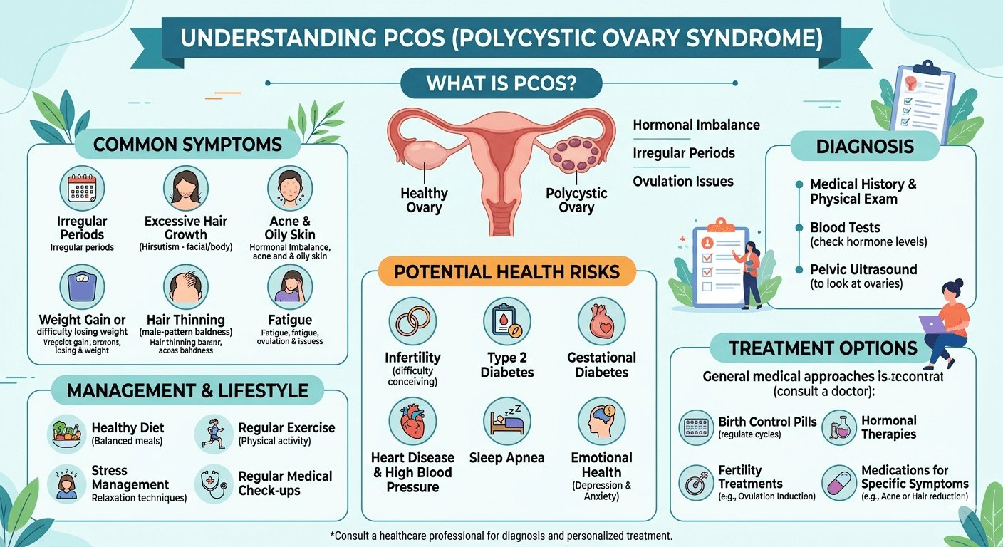
    Inflammatory Bowel Disease A Complete Medical Guide

    Understanding inflammatory bowl disease

    Introduction

    Inflammatory Bowel Disease (IBD) refers to a group of long-term disorders that cause inflammation within the digestive system. The two primary types are Crohn’s disease and ulcerative colitis. These conditions can greatly impact daily life by leading to symptoms such as abdominal discomfort, diarrhea, tiredness, weight reduction, and poor nutrient absorption.

    Learn about Inflammatory Bowel Disease, including its causes, symptoms, diagnosis, complications, and treatment options in this complete medical guide.

    IBD should not be confused with Irritable Bowel Syndrome (IBS). IBS is a functional digestive disorder without intestinal inflammation, whereas IBD involves chronic inflammation that may damage the bowel lining and result in severe complications.

    Although the exact cause remains uncertain, experts believe IBD develops from an abnormal immune reaction influenced by genetics, environmental exposures, and gut microorganisms. The disease commonly alternates between active flare-ups and periods of remission.

    Types of Inflammatory Bowel Disease

    1. Crohn’s Disease

    Crohn’s disease may affect any section of the gastrointestinal tract, from the mouth to the anus. However, it most often occurs in the lower small intestine and the beginning of the large intestine. Inflammation can involve all layers of the bowel wall.

    Main Characteristics

    • Patchy inflamed areas known as “skip lesions”
    • Deep ulcer formation and thickened bowel walls
    • Risk of fistulas, abscesses, and intestinal narrowing
    • Possible effects on the skin, eyes, and joints

    2. Ulcerative Colitis

    Ulcerative colitis primarily affects the colon and rectum. The inflammation usually spreads continuously from the rectum and mainly involves the inner lining of the intestine.

    Main Characteristics

    • Continuous inflammation starting in the rectum
    • Frequent bloody diarrhea
    • Higher long-term risk of colon cancer
    • More superficial inflammation than Crohn’s disease
    Understanding inflammatory bowl disease IBD its Anatomy and Pathology

    Pathophysiology of IBD

    IBD develops through a complicated interaction involving immune dysfunction, intestinal bacteria, inherited factors, and environmental influences.

    1. Immune System Abnormalities

    Normally, the immune system protects against harmful organisms while tolerating healthy gut bacteria. In IBD, the immune system mistakenly attacks intestinal tissue.

    This can result in:

    • Persistent inflammation
    • Increased inflammatory chemicals
    • Ulceration and tissue injury
    • Reduced healing ability

    2. Genetic Predisposition

    Research has identified multiple genes linked to IBD, especially Crohn’s disease. Individuals with affected family members are more likely to develop the condition.

    Genetic changes may:

    • Disrupt immune regulation
    • Damage intestinal barrier function
    • Alter gut bacteria interactions

    3. Gut Microbiome Disturbance

    The intestines contain beneficial microorganisms important for digestion and immunity. In IBD, imbalance in these bacteria may trigger excessive immune activity.

    4. Environmental Influences

    Environmental exposures may activate the disease in genetically susceptible individuals.

    Potential triggers include:

    • Smoking
    • Certain medications
    • Diets high in processed foods
    • Stress
    • Infections
    • Pollution

    5. Intestinal Injury

    Ongoing inflammation may damage the bowel lining and lead to:

    • Ulcers
    • Bleeding
    • Scar tissue
    • Narrowed intestines
    • Poor nutrient absorption
    • Fistulas in Crohn’s disease

    Causes of Inflammatory Bowel Disease

    The precise cause of IBD is unknown, but several contributing factors have been identified.

    1. Genetic Factors

    • Family history of IBD
    • Inherited gene mutations
    • Increased risk among close relatives

    2. Autoimmune Reactions

    The immune system may wrongly attack healthy intestinal tissue, causing chronic inflammation.

    3. Environmental Factors

    • Smoking raises Crohn’s disease risk
    • Urban lifestyles may contribute
    • Diets rich in unhealthy fats and processed foods
    • Pollutant exposure

    4. Microbial Changes

    Alterations in gut bacteria may stimulate abnormal immune responses.

    5. Psychological Stress

    Stress does not directly cause IBD, but it may worsen symptoms and provoke flare-ups.

    Symptoms of Inflammatory Bowel Disease

    Symptoms differ depending on the location and severity of inflammation.

    Digestive Symptoms

    • Chronic diarrhea
    • Abdominal cramps and pain
    • Blood in stool
    • Urgent bowel movements
    • Rectal bleeding
    • Weight loss
    • Reduced appetite
    • Bloating
    • Nausea

    General Symptoms

    • Fatigue
    • Fever
    • Weakness
    • Malnutrition
    • Delayed growth in children

    Effects Outside the Intestines

    Skin

    • Erythema nodosum
    • Pyoderma gangrenosum

    Joints

    • Arthritis
    • Joint swelling and pain

    Eyes

    • Uveitis
    • Episcleritis

    Liver and Bile Ducts

    Diagnosis of IBD

    Diagnosis involves medical evaluation, laboratory testing, imaging, and endoscopic procedures.

    1. Medical History and Examination

    Doctors evaluate:

    • Symptom duration and intensity
    • Family history
    • Weight changes
    • Nutritional condition

    2. Blood Tests

    Blood work may reveal:

    • Anemia
    • Elevated inflammatory markers such as ESR and CRP
    • Electrolyte disturbances
    • Nutritional deficiencies

    3. Stool Testing

    Stool tests help exclude infections and identify signs of intestinal inflammation.

    4. Colonoscopy

    Colonoscopy is a major diagnostic procedure that allows physicians to:

    • Examine intestinal inflammation
    • Detect ulcers and bleeding
    • Collect biopsy samples

    5. Imaging Techniques

    • CT scans
    • MRI enterography
    • Abdominal ultrasound

    These tests help identify complications such as fistulas, abscesses, and narrowed bowel segments.

    6. Biopsy

    Tissue samples taken during endoscopy help confirm the diagnosis and differentiate Crohn’s disease from ulcerative colitis.

    Risk Factors for IBD

    1. Family History

    Having a close relative with IBD significantly increases the likelihood of developing the condition.

    2. Age

    IBD often appears between ages 15 and 35, although it can occur at any stage of life.

    3. Smoking

    Smoking is strongly linked to Crohn’s disease and may worsen its severity.

    4. Diet

    Highly processed foods and unhealthy fats may contribute to inflammation.

    5. Environmental Factors

    • Urban living
    • Pollution
    • Westernized lifestyle

    6. Ethnicity

    Certain ethnic populations experience higher rates of IBD.

    7. Medications

    Some medications associated with increased risk include:

    • Nonsteroidal anti-inflammatory drugs (NSAIDs)
    • Antibiotics
    • Oral contraceptives

    When to Seek Medical Attention

    A doctor should be consulted if any of the following occur:

    • Diarrhea lasting several weeks
    • Blood in stool
    • Severe abdominal pain
    • Unexplained weight loss
    • Fever with digestive symptoms
    • Extreme fatigue
    • Persistent rectal bleeding
    • Difficulty eating due to nausea or pain

    Immediate medical care is necessary for severe dehydration, intestinal blockage, or heavy bleeding.

    Prognosis of IBD

    IBD is a chronic lifelong illness, but many individuals can successfully manage symptoms with proper treatment and healthy lifestyle changes.

    Factors Influencing Prognosis

    • Severity of inflammation
    • Frequency of flare-ups
    • Treatment response
    • Presence of complications
    • Smoking habits

    Potential Complications

    Crohn’s Disease

    • Fistulas
    • Intestinal strictures
    • Abscesses
    • Malnutrition

    Ulcerative Colitis

    • Severe bleeding
    • Toxic megacolon
    • Higher colorectal cancer risk

    Long-Term Outlook

    With early diagnosis and effective treatment:

    • Many patients achieve remission
    • Quality of life improves
    • Complications are reduced
    • Nutritional status can be maintained

    Ongoing medical follow-up and treatment adherence are important for long-term disease management.

    Lifestyle and Dietary Management

    Dietary Tips

    • Eat smaller, frequent meals
    • Maintain good hydration
    • Avoid foods that trigger symptoms
    • Reduce processed food intake
    • Limit dairy if intolerant
    • Choose nutrient-rich foods

    Stress Reduction

    • Meditation
    • Yoga
    • Counseling
    • Adequate sleep

    Smoking Cessation

    Stopping smoking is particularly important for individuals with Crohn’s disease.

    Conclusion

    Inflammatory Bowel Disease is a chronic condition involving inflammation of the digestive tract, mainly in the form of Crohn’s disease and ulcerative colitis. The disorder develops due to a combination of immune abnormalities, genetic factors, environmental triggers, and disturbances in gut bacteria.

    IBD can produce significant digestive symptoms and may also affect other body systems, including the skin, joints, and eyes. Early diagnosis and proper treatment are essential to minimize complications and improve quality of life.

    While there is currently no permanent cure, modern therapies, dietary strategies, and lifestyle modifications allow many people with IBD to maintain long-term remission and live productive lives. Consistent medical care, medication adherence, and healthy daily habits remain essential for successful management.

    Disclaimer: The information provided in this blog post is for educational and informational purposes only and should not be considered medical advice. It is not intended to replace professional medical consultation, diagnosis, or treatment.