
Splenomegaly FAQ: Everything You Need to Know

Understanding splenomegaly its pathogenesis, causes, and consequences

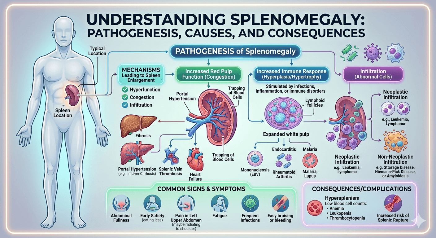
Splenomegaly refers to an abnormal enlargement of the spleen, an organ located in the upper left part of the abdomen that plays a key role in filtering blood and supporting the immune system. It is not a disease itself but a sign of underlying conditions such as infections (like malaria or mononucleosis), liver diseases (such as cirrhosis), blood disorders, or cancers like leukemia and lymphoma. Symptoms may include a feeling of fullness in the left upper abdomen, fatigue, anemia, or frequent infections. Diagnosis typically involves physical examination and imaging tests, while treatment focuses on managing the underlying cause.

1. What is splenomegaly?

Splenomegaly means enlargement of the spleen, an organ located in the upper left side of the abdomen, just below the ribs. Normally, the spleen is about the size of a fist, but in certain diseases it can become significantly larger. An enlarged spleen is not a disease itself  it’s usually a sign of another underlying condition.

2. What does the spleen do in the body?

The spleen has several vital functions:

  • Filters old or damaged blood cells from circulation.
  • Stores blood components, especially platelets and white blood cells.
  • Supports the immune system by producing antibodies and fighting infections.
  • Helps recycle iron from red blood cells.
    Because it is highly vascular (contains lots of blood), the spleen can enlarge when these functions are overactive or disrupted.

3. What are the common causes of splenomegaly?

Splenomegaly can result from a wide range of conditions. The main categories include:

CategoryExamples
InfectionsViral (EBV/mononucleosis, CMV, HIV), Bacterial (endocarditis, brucellosis), Parasitic (malaria, leishmaniasis)
Blood disordersHemolytic anemias, thalassemia, sickle cell disease (early stages)
CancersLymphomas, leukemias, myelofibrosis, chronic myeloid leukemia
Liver and vascular diseasesCirrhosis with portal hypertension, splenic vein thrombosis
Autoimmune or inflammatory diseasesSystemic lupus erythematosus, rheumatoid arthritis (Felty’s syndrome)
Metabolic or storage disordersGaucher disease, Niemann–Pick, Amyloidosis

4. What symptoms can splenomegaly cause?

Many people have no symptoms until the spleen becomes very large. Common symptoms include:

  • A feeling of fullness or discomfort in the upper left abdomen.
  • Early satiety (feeling full quickly) due to stomach compression.
  • Pain in the left shoulder or upper abdomen.
  • Fatigue, pallor, or frequent infections (from anemia or low white cell counts).
  • Easy bruising or bleeding (from low platelets).

5. How is splenomegaly diagnosed?

Diagnosis usually starts with a clinical examination and is confirmed with tests such as:

  • Physical exam: Palpation of the spleen below the left rib cage.
  • Ultrasound or CT scan: Confirms size and appearance of the spleen.
  • Blood tests (CBC, liver tests): To check for anemia, infection, or blood cell abnormalities.
  • Bone marrow or serologic tests: If cancer, infection, or autoimmune causes are suspected.

6. What is hypersplenism, and how is it related?

Hypersplenism refers to an overactive spleen that destroys blood cells too rapidly. It often occurs in chronic splenomegaly. The result can be:

  • Anemia (low red cells)
  • Leucopenia (low white cells)
  • Thrombocytopenia (low platelets)
    Treatment focuses on managing the underlying cause, and in severe cases, partial or total splenectomy may be required.

7. Is splenomegaly dangerous?

An enlarged spleen can be dangerous because:

  • It can rupture easily, especially after trauma, causing internal bleeding.
  • It may trap and destroy blood cells, leading to low counts and complications.
  • The underlying disease (infection, cancer, liver disease) can be serious.
    Patients with splenomegaly should avoid contact sports or heavy trauma until the cause is identified and treated.

8. How is splenomegaly treated?

Treatment depends on the underlying cause, not the size of the spleen itself. Examples include:

  • Infections: Appropriate antibiotics, antivirals, or antiparasitic therapy.
  • Autoimmune diseases: Corticosteroids or immunosuppressants.
  • Blood cancers: Chemotherapy, targeted therapy, or JAK inhibitors (for myelofibrosis).
  • Portal hypertension: Management of liver disease and control of varices.
  • Surgical option (splenectomy): For trauma, severe hypersplenism, or specific hematologic disorders (e.g., hereditary spherocytosis, refractory immune thrombocytopenia).

9. When is splenectomy (spleen removal) needed?

Splenectomy is recommended when:

  • The spleen is causing severe pain, early satiety, or compression symptoms.
  • Cytopenias (low blood counts) due to hypersplenism are severe or unresponsive to medical therapy.
  • Certain diseases (e.g., hereditary spherocytosis, ITP, lymphoma) require removal for cure or control.
    However, splenectomy increases infection risk, so it’s only done when clearly indicated.

10. What are the risks after splenectomy?

Without a spleen, the body becomes more susceptible to infections by encapsulated bacteria such as:

  • Streptococcus pneumoniae
  • Haemophilus influenzae type b
  • Neisseria meningitidis
    This can lead to overwhelming post-splenectomy infection (OPSI), which is rare but life-threatening. Preventive strategies include vaccination and prompt treatment of any fever.

11. What vaccines are needed after spleen removal or dysfunction?

Vaccinations should be given before or shortly after splenectomy, and repeated as needed:

  1. Pneumococcal vaccines  PCV13/15/20 and PPSV23 boosters.
  2. Meningococcal vaccines  both ACWY and B types.
  3. Haemophilus influenzae type b (Hib) vaccine.
  4. Annual influenza vaccine.
    These vaccines significantly reduce infection risk in asplenic or hyposplenic patients.

12. What precautions should I take if I have an enlarged spleen?

  • Avoid contact sports or heavy lifting to prevent rupture.
  • Seek medical advice for any fever, sore throat, or infection symptoms.
  • Stay up to date with vaccinations.
  • Inform healthcare providers about your splenic condition before surgeries or medical treatments.
  • Wear a medical alert bracelet if you have no spleen or functional asplenia.

13. Can the spleen return to normal size?

Yes   if the underlying condition is successfully treated (e.g., infection cleared, inflammation resolved, blood disorder managed), the spleen may shrink back to its normal size. However, in chronic or infiltrative diseases (like myelofibrosis or storage disorders), enlargement may persist.

14. What happens if splenomegaly is left untreated?

Ignoring splenomegaly can lead to serious complications:

  • Splenic rupture (medical emergency with internal bleeding).
  • Severe anemia or thrombocytopenia (risk of bleeding or infection).
  • Progression of the underlying disease (cancer, infection, liver failure).
    Early diagnosis and management are essential to prevent long-term complications.

15. Which doctor should I see for splenomegaly?

Initial evaluation can be done by a primary care physician, but depending on the cause, you may be referred to:

  • Hematologist – for blood disorders or malignancies.
  • Gastroenterologist / Hepatologist  for liver disease and portal hypertension.
  • Infectious disease specialist  for parasitic or chronic infections.
  • Surgeon  if splenectomy or biopsy is required.

16. What is the outlook for people with splenomegaly?

The prognosis depends entirely on the underlying cause:

  • Infectious causes (like mononucleosis or malaria) often resolve completely.
  • Chronic hematologic or neoplastic causes require ongoing management.
  • Patients without a spleen can live normal, healthy lives with proper vaccinations and preventive care.

Key Takeaway

Splenomegaly is a symptom of an underlying condition, not a standalone disease.
Early evaluation, accurate diagnosis, and preventive care  especially against infections are essential for long-term health and safety.

Disclaimer: The information provided in this blog post is for educational and informational purposes only and should not be considered medical advice.
Why Does Splenomegaly Occur in Cirrhosis?

Enlarged spleen in liver disease

(Pathophysiology, Causes, Symptoms, and Management Explained)

Introduction

Splenomegaly, or enlargement of the spleen, is a common clinical finding in patients with cirrhosis. It is not just an incidental feature but an important marker of disease progression and portal hypertension. Understanding why splenomegaly occurs in cirrhosis helps clinicians and patients better manage complications and improve outcomes.

What is Splenomegaly?

Splenomegaly is a medical term for an enlarged spleen, an organ located in the upper left side of the abdomen. The spleen plays a key role in filtering blood, removing old or damaged blood cells, and supporting the immune system. When it becomes enlarged, it is usually a sign of an underlying condition such as infections, liver disease (like cirrhosis), blood disorders, or inflammatory diseases. Splenomegaly may be asymptomatic in early stages, but larger enlargement can cause abdominal discomfort, early satiety, and low blood cell counts due to increased sequestration within the spleen.

What is Cirrhosis?

Cirrhosis is a chronic liver disease in which normal liver tissue is gradually replaced by scar tissue (fibrosis), leading to loss of liver function. This scarring disrupts blood flow through the liver and impairs its ability to perform vital tasks such as detoxification, protein synthesis, and bile production. Common causes include long-term alcohol use, chronic viral hepatitis (B and C), and non-alcoholic fatty liver disease. Early stages may have few symptoms, but advanced cirrhosis can lead to complications like jaundice, ascites, portal hypertension, and organ failure.

Pathophysiology: Why Does Splenomegaly Occur in Cirrhosis?

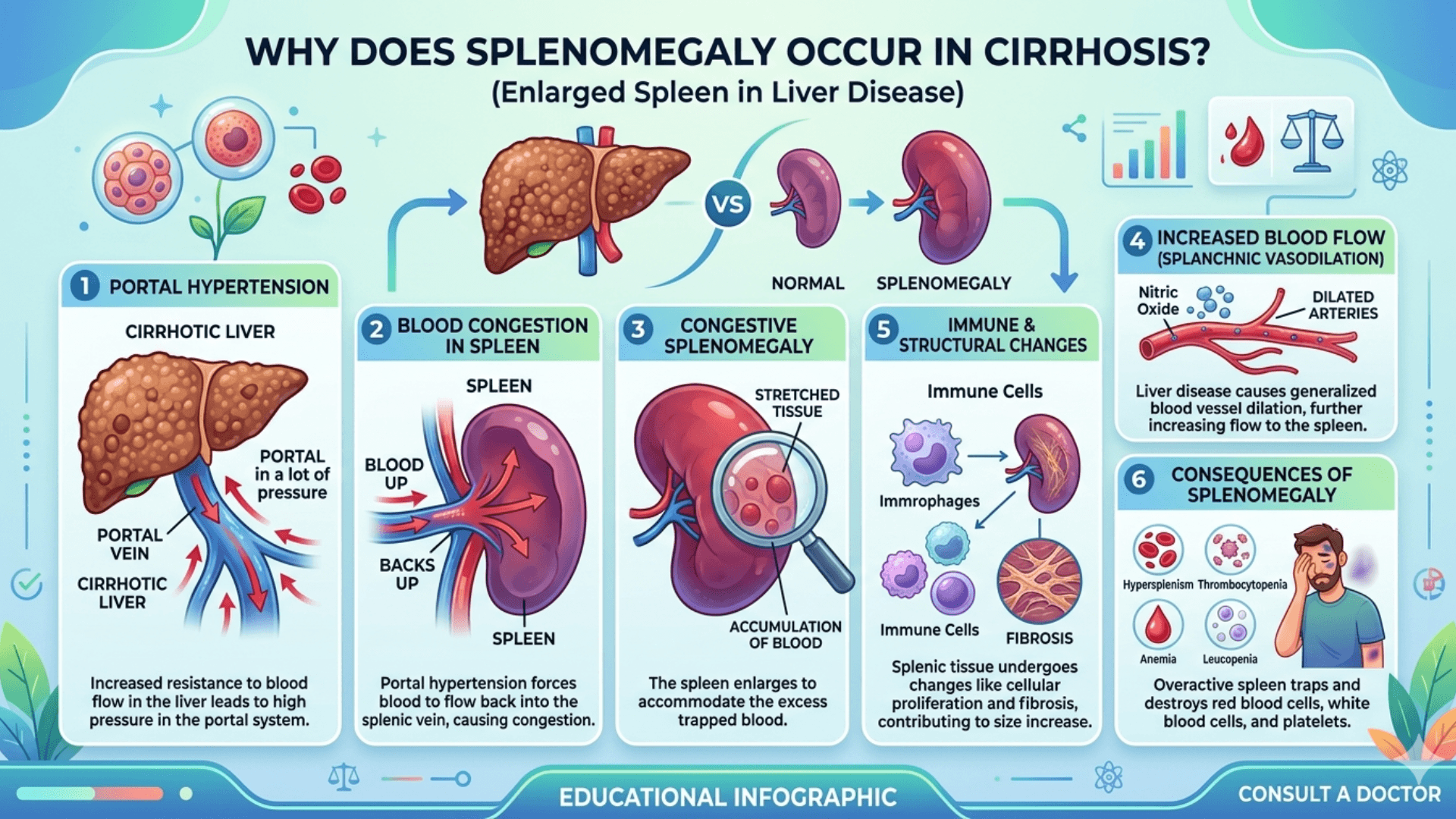
1. Portal Hypertension (Primary Mechanism)

The most important cause of splenomegaly in cirrhosis is portal hypertension.

  • Cirrhosis causes scarring and fibrosis of liver tissue
  • This increases resistance to portal blood flow
  • Pressure builds up in the portal vein
  • Blood backs up into the spleen via the splenic vein

 This leads to congestive splenomegaly

2. Splenic Congestion and Sequestration

Due to increased pressure:

  • The spleen becomes engorged with blood
  • It traps (sequesters) blood cells like platelets, RBCs, and WBCs
  • This results in hypersplenism

3. Hypersplenism

Hypersplenism leads to:

  • Thrombocytopenia (low platelets)
  • Leucopenia (low white blood cells)
  • Anemia

This is a hallmark feature of cirrhosis-associated splenomegaly.

4. Increased Reticuloendothelial Activity

The spleen is part of the immune system. In cirrhosis:

  • Chronic inflammation stimulates splenic immune function
  • This contributes further to splenic enlargement

Causes of Splenomegaly in Cirrhosis

Although portal hypertension is the main cause, underlying etiologies of cirrhosis also contribute:

Common Causes of Cirrhosis Leading to Splenomegaly

  • Chronic alcohol use
  • Chronic viral hepatitis (Hepatitis B and C)
  • Non-alcoholic fatty liver disease (NAFLD)
  • Autoimmune hepatitis
  • Biliary cirrhosis

Additional Contributing Factors

  • Portal vein thrombosis
  • Increased splenic blood flow
  • Collateral circulation formation

Signs and Symptoms of Splenomegaly in Cirrhosis

“Key symptoms include abdominal fullness, early satiety, fatigue, anemia, and increased bleeding risk.”

Often Asymptomatic Initially

Many patients do not notice symptoms early on.

Common Symptoms

  • Fullness or discomfort in the left upper abdomen
  • Early satiety (feeling full quickly)
  • Fatigue (due to anemia)

Clinical Signs

  • Palpable enlarged spleen
  • Thrombocytopenia (easy bruising, bleeding)
  • Leucopenia (increased infections)
  • Anemia (weakness, pallor)

Associated Features of Cirrhosis

  • Ascites
  • Jaundice
  • Spider angiomas
  • Esophageal varices

Diagnosis

Clinical Examination

  • Palpation of spleen below left costal margin

Laboratory Findings

  • Low platelet count (often the earliest sign)
  • Pancytopenia in advanced cases

Imaging Studies

  • Ultrasound abdomen (first-line)
  • CT scan or MRI (for detailed evaluation)

Additional Tests

  • Liver function tests
  • Endoscopy (to detect varices)

Line of Management

Management of portal hypertension in cirrhosis focuses on reducing elevated pressure in the portal venous system and preventing life-threatening complications like variceal bleeding. First-line treatment includes non-selective beta-blockers (such as propranolol) to lower portal pressure. Endoscopic therapies, especially variceal ligation, are used to prevent or control bleeding. In more severe cases, procedures like TIPS (Transjugular Intrahepatic Portosystemic Shunt) help divert blood flow and reduce pressure. Alongside these, managing the underlying liver disease, avoiding alcohol, and monitoring for complications like ascites are essential parts of care.

“Management focuses on treating cirrhosis, reducing portal pressure, and managing complications.”

1. Treat Underlying Cirrhosis

  • Alcohol cessation
  • Antiviral therapy for hepatitis
  • Weight loss and lifestyle changes for NAFLD
  • Immunosuppressive therapy for autoimmune causes

2. Management of Portal Hypertension

  • Non-selective beta-blockers (e.g., propranolol)
  • Endoscopic variceal ligation
  • TIPS (Transjugular Intrahepatic Portosystemic Shunt) in severe cases

3. Management of Hypersplenism

  • Monitoring blood counts
  • Platelet transfusion (if severe thrombocytopenia)
  • Growth factors in select cases

4. Splenectomy (Rare Cases)

  • Considered only in severe hypersplenism
  • Risky in cirrhotic patients
  • Reserved for selected cases

5. Liver Transplantation

  • Definitive treatment for advanced cirrhosis
  • Resolves portal hypertension and splenomegaly

Complications of Splenomegaly in Cirrhosis

  • Severe thrombocytopenia  bleeding risk
  • Increased infection risk
  • Worsening anemia
  • Portal hypertension complications

Summary

Splenomegaly in cirrhosis occurs mainly due to portal hypertension, a condition where scarring of the liver increases resistance to blood flow. As pressure builds in the portal vein, blood backs up into the spleen through the splenic vein, causing congestion and enlargement. Over time, the enlarged spleen begins to trap and destroy blood cells, leading to hypersplenism, which results in low platelet count, anemia, and reduced white blood cells. This makes splenomegaly an important clinical indicator of advanced liver disease and portal hypertension.

Disclaimer: The information provided in this blog post is for educational and informational purposes only and should not be considered medical advice. It is not intended to replace professional medical consultation, diagnosis, or treatment. Always seek the guidance of a qualified healthcare professional regarding any medical condition or health-related concerns. The author and publisher are not responsible for any actions taken based on the information presented in this article.
Early Warning Signs of Metabolic Dysfunction

Early Warning Signs of metabolic dysfunction

Discover the early warning signs of metabolic dysfunction, including fatigue, weight gain, blood sugar imbalance, and insulin resistance. Learn early warning symptoms and ways to improve metabolic health naturally.

Liver disease often develops quietly, especially when it is related to metabolic dysfunction. Metabolic Dysfunction–Associated Steatohepatitis (MASH) is an advanced stage of Metabolic Dysfunction–Associated Steatotic Liver Disease (MASLD), a condition in which excess fat accumulates in the liver.

In MASH, fat accumulation triggers inflammation and liver cell injury. Over time, this ongoing damage can lead to fibrosis, Cirrhosis, or even Hepatocellular Carcinoma.

What makes MASH particularly concerning is that many early symptoms are subtle and easy to dismiss. Recognizing these warning signs can help individuals seek medical attention before serious liver damage occurs.

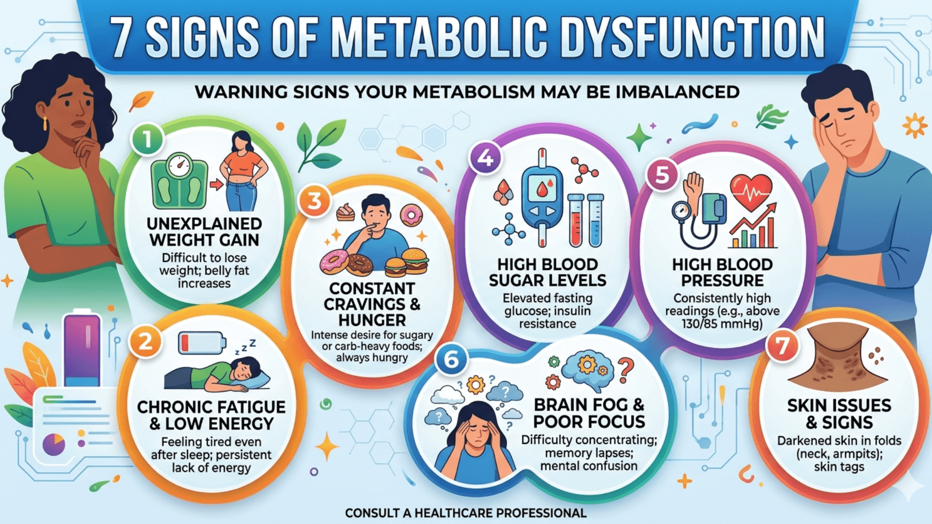
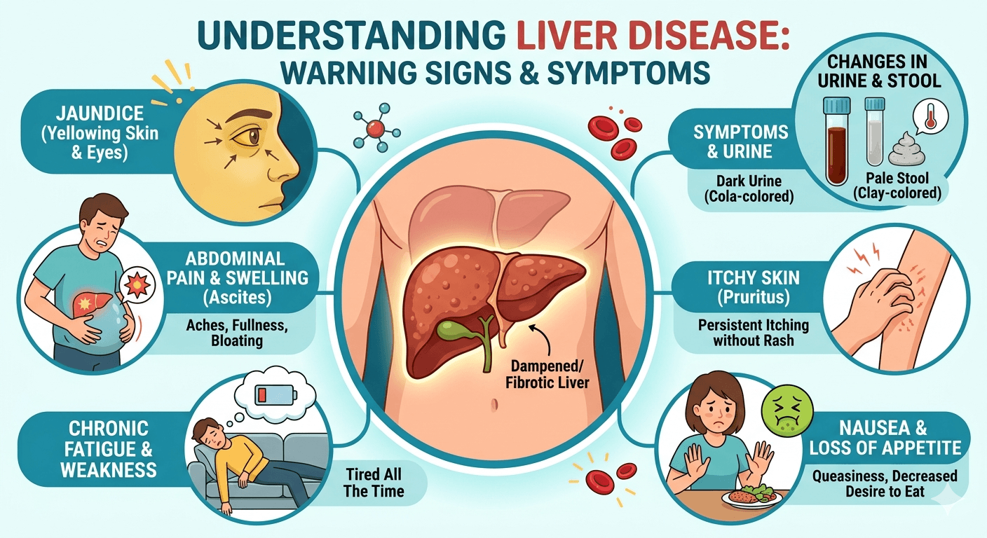
1. Persistent Fatigue

One of the most frequently reported symptoms of MASH is unexplained fatigue.

People often describe feeling tired throughout the day, even after adequate sleep. This occurs because liver inflammation disrupts normal metabolism and energy regulation. The liver plays a critical role in processing nutrients and producing energy, so when its function is impaired, fatigue can become persistent.

Because tiredness is common in modern lifestyles, many people overlook this sign.

2. Discomfort in the Upper Right Abdomen

The liver is located in the upper right part of the abdomen, just beneath the rib cage. When inflammation develops, the liver may enlarge slightly.

This can cause:

  • A dull ache below the right ribs
  • A feeling of pressure or fullness
  • Mild pain that worsens after large meals

Although the discomfort is usually mild, persistent pain in this area may indicate underlying liver inflammation.

3. Gradual Abdominal Weight Gain

MASH is closely linked to metabolic disorders such as Obesity, Type 2 Diabetes, and Metabolic Syndrome.

Many people with metabolic liver disease develop central obesity, meaning fat accumulates primarily around the abdomen.

Excess abdominal fat contributes to insulin resistance, which increases the delivery of fatty acids to the liver and accelerates liver fat accumulation.

4. Yellowing of the Skin or Eyes (Jaundice)

As liver function declines, the body may struggle to process bilirubin, a pigment produced during the breakdown of red blood cells.

When bilirubin accumulates in the bloodstream, it causes jaundice, which appears as:

  • Yellowing of the skin
  • Yellow discoloration of the eyes
  • Dark urine or pale stools

Jaundice is often a sign of more advanced liver dysfunction and requires prompt medical evaluation.

5. Swelling in the Abdomen or Legs

Swelling in the abdomen or legs is a sign of advanced liver involvement. Abdominal swelling, known as Ascites, occurs when fluid builds up in the abdominal cavity. Swelling in the legs and feet, called Peripheral Edema, results from fluid accumulation in the tissues.

These symptoms develop due to portal hypertension (increased pressure in liver blood vessels) and reduced production of albumin, a protein that helps keep fluid within blood vessels. As liver function declines, fluid leaks into surrounding tissues, leading to visible swelling.

Patients may notice

  • Tight or distended abdomen
  • Rapid weight gain
  • Puffiness in the ankles or legs

especially by the end of the day.

6. Easy Bruising or Bleeding

The liver produces many proteins involved in blood clotting. When liver function declines, the production of these clotting factors decreases.

As a result, people with MASH may notice:

  • Frequent bruising
  • Bleeding that takes longer to stop
  • Occasional nosebleeds

These symptoms suggest impaired liver function and should not be ignored.

7. Brain Fog and Difficulty Concentrating

In Metabolic Dysfunction-Associated Steatotic Liver Disease, brain fog and difficulty concentrating are increasingly recognized as subtle but impactful symptoms. Patients may experience

  • mental fatigue
  • poor focus
  • forgetfulness
  • slowed thinking

even in early stages of the disease.

These cognitive changes are thought to result from chronic inflammation, insulin resistance, and disruptions in the gut–liver–brain axis. Accumulation of toxins and altered metabolism can affect neurotransmitter balance and brain function, leading to reduced mental clarity.

Severe cases may progress to Hepatic Encephalopathy, a neurological complication of advanced liver disease.

When to See a Doctor

Many people with metabolic liver disease do not experience noticeable symptoms until the condition has progressed. For this reason, individuals with metabolic risk factors should consider regular screening.

Medical evaluation may include:

  • Liver enzyme blood tests
  • Ultrasound or liver elastography
  • Additional imaging or biopsy if necessary

Early detection can help slow or even reverse liver damage.

Final Thoughts

MASH is a progressive condition, but it often develops silently. Symptoms such as fatigue, abdominal discomfort, unexplained weight gain, and jaundice may seem minor at first, yet they can signal underlying liver inflammation.

Recognizing these warning signs and addressing metabolic risk factors early can significantly reduce the risk of serious complications such as cirrhosis or liver cancer.

Disclaimer: The information provided in this blog post is for educational and informational purposes only and should not be considered medical advice. It is not intended to replace professional medical consultation, diagnosis, or treatment. Always seek the guidance of a qualified healthcare professional regarding any medical condition or health-related concerns. The author and publisher are not responsible for any actions taken based on the information presented in this article.
Fatty Liver Disease

Understanding liver disease, its warning signs and symptoms

Fatty Liver Disease

Fatty liver disease, also called hepatic steatosis, results when the liver accumulates too much fat. Although the liver should normally have small amounts of fat, too much fat accumulation may result in inflammation, damage to the liver, and even liver failure in advanced conditions. Fatty liver has become widespread globally, particularly in industrialized nations, and has close associations with contemporary lifestyle and diet.

Learn about fatty liver disease, including its causes, symptoms, diagnosis, treatment, and prevention. Discover early warning signs and effective lifestyle changes to protect your liver health.

There are two primary forms of fatty liver disease:

Non-Alcoholic Fatty Liver Disease (NAFLD)

Alcoholic Fatty Liver Disease (AFLD)

Even though both consist of fat deposition in the liver, their causes are quite different.

Causes of Non-Alcoholic Fatty Liver Disease (NAFLD)

NAFLD is the most prevalent condition of fatty liver disease and affects people who drink little or no alcohol. The progression of NAFLD is closely linked with metabolic health and lifestyle factors.

Chief Causes and Risk Factors:

 Obesity

Increased body fat, particularly visceral fat in the abdominal area, leads to the deposition of fat in the liver. Research indicates that as many as 90% of obese individuals have some degree of fatty liver.

 Type 2 Diabetes and Insulin Resistance

The insulin resistance results in high blood sugar and fat, favoring the storage of fat in the liver. Most individuals with type 2 diabetes also have NAFLD.

 High Cholesterol and Triglycerides

Dyslipidemia (abnormal lipid content in the blood) results in fat buildup in organs, including the liver.

 Poor Diet

Diets rich in refined carbohydrates, sweetened beverages, saturated fats, and highly processed foods are leading contributors to fat accumulation in the liver.

 Sedentary Lifestyle

Inactivity decelerates metabolism, which leads to weight gain and insulin resistance, both of which have been associated with fatty liver.

 Polycystic Ovary Syndrome (PCOS)

Women with PCOS tend to have insulin resistance and are more likely to have NAFLD.

 Sleep Apnea

This condition is associated with decreased oxygen levels in sleep, which can exacerbate fat accumulation and liver inflammation.

Causes of Alcoholic Fatty Liver Disease (AFLD)

AFLD is causally linked to excess and prolonged alcohol use. Alcohol is metabolized in the liver, and excessive consumption interferes with normal fat metabolism, resulting in fat accumulation.

Key Contributing Factors:

 Chronic Alcohol Use

Recurrent heavy alcohol use compromises the liver’s capacity to metabolize fats, resulting in their accumulation.

 Genetic Susceptibility

Genetic predisposition to liver injury from alcohol varies among individuals.

 Malnutrition

Alcoholics tend to have poor diets, which can worsen liver injury and compromise fat metabolism.

 Other Less Usual Causes of Fatty Liver

Starvation or rapid weight loss

Some medications (e.g., Tamoxifen, Methotrexate, corticosteroids)

Viral hepatitis

Genetic metabolic disorders (e.g., Wilson’s disease)

Conclusion

Fatty liver disease is an increasing global health issue, frequently progressing without symptom awareness. Awareness of the underlying causes specifically modifiable lifestyle habits such as diet, weight, and alcohol use is essential for prevention and treatment. Intervention with healthy lifestyle modifications early in the course of fatty liver often results in its reversal and avoids complications like cirrhosis, liver failure, or liver cancer.

Major Strategies to manage Fatty Liver Disease

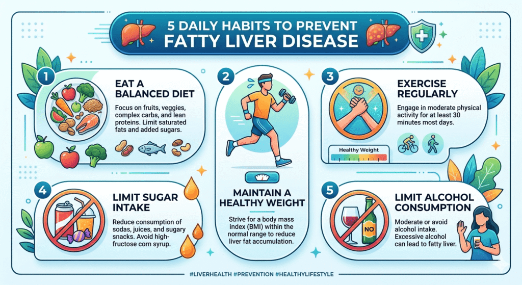
Daily Habits to Prevent Fatty Liver Disease

1. Lifestyle Changes

a. Weight Reduction.

Goal: 5–10% of body weight decrease

Even 5% body weight loss decreases liver fat.

10% loss can reverse inflammation and fibrosis in established disease.

b. Balanced Diet.

Emphasize:

Fresh fruits and vegetables

Whole grains

 Lean proteins (e.g., fish, chicken, legumes)

Healthy fats (e.g., nuts, olive oil, avocado)

Avoid:

 Sugar-sweetened beverages (e.g., soda, juice)

Refined carbohydrates (e.g., white bread, pastries)

Fried and processed foods

High fructose corn syrup (present in most packaged foods)

c. Regular Physical Activity.

Target: A minimum of 150 minutes of weekly moderate-intensity aerobic physical activity (e.g., brisk walking, swimming)

Regular strength training 2–3 times a week also enhances insulin sensitivity.

 Alcohol Management

AFLD: Total abstinence is required to avoid additional liver damage.

NAFLD: Even modest use of alcohol can exacerbate liver disease; abstinence or very strong restriction is advised.

 Control of Blood Sugar and Cholesterol

Control diabetes or prediabetes by diet, medications, and exercise.

Manage cholesterol and triglyceride levels with diet and medication if necessary.

 Medications (Only as Prescribed)

 Vitamin E: Potentially decreases inflammation in the liver in non-diabetics with NASH.

 Statins: May safely be used in most individuals with fatty liver to control cholesterol.

 Important: Always consult a medical professional before taking supplements or medication for fatty liver.

 Regular Monitoring and Medical Care

Liver function tests and ultrasound tests can be performed periodically to monitor progress.

FibroScan or liver biopsy can be necessary in advanced or doubtful cases.

 Treatment of Underlying Conditions

Hypothyroidism, sleep apnea, or polycystic ovary syndrome (PCOS) are conditions that can lead to fatty liver and must be treated accordingly.

Can Fatty Liver Be Reversed ?

Yes especially in the initial period, fatty liver can generally be reversed by losing weight, healthy diet, and regular exercise. Even late-stage cases may be stabilized or improved with proper care.